
Anatomy and physiology of nose
anatomy of nose nasal cavity diagram
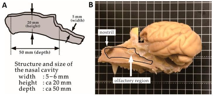
This composite educational image illustrates the comparative anatomy and morphometry of the primate nasal cavity and its spatial relationship to the brain, using a cynomolgus monkey model. Panel A is a schematic diagram detailing the dimensions of the nasal cavity, specifying a width of 5-6 mm, a height of approximately 20 mm, and a depth of approximately 50 mm. Panel B shows a gross anatomical sagittal cross-section of a monkey skull on a grid background. The nasal cavity is outlined in black, with the nostril labeled at the anterior aspect. A white dotted circle identifies the olfactory region, located approximately 40 mm posterior to the nostril near the cribriform plate. The brain is positioned superior and slightly posterior to the nasal cavity, demonstrating the proximity of the olfactory bulb to the nasal mucosa. This visualization is clinically relevant for studying direct nose-to-brain (DNTB) drug delivery pathways and understanding the anatomical barriers and distances involved in targeting the olfactory region.
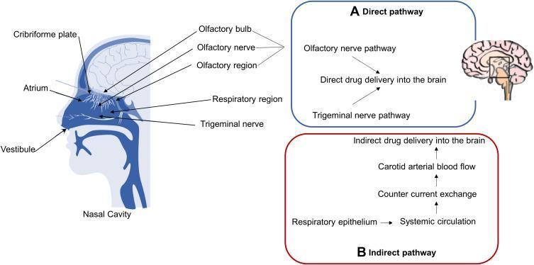
An anatomical and pathophysiological diagram illustrating the nose-to-brain drug delivery pathways. The left side features a sagittal cross-section of the human head, specifically detailing the nasal cavity anatomy. Labeled landmarks include the vestibule, atrium, and respiratory region, as well as the superior olfactory region adjacent to the cribriform plate and olfactory bulb. The diagram highlights two distinct mechanisms of drug transport to the central nervous system (CNS). (A) The 'Direct pathway' (blue box) shows intranasal drugs bypassing the blood-brain barrier (BBB) via the olfactory and trigeminal nerve pathways. (B) The 'Indirect pathway' (red box) describes systemic absorption through the respiratory epithelium into the circulation, involving countercurrent exchange and carotid arterial blood flow to reach the brain. This schematic is designed for pharmacology and neurology education, focusing on therapeutic delivery strategies for neurodegenerative conditions like Alzheimer's disease.
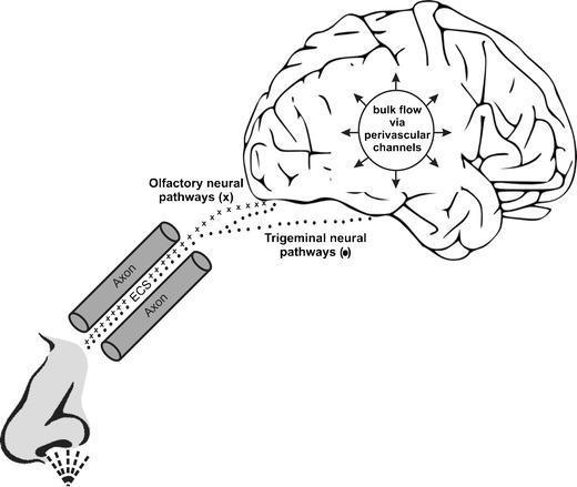
A pathophysiology diagram illustrating the pathways for nose-to-brain drug delivery. The schematic shows a human nasal cavity at the base, leading to two primary neural pathways: the olfactory and trigeminal nerves. The diagram highlights both intracellular transport via axons and extracellular transport through the extracellular space (ECS) between axons. The olfactory neural pathways (marked with 'x') are shown entering the anterior brain region. In contrast, the trigeminal neural pathways (marked with dots) provide broader access, entering both anterior and posterior regions, including the pons and cribriform plate areas. Once inside the central nervous system, a central circular annotation indicates the distribution of substances through 'bulk flow via perivascular channels,' characterized by outward-pointing arrows. This illustration serves as an educational model for neuro-pharmacology and intranasal therapeutics, emphasizing the mechanisms by which CNS-targeted drugs bypass the blood-brain barrier via cranial nerve pathways.
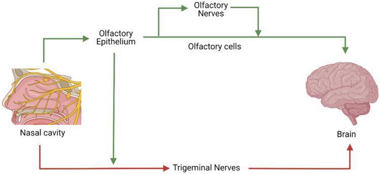
This medical schematic illustrates the anatomical and functional pathways for nose-to-brain (N-to-B) transport, specifically highlighting direct neural routes that bypass the blood-brain barrier. The diagram features a sagittal anatomical illustration of the human nasal cavity on the left and a representation of the brain on the right, connected by two primary signaling pathways. 1. Olfactory Pathway (Green): This route originates from the nasal cavity, proceeding to the olfactory epithelium. It shows subsequent transport through olfactory cells and olfactory nerves, which serve as a direct conduit to the central nervous system. 2. Trigeminal Pathway (Red): This alternative route illustrates the connection from the nasal cavity to the brain via the trigeminal nerves, which innervate both the respiratory and olfactory mucosa. The illustration serves as an educational tool for pharmacokinetics and neurobiology, demonstrating how therapeutic agents, such as biomacromolecules or nanoparticles, can be delivered to the brain through cranial nerve innervation points located within the nasal passages.
nasal turbinates lateral wall nose anatomy
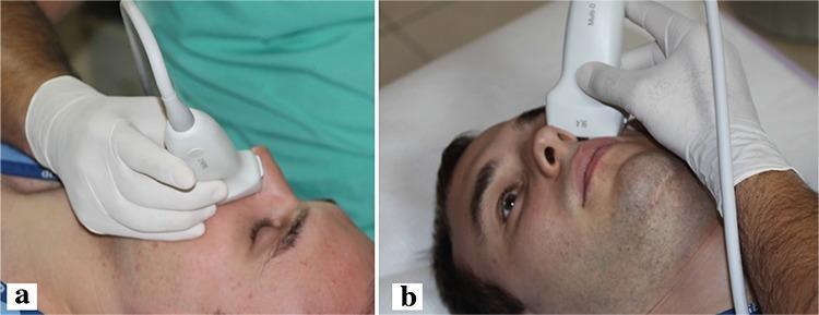
This clinical photograph consists of two panels (a and b) demonstrating the technique for performing ultrasound elastography of the lateral nasal wall. A patient is shown in a supine position while a radiologist, wearing protective medical gloves, applies a high-frequency linear ultrasound probe to the external lateral aspect of the nose. Panel 'a' provides a lateral profile view, showing the probe's contact point relative to the nasal bridge and cheek. Panel 'b' offers a superior-frontal perspective, illustrating the precise placement of the transducer over the area corresponding to the internal nasal turbinates. The procedure is used to measure tissue stiffness via the propagation speed of sound waves (m/s), which serves as a diagnostic tool in assessing conditions such as allergic rhinitis (AR). This visual serves as an educational guide for standardized probe positioning in rhinology-focused ultrasound imaging and elastographic evaluation of nasal mucosa.
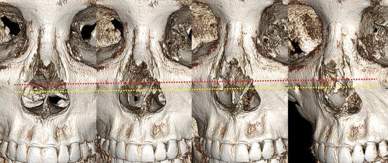
This diagnostic image consists of a series of four 3D computed tomography (CT) reconstructions of a human skull, focusing on the midface and nasal region. The panels illustrate the spatial relationship between rhinoplasty surgical landmarks and internal nasal anatomy. A red dotted line represents the lateral osteotomy path along the frontal process of the maxilla and nasal bones. A yellow dotted line indicates the anatomical attachment level of the inferior turbinates to the lateral nasal wall. In all views, the lateral osteotomy line (red) is positioned superior to the inferior turbinate attachment line (yellow). The images highlight the 'safe zone' for osteotomies relative to the pyriform aperture and the nasal cavity's internal structures. This comparison is clinically significant for surgical planning to avoid inferior turbinate medialization or airway compromise during nasal bone narrowing. Key structures visible include the orbits, nasal aperture, maxilla with dental arch, and the internal bony framework of the nasal passage.
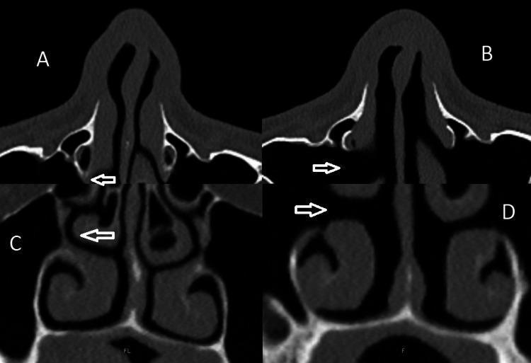
This diagnostic imaging set consists of four paranasal sinus Computed Tomography (CT) scans comparing preoperative and postoperative states following pyriform turbinoplasty (PT) and lateral nasal wall lateralization (LNWL). Panel A (axial) and Panel C (coronal) show the preoperative baseline, where the inferior turbinates (IT) contribute to a relatively narrow nasal airway. White arrows point to the IT position. Panel B (axial) and Panel D (coronal) display the postoperative results, characterized by a significant widening of the nasal cavity. The visual evidence shows the inferior turbinates have been lateralized, increasing the cross-sectional area of the nasal airway and the space between the nasal septum and the lateral nasal wall. The images illustrate the structural changes in the lateral nasal wall and the repositioning of the IT 'shoulder' to alleviate nasal obstruction. This comparison serves as an educational example for otolaryngologists and radiology students to visualize the surgical outcomes of functional nasal procedures on paranasal sinus anatomy.
Anatomy and Physiology of the Nose
1. Overview
- Scott-Brown's Otorhinolaryngology Head & Neck Surgery
- Bailey and Love's Short Practice of Surgery, 28th Edition
2. External Nose - Structural Framework
Bony Framework
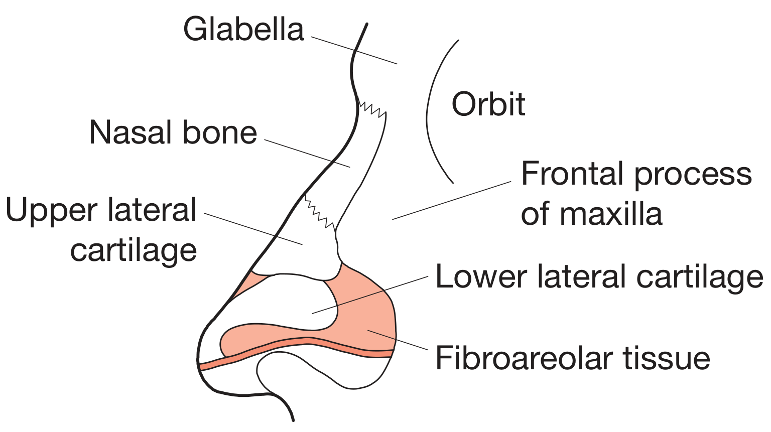
- Nasal bones - paired bones articulating superiorly with the frontal bone at the nasofrontal suture and laterally with the frontal process of the maxilla
- Frontal process of the maxilla - forms the lateral bony buttress
Cartilaginous Framework
- Upper lateral cartilages (ULC) - paired cartilages forming the middle third; they fuse with the septal cartilage in the midline
- Lower lateral cartilages (LLC) - paired "alar" cartilages forming the nasal tip and nostril rims; each has a medial crus, intermediate crus, and lateral crus
- Septal cartilage (quadrilateral cartilage) - central support
- Sesamoid cartilages - smaller accessory cartilages at the lateral alar margins
- Fibroareolar tissue - fills the alar lobule lateral to the LLC
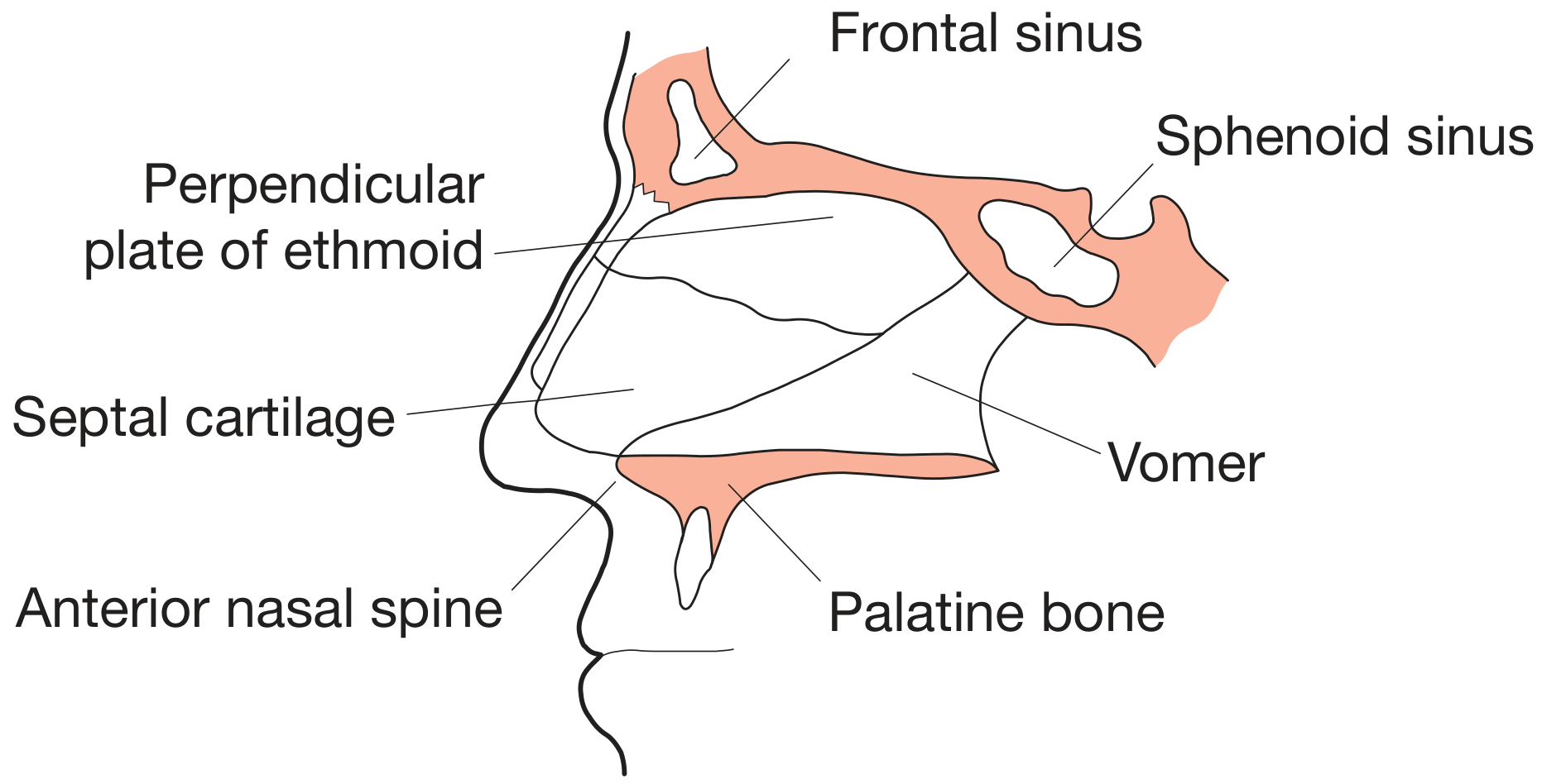
3. Nasal Septum
| Component | Type | Position |
|---|---|---|
| Quadrilateral (septal) cartilage | Hyaline cartilage | Anterior-central |
| Perpendicular plate of ethmoid | Bone | Posterosuperior |
| Vomer | Bone | Posteroinferior |
| Anterior nasal spine | Bone | Inferoanterior |
| Palatine bone | Bone | Posterior floor |
4. Nasal Cavity - Internal Anatomy
Walls of the Nasal Cavity
- Floor - formed by the hard palate (palatine process of maxilla + horizontal plate of palatine bone)
- Roof - the narrowest part; formed anteriorly by nasal bones, centrally by the cribriform plate of the ethmoid (transmitting olfactory nerve fibres), and posteriorly by the body of the sphenoid
- Medial wall - the nasal septum
- Lateral wall - the most complex wall, bearing the turbinates (conchae)
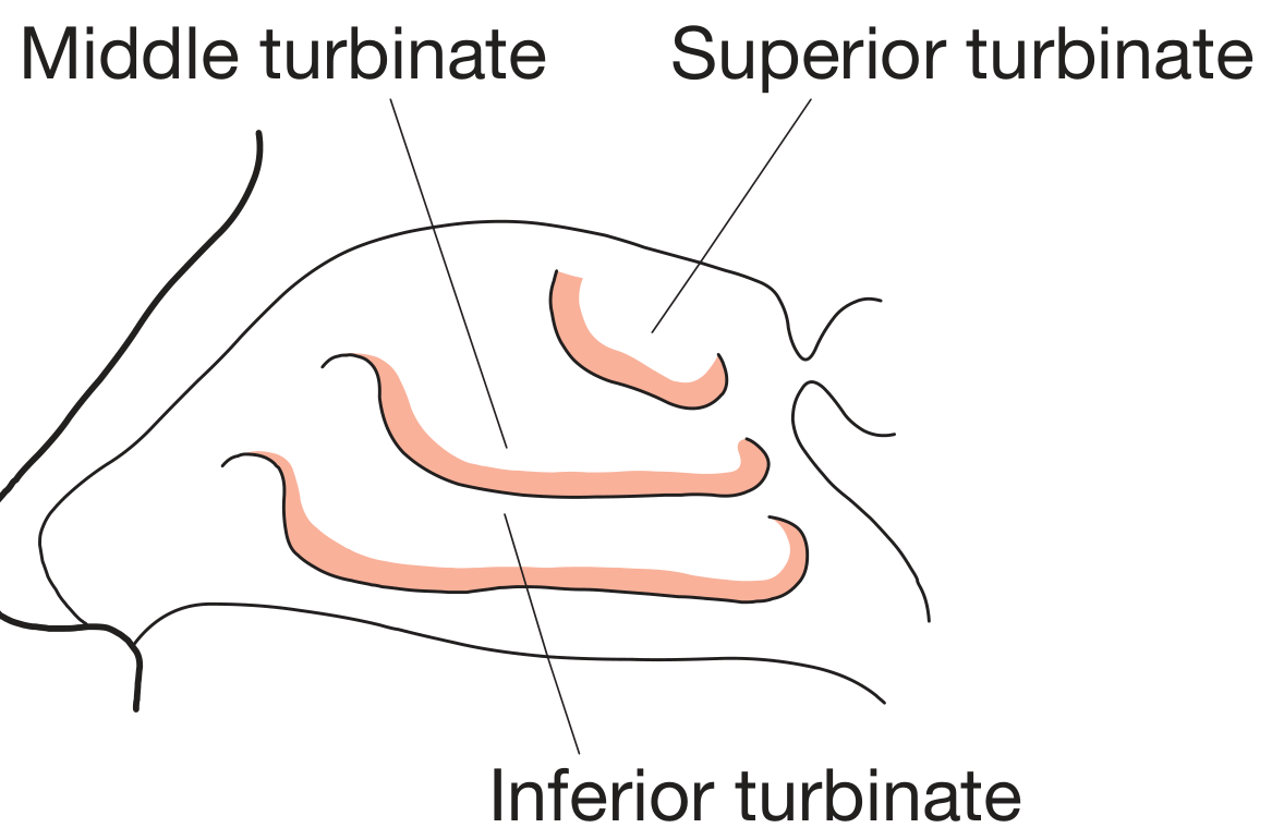
Turbinates (Conchae)
| Turbinate | Origin | Meatus Below | Key Drainage |
|---|---|---|---|
| Superior | Ethmoid bone | Superior meatus | Posterior ethmoid sinuses; sphenoid sinus drains into sphenoethmoidal recess |
| Middle | Ethmoid bone | Middle meatus | Frontal, maxillary, anterior ethmoid sinuses |
| Inferior | Independent bone | Inferior meatus | Nasolacrimal duct |
Regions of the Nasal Cavity
- Vestibule - the most anterior part, lined by skin bearing vibrissae (nasal hairs) that trap large particles
- Atrium - the transition zone just inside the vestibule
- Respiratory region - the bulk of the cavity; lined by pseudostratified ciliated columnar (respiratory) epithelium with goblet cells
- Olfactory region - the roof and upper part of the lateral wall and septum, lined by specialized olfactory mucosa
5. Blood Supply
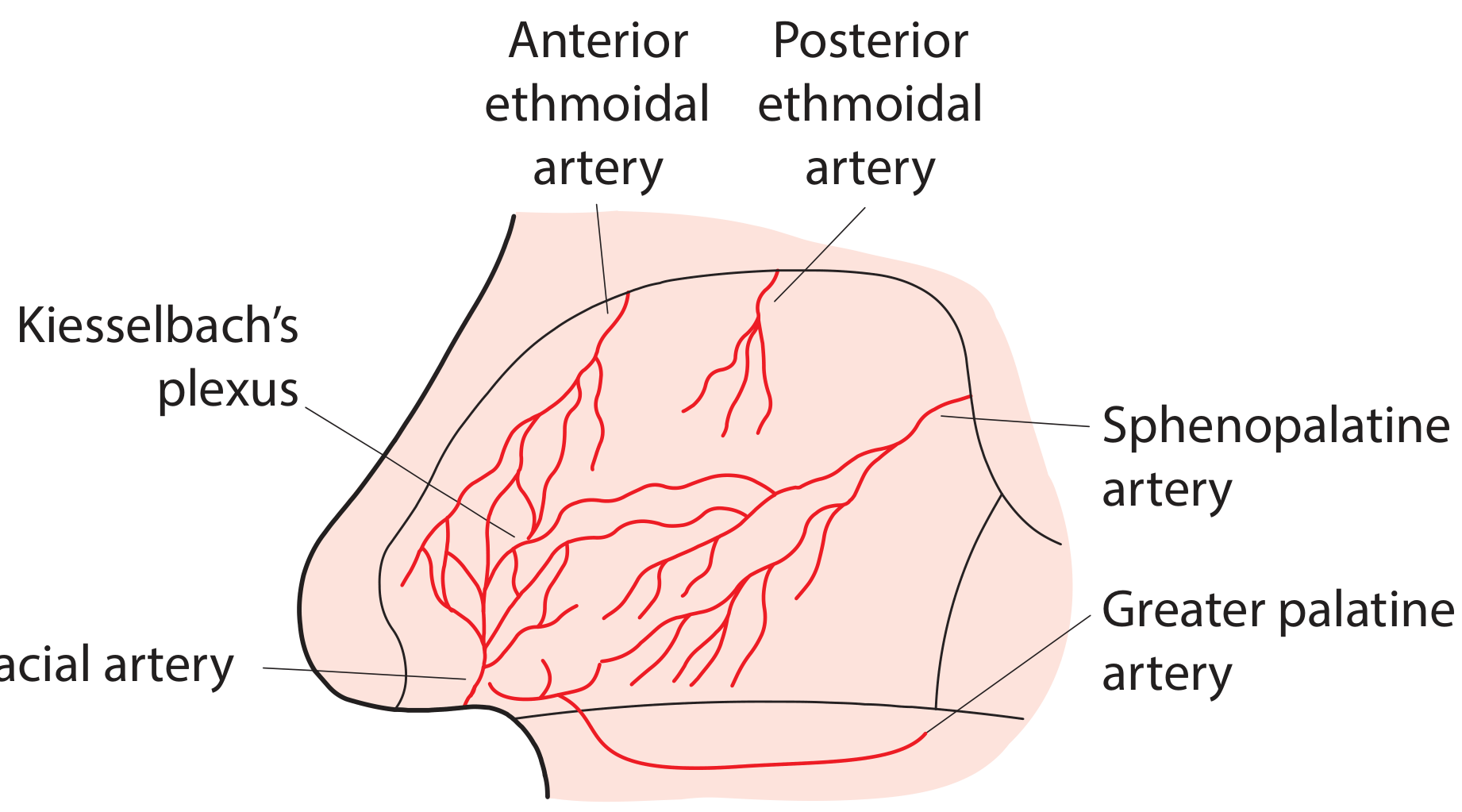
Arterial Supply
- Sphenopalatine artery (branch of maxillary artery) - the dominant blood supply, entering via the sphenopalatine foramen; supplies the lateral nasal wall and septum
- Greater palatine artery - supplies the anteroinferior septum via the incisive canal
- Facial artery - gives the superior labial artery, supplying the anterior septum
- Anterior and posterior ethmoidal arteries (branches of the ophthalmic artery) - supply the upper septum and roof
Kiesselbach's Plexus (Little's Area)
Venous Drainage
-
Via the ophthalmic and facial veins and the pterygoid and pharyngeal plexuses
-
Intracranial drainage into the cavernous sinus via the ophthalmic vein is clinically important - it creates a route for intracranial spread of nasal sepsis (the "danger area" of the face)
-
Bailey and Love's Short Practice of Surgery, 28th Edition
6. Nerve Supply
Sensory Innervation
- Olfactory nerve (CN I) - olfactory filaments pass through the cribriform plate to the olfactory bulb; serves smell
- Ophthalmic division of trigeminal (V1) - anterior ethmoidal nerve (external and internal nasal branches), supplying the anterior septum, roof, and dorsal skin of the nose tip
- Maxillary division of trigeminal (V2) - via the sphenopalatine ganglion; the nasopalatine nerve supplies the posterior septum and the posterior lateral nasal branches supply the turbinates
- Nasal branches of the infraorbital nerve - skin of the nasal alae and vestibule
Autonomic Innervation
- Parasympathetic - via the vidian nerve (nerve of the pterygoid canal) and sphenopalatine ganglion; mediates mucus secretion and vasodilatation
- Sympathetic - via the deep petrosal nerve → vidian nerve → sphenopalatine ganglion; mediates vasoconstriction (nasal decongestant effect)
7. Lymphatic Drainage
- Anterior nasal cavity drains to submandibular nodes
- Posterior nasal cavity drains to retropharyngeal and deep cervical nodes
8. Nasal Mucosa - Histology
Respiratory Mucosa
- Ciliated columnar cells
- Goblet cells (mucus secreting)
- Basal cells
- Brush cells
Olfactory Mucosa
| Cell Type | Function |
|---|---|
| Olfactory receptor cells (bipolar neurons) | Bear 10-23 long, nonmotile cilia on the olfactory vesicle; contain odorant receptor proteins; axons pass through the cribriform plate as CN I |
| Supporting (sustentacular) cells | Columnar; span the full epithelial thickness; provide metabolic support; synthesize odorant-binding proteins |
| Basal cells | Stem cells; renew the olfactory receptor cells and supporting cells throughout life |
- Histology: A Text and Atlas with Correlated Cell and Molecular Biology
9. Physiology of the Nose
9.1 Air Conditioning (Warming and Humidification)
- Inspired air is warmed to near 37°C by the highly vascular turbinate mucosa
- Relative humidity is raised to near 100% by evaporation from the mucosal surface
- Vibrissae and the mucociliary apparatus trap particles
9.2 Mucociliary Clearance
- Periciliary (sol) layer - watery, low-viscosity layer in which cilia beat freely
- Gel (mucus) layer - the sticky outer layer that traps particles
9.3 The Nasal Cycle
9.4 Olfaction (Sense of Smell)
- Odorant molecules dissolve in the mucus overlying the olfactory epithelium
- They bind to odorant-binding proteins (secreted by Bowman's glands) and are presented to olfactory receptor proteins on the cilia
- Receptor activation triggers a cAMP-mediated second messenger cascade, depolarizing the olfactory receptor cell
- Action potentials travel along the olfactory nerve (CN I) through the cribriform plate to the olfactory bulb and onward to the olfactory cortex (piriform cortex, amygdala, entorhinal cortex)
- Basal cells provide ongoing neurogenesis - olfactory receptor neurons are one of the few neuron populations that are continually replaced throughout life
9.5 Nasal Blood Flow and Turbinance
- Thermoregulation of inspired air
- Nasal congestion in response to allergens, infection, or cold air (parasympathetic-mediated vasodilatation)
- Decongestion via sympathetic-mediated vasoconstriction (mechanism of action of topical decongestants)
9.6 Functions of the Paranasal Sinuses
- Vocal resonance
- Reduction of skull weight
- Contribution to humidification and air conditioning
- Physical buffer against facial injury
- Heat insulation for the brain
10. Embryology (Summary)
-
Development begins at week 4 of gestation
-
Neural crest cells form nasal placodes
-
Medial and lateral nasal processes fuse to form the philtrum, columella, and nose
-
The nasobuccal membrane ruptures by week 10, creating nasal-nasopharyngeal communication
-
The nasal septum fuses with the palate: anterior by week 9, complete by week 12
-
Cummings Otolaryngology Head and Neck Surgery
- Bailey and Love's Short Practice of Surgery, 28th Edition - Chapter 51
- Cummings Otolaryngology Head and Neck Surgery - Chapter 29
- Scott-Brown's Otorhinolaryngology Head & Neck Surgery, Vol. 1
- Miller's Anesthesia, 10th Edition - Chapter 66
- Histology: A Text and Atlas with Correlated Cell and Molecular Biology - Chapter 19