
Contact inhibition
Contact inhibition in tympanic membrane
contact inhibition tympanic membrane keratinocyte migration
"contact inhibition" tympanic membrane cholesteatoma
tympanic membrane epithelial migration diagram
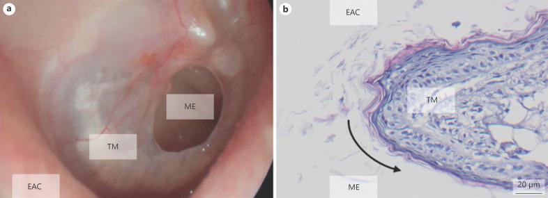
Educational visual regarding tympanic membrane perforation (TMP) and epithelialization, featuring clinical imaging and histology. Panel (a) is an otoscopic/microscopic clinical photograph showing a clear, oval-shaped perforation in the tympanic membrane (TM). The middle ear (ME) is visible through the defect. The margins of the perforation are distinct and rounded, demonstrating characteristic epithelialization. Panel (b) presents a high-magnification histology section stained with Hematoxylin and Eosin (H&E), showing the TM cross-section at the perforation edge. It illustrates the overgrowth of keratinizing squamous epithelium from the external auditory canal (EAC) side, curling around the defect margin toward the middle ear (ME) surface. A black arrow indicates the direction of epithelial migration, which inhibits spontaneous closure by creating a continuous skin layer over the wound edge. This composite serves as a diagnostic and pathological reference for chronic tympanic membrane defects and wound healing failure in otolaryngology.
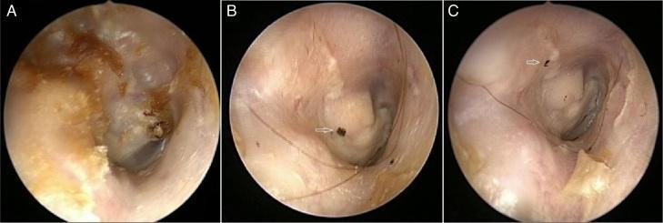
This composite of clinical endoscopic photographs illustrates a comparative study of the right ear canal and epithelial migration. Image A shows the pre-operative ear canal, characterized by the presence of cerumen or pathological debris (dark brown/black mass) obstructing the view of the tympanic membrane. Image B displays the post-operative state, showing a clearer canal with surgical changes and a visible ink marker used for tracking. Image C demonstrates the epithelial migration pattern in a posterior-superior direction, evidenced by the movement of dark ink markings (indicated by a white arrow) along the canal wall. The series serves to educate on otologic surgical outcomes and the physiological process of epithelial self-cleansing in the external auditory canal. Key concepts include postoperative monitoring of ear canal health and the quantification of migration rates (linear movement measured in mm/week) to assess physiological recovery.
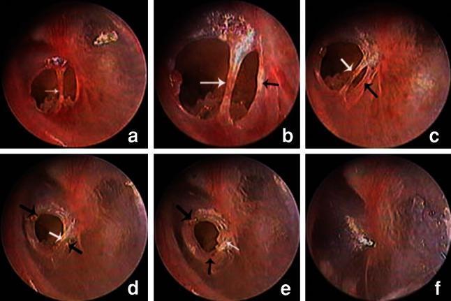
This series of six otoendoscopic images (a–f) illustrates the chronological stages of spontaneous tympanic membrane healing over a 49-day period. The initial images (a, b) show a traumatic eardrum perforation bisected by an 'eardrum bridge' (white arrow), which is a strip of intact tissue spanning the defect. Subsequent frames (c–e) document the process of stratified epithelial migration. Proliferating epithelium at the perforation margins (black arrows) is seen moving centripetally to close the gap. During this progression, the eardrum bridge serves as a scaffold; it gradually incorporates into the regenerating tissue or is replaced by newly formed epithelium. By day 49 (f), the perforation has achieved complete closure, the eardrum bridge is no longer a distinct structure, and the tympanic membrane shows a restored, uniform surface with minor residual scarring. This series demonstrates key concepts in otolaryngology, specifically the natural history of traumatic perforation repair and the role of epithelial hyperplasia in secondary intention healing of the tympanic membrane.
Contact Inhibition in the Tympanic Membrane
What Is Contact Inhibition?
Normal Epithelial Migration of the Tympanic Membrane
- Keratinocytes originate at the umbo (centre of the TM) and migrate radially outward toward the annulus, then down the external auditory canal.
- This self-cleaning "conveyor belt" removes debris, keratin flakes, and foreign material from the drum surface.
- This migration is tightly regulated and normally proceeds in an orderly, controlled fashion.
Role of Contact Inhibition in TM Repair
"Loss of contact inhibition of the squamous epithelial cells is postulated to be the driver for repair of acute injuries to the membrane." - Scott-Brown's Otorhinolaryngology, Vol 2
- With loss of contact between cells at the wound edge, squamous epithelial cells are "released" from inhibition and begin proliferating and migrating to re-epithelialize the defect.
- 70-80% of acute perforations heal spontaneously within 30 days via this mechanism.
- Once healing is complete and cells meet again at the wound centre, contact inhibition is re-established, stopping further migration.
When Healing Fails: The Chronic Perforation
"When a perforation becomes chronic, the margin of the perforation can be seen to be stable with the squamous epithelial cells coming into contact with the epithelial cells of the mucosal layer, creating a barrier against further vascular proliferation." - Scott-Brown's Otorhinolaryngology, Vol 2
Pathological Loss of Contact Inhibition: Cholesteatoma Formation
"In some cases, epithelial cells migrate medially through the perforation rather than stopping at the edge. The cause of this migration is not known, but it does have significant clinical implications." - Shambaugh Surgery of the Ear
- The appearance of squamous lining in the middle-ear space (which normally has only mucosal epithelium) is pathological.
- If infection supervenes, migration ceases but squamous hyperplasia and keratin accumulation begin, leading to cholesteatoma formation.
- Surgically, if migrated epithelial cells are not removed from the middle ear before TM repair, an iatrogenic cholesteatoma can result.
Molecular Basis: The NF2/Merlin Pathway
- At high cell density (cells in contact): merlin is hypophosphorylated and inhibits cell growth via interactions with CD44 and hyaluronate (extracellular matrix components).
- At low cell density (cells not in contact, e.g., at a wound edge): merlin is phosphorylated, releasing the growth inhibition and allowing migration and proliferation.
- Loss-of-function mutations in NF2/merlin result in loss of contact inhibition, contributing to tumorigenesis (e.g., schwannomas, vestibular schwannomas).
Summary Table
| Situation | Contact Inhibition Status | Clinical Result |
|---|---|---|
| Normal TM, cells in contact | Active | Orderly centrifugal migration only |
| Acute perforation | Transiently lost | Cell proliferation/migration to heal defect |
| Healed perforation | Re-established | Migration stops, drum closed |
| Chronic perforation edge | Re-established (squamous meets mucosa) | Stable margin, no healing |
| Abnormal: cells migrate through perforation | Lost/absent | Squamous lining in middle ear, potential cholesteatoma |
| Infection + aberrant migration | Migration ceases, hyperplasia begins | Keratin accumulation, cholesteatoma |
- Scott-Brown's Otorhinolaryngology, Vol 2, pp. 1025-1026, 1065
- Shambaugh Surgery of the Ear, pp. 454-455
- Robbins & Kumar Basic Pathology - TGF-β, Contact Inhibition, and APC-β-Catenin Pathways