
Draw a gastric gland (cross-section): · Top (Pit): Mucous neck cells (secrete mucus). · Middle (Neck & Body): · Parietal cells (large, round, eosinophilic) – label "Have canaliculi with H⁺/K⁺ ATPase". · Chief cells (basophilic, zymogen granules) – label "Secrete Pepsinogen". · Base (Antrum): · G-cells (open to lumen) – label "Secrete Gastrin into blood". · Lumen: Label "HCl + Pepsinogen + Mucus".
Detailed scientific cross-section diagram of a gastric gland, vertically oriented, showing three regions clearly labeled: TOP REGION - GASTRIC PIT: Label "Gastric Pit (Lumen Opening)" at the very top. Show a central open lumen labeled "HCl + Pepsinogen + Mucus". Line the pit with tall columnar mucous neck cells labeled "Mucous Neck Cells – secrete mucus" with pale cytoplasm. MIDDLE REGION - NECK AND BODY: Show two cell types alternating along the gland walls: 1. Parietal cells: large, round, eosinophilic (bright pink), abundant cytoplasm, central nucleus. Label with arrow: "Parietal Cells – Have canaliculi with H⁺/K⁺ ATPase" 2. Chief cells: smaller, basophilic (blue-purple), basal nucleus, apical zymogen granules. Label with arrow: "Chief Cells – Secrete Pepsinogen" BOTTOM REGION - BASE / ANTRUM: Show G-cells lining the base, open to the lumen side, triangular/flask-shaped. Label with arrow: "G-Cells – Secrete Gastrin into blood". Show small arrows pointing toward capillary on the basal side. Style: clean medical textbook illustration, white background, color-coded cells, clear black label lines with arrows, anatomical cross-section style, H&E-like coloring (pink parietal, blue chief, pale mucous, triangular G-cells). Include a vertical bracket on the side labeling the three zones: "Pit", "Neck & Body", "Base (Antrum)".
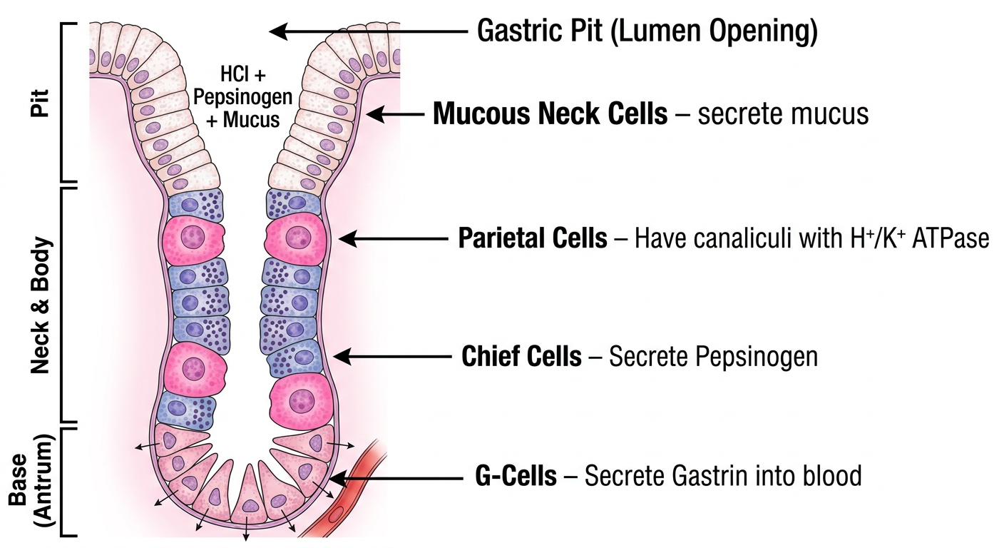
- Gastric Pit (Top): Mucous neck cells lining the pit opening, with the central lumen labeled "HCl + Pepsinogen + Mucus"
- Neck & Body (Middle):
- Parietal cells - large, round, eosinophilic (pink), labeled with their intracellular canaliculi and H⁺/K⁺ ATPase proton pump
- Chief cells - basophilic (blue-purple), with apical zymogen granules, labeled for pepsinogen secretion
- Base/Antrum (Bottom): G-cells open to the lumen, with secretion arrows pointing toward the basal capillary, labeled for gastrin release into blood