
Reason ferritin in low in body 23 y old girl its circumstances
causes of low ferritin young women 23 year old iron deficiency circumstances
ferritin iron deficiency young women causes
iron deficiency anemia low ferritin causes pathophysiology diagram
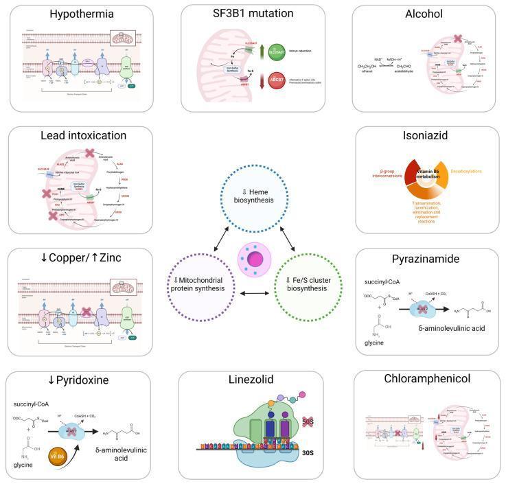
A multi-panel pathophysiology infographic illustrating the main causes of acquired sideroblastic anemia through mitochondrial dysfunction. The central diagram depicts an erythroblast surrounded by three key impaired processes: Heme biosynthesis, Mitochondrial protein synthesis, and Fe/S (iron-sulfur) cluster biosynthesis. Ten surrounding panels detail specific etiologies including Hypothermia, SF3B1 mutation (Myelodysplastic Syndrome), Alcohol, Lead intoxication, Isoniazid, Copper deficiency/Zinc excess, Pyrazinamide, Pyridoxine (Vitamin B6) deficiency, Linezolid, and Chloramphenicol. Each panel uses molecular models to show specific inhibitory sites: for example, the inhibition of ALA-synthase by lead and vitamin B6 deficiency, or the disruption of the mitochondrial ribosome (30S and 50S subunits) by antibiotics like linezolid. The visual highlights how diverse toxic, nutritional, and genetic factors converge on mitochondrial metabolism, leading to iron overload in erythroblasts and the subsequent formation of ring sideroblasts.
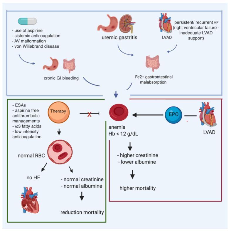
This pathophysiology diagram illustrates the multifactorial causes and consequences of anemia in patients with heart failure (HF) and a left ventricular assist device (LVAD). The top section identifies contributors to anemia, including chronic gastrointestinal (GI) bleeding secondary to aspirin use, systemic anticoagulation, arteriovenous (AV) malformations, and von Willebrand disease. Other contributing factors shown are uremic gastritis, iron (Fe2+) malabsorption, and persistent right ventricular failure. Central to the diagram is the manifestation of anemia (defined as Hemoglobin < 12 g/dL), which correlates with elevated creatinine, decreased albumin, and increased mortality. The diagram also highlights therapeutic interventions such as erythropoiesis-stimulating agents (ESAs), aspirin-free antithrombotic management, omega-3 fatty acids, and low-intensity anticoagulation. Successful therapy is depicted as leading to normal red blood cell (RBC) counts, resolution of heart failure symptoms, and reduced mortality. The role of erythropoietin (EPO) is also noted in the context of LVAD-related anemia management.
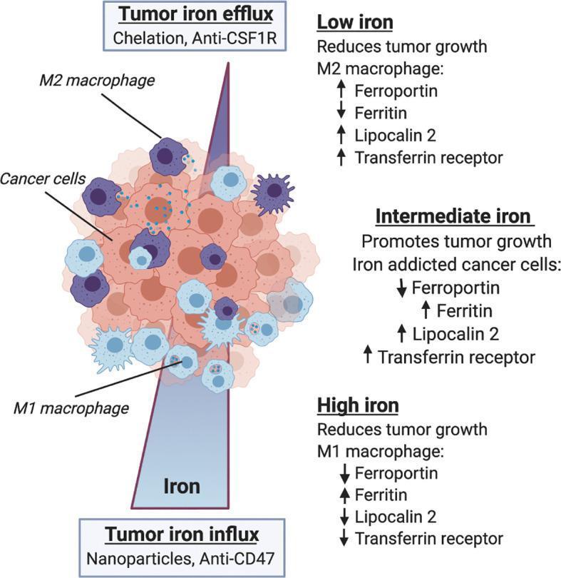
This pathophysiology diagram illustrates the relationship between iron metabolism, macrophage polarization, and tumor growth within the tumor microenvironment (TME). A central cellular cluster depicts cancer cells (light orange), M1 macrophages (light blue, smooth), and M2 macrophages (purple, spiky). A vertical gradient represents iron concentration, influenced by 'Tumor iron influx' (bottom, associated with nanoparticles and anti-CD47) and 'Tumor iron efflux' (top, associated with chelation and anti-CSF1R). The diagram categorizes the TME into three metabolic regimes. 1) Low iron: favors M2 macrophages, characterized by increased ferroportin, lipocalin 2, and transferrin receptor but reduced ferritin, leading to reduced tumor growth. 2) Intermediate iron: characterized by 'iron-addicted' cancer cells with low ferroportin and high levels of ferritin, lipocalin 2, and transferrin receptor, which promotes tumor growth. 3) High iron: favors M1 macrophages, characterized by increased ferritin and reduced ferroportin, lipocalin 2, and transferrin receptor, resulting in reduced tumor growth. This educational infographic demonstrates how iron availability modulates immune cell phenotype and clinical outcomes in oncology.
Why Is Ferritin Low in a 23-Year-Old Woman?
What Is "Low" Ferritin?
- Normal range for women: 12-150 ng/mL (varies by lab)
- Below 12 ng/mL = definite iron deficiency
- Below 30 ng/mL = strongly suggestive of iron deficiency in symptomatic patients
- Iron deficiency can exist without anemia (ferritin low, hemoglobin still normal) - a very common scenario in young women
The Two Master Causes
- Increased iron loss (blood loss)
- Reduced iron absorption or intake
Circumstance 1 - Menstrual Blood Loss (Most Common Cause)
- The average menstrual period causes loss of 35 mL of blood and 16 mg of iron
- Women require 1-3 mg of iron/day, rising significantly with heavy periods
- Heavy menstrual bleeding (>80 mL/cycle) - caused by fibroids, endometriosis, PCOS, or coagulation disorders - rapidly depletes stores
- Studies show bone marrow iron stores are absent in 25% of healthy college-age women and reduced in another 33% - Goldman-Cecil Medicine
- A JAMA study found nearly 40% of women ages 12-21 have low iron levels
Circumstance 2 - Inadequate Dietary Iron Intake
- Vegetarian/vegan diet: Non-heme iron (from plants) is in the ferric (Fe³+) form, which absorbs far less efficiently than heme iron from meat. Lactovegetarians are at even higher risk - Goldman-Cecil Medicine
- Calorie-restricted diets / disordered eating (common in young women)
- Skipping meals, poor overall nutrition, reliance on processed foods low in iron
- Iron-rich foods such as red meat, organ meats, legumes, and leafy greens are often under-consumed
Circumstance 3 - Poor Iron Absorption (Malabsorption)
| Condition | Mechanism |
|---|---|
| Celiac disease | Duodenal villous atrophy impairs absorption; very common in young women |
| H. pylori infection | Causes achlorhydria (low stomach acid), reducing Fe³+ → Fe²+ conversion; also causes ulcer bleeding |
| Inflammatory bowel disease (Crohn's/UC) | Mucosal damage + chronic GI blood loss |
| Autoimmune gastritis | Impairs iron and later B12 absorption |
| Bariatric surgery | Up to 50% of post-bariatric patients become iron deficient |
| Proton pump inhibitors / antacids | Reduce stomach acid needed for iron conversion |
Circumstance 4 - Increased Iron Demand
- Pregnancy: Requires ~900 mg total iron; about 6 mg/day in the third trimester (relevant even in young women)
- Breastfeeding: Iron transfer to infant
- Intense exercise / athletic training: Hemolysis from foot-strike, increased red cell turnover, sweat losses - a major cause in young female athletes
Circumstance 5 - GI Blood Loss (Non-menstrual)
- Peptic ulcers / gastritis (especially from NSAIDs or H. pylori)
- Hemorrhoids
- Occult GI bleeding from any source
Circumstance 6 - Obesity / Elevated Hepcidin
- Adipose tissue promotes chronic low-grade inflammation, raising hepcidin levels
- Hepcidin degrades ferroportin, the iron export channel in the gut, thereby blocking iron absorption
- About 8% of obese patients have elevated hepcidin as a marker of reduced iron absorption - Goldman-Cecil Medicine
- A 2024 systematic review (PMID 39324337) confirmed the link between overweight/obesity and iron deficiency anaemia in reproductive-age women
Circumstance 7 - Hypothyroidism
- Thyroid hormones are needed for normal stomach acid secretion and intestinal motility
- Hypothyroidism (more common in young women) reduces hydrochloric acid production, impairing iron absorption
- Fatigue from hypothyroidism can mimic or worsen iron deficiency symptoms
Circumstance 8 - Blood Donation
Symptoms of Low Ferritin (Even Without Anemia)
- Fatigue and low energy (most common complaint)
- Brain fog, poor concentration
- Hair loss / thinning
- Brittle nails, pale skin
- Headaches, lightheadedness
- Cold hands and feet
- Restless leg syndrome
- Reduced athletic performance
- Shortness of breath on exertion
Summary Table
| Category | Specific Cause |
|---|---|
| Blood loss | Heavy periods, GI bleeding, blood donation |
| Diet | Vegetarian/vegan, calorie restriction, poor iron intake |
| Malabsorption | Celiac disease, H. pylori, IBD, low stomach acid, PPI use |
| Increased demand | Pregnancy, breastfeeding, intense exercise |
| Hormonal/metabolic | Hypothyroidism, obesity (elevated hepcidin) |
Sources: Goldman-Cecil Medicine (Iron Deficiency chapter), Tietz Textbook of Laboratory Medicine, Symptom to Diagnosis (4th Ed.), Franciscan Health, Nebraska Medicine
Tyroid 2.29 in same patient
TSH 2.29 normal range interpretation young woman thyroid function
TSH = 2.29 mIU/L in a 23-Year-Old Woman
Verdict: NORMAL
Where Does 2.29 Sit Exactly?
| TSH Zone | Range | This Patient |
|---|---|---|
| Hyperthyroid | < 0.4 mIU/L | No |
| Normal (euthyroid) | 0.4 - 4.0 mIU/L | Yes - 2.29 |
| Subclinical hypothyroid | 4.0 - 10 mIU/L | No |
| Overt hypothyroidism | > 10 mIU/L | No |
Does TSH 2.29 Explain Her Low Ferritin?
- Menstrual blood loss (most likely)
- Dietary iron deficiency (vegetarian diet, poor intake)
- Malabsorption (celiac, H. pylori - worth checking)
- Increased demand (exercise, etc.)
One Important Note - Functional Medicine Perspective
What Should Be Done Next?
- Treat the low ferritin - oral iron supplementation (ferrous sulfate 150-200 mg elemental iron/day)
- Investigate heavy menstrual bleeding if present
- Check dietary history for iron intake
- Consider celiac screen (tTG-IgA) if GI symptoms exist or ferritin remains low despite supplementation
- Recheck ferritin in 3 months after supplementation