
Inspection of respiratory system general medicine
inspection respiratory system chest examination findings
![Respiratory examination findings
<table><thead><tr><th>Symptom and signs</th><th>Possible cancer</th><th>Recommendation</th></tr></thead><tbody><tr><td>Chest signs consistent with lung cancer, 40 and over</td><td>Lung</td><td>Consider an urgent chest X-ray (to be done within 2 weeks) [1.1.3]</td></tr><tr><td>Chest signs compatible with pleural disease, 40 and over</td><td>Mesothelioma</td><td>Consider an urgent chest X-ray (to be done within 2 weeks) [1.1.6]</td></tr><tr><td>Finger clubbing, 40 and over</td><td>Lung or mesothelioma</td><td>Consider an urgent chest X-ray (to be done within 2 weeks) [1.1.3] [1.1.6]</td></tr></tbody></table>](https://cdn.orris.care/cdss_images/GLGCA_4543562_1766478019695_1574605f-25f8-4d87-a88f-bc54041ac4c6_a5b9af6e-2baa-42d3-b397-ece6c8b4ac89.png)
Respiratory examination findings <table><thead><tr><th>Symptom and signs</th><th>Possible cancer</th><th>Recommendation</th></tr></thead><tbody><tr><td>Chest signs consistent with lung cancer, 40 and over</td><td>Lung</td><td>Consider an urgent chest X-ray (to be done within 2 weeks) [1.1.3]</td></tr><tr><td>Chest signs compatible with pleural disease, 40 and over</td><td>Mesothelioma</td><td>Consider an urgent chest X-ray (to be done within 2 weeks) [1.1.6]</td></tr><tr><td>Finger clubbing, 40 and over</td><td>Lung or mesothelioma</td><td>Consider an urgent chest X-ray (to be done within 2 weeks) [1.1.3] [1.1.6]</td></tr></tbody></table>
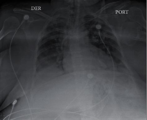
This diagnostic image is a portable anteroposterior (AP) chest X-ray of an adult patient. The primary radiographic findings include extensive, bilateral interstitial and alveolar infiltrates across both lung fields, consistent with Acute Respiratory Distress Syndrome (ARDS) or multifocal pneumonia. There is a dense, focal area of consolidation in the right basal lung region, likely representing localized infectious consolidation or atelectasis. Multiple medical devices are visible: a port catheter (labeled 'PORT') is present in the left upper chest with its tip directed centrally, and a separate lead system (marked 'DER' for right) with surface electrodes is visible for cardiac monitoring. The diaphragmatic contours are somewhat obscured by the basal opacities, and the cardiac silhouette appears enlarged, though evaluation is limited by the AP technique. Clinically, these findings are highly relevant for diagnosing severe lower respiratory tract involvement in systemic viral or bacterial infections, such as SARS-CoV-2 pneumonia complicated by superinfection.
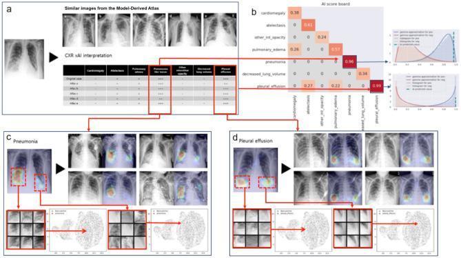
This infographic illustrates the workflow of an explainable Artificial Intelligence (xAI) system for chest X-ray (CXR) interpretation in respiratory infections. Section (a) displays a test CXR alongside similar images from a 'Model-Derived Atlas' and a probability table for various findings. Section (b) shows an 'AI scoreboard' quantifying confidence scores, highlighting high probabilities for pneumonia (0.96) and pleural effusion (0.99). Sections (c) and (d) provide visual evidence via Gradient-weighted Class Activation Mapping (Grad-CAM). For pneumonia (c), heat maps identify focal opacities in the lower lung fields. For pleural effusion (d), the Grad-CAM highlights the costophrenic angles, where blunting or increased density is visible. Both sections include UMAP dimensionality reduction plots and high-resolution feature patches, demonstrating how the model clusters the test image's pathological features with known reference cases from the atlas. The content is designed for intermediate to advanced medical imaging informatics, focusing on diagnostic transparency and lesion localization in radiology.

This medical illustration demonstrates maneuvers used for the local modification of chest wall compliance in patients with severe respiratory distress, such as late-phase COVID-19 ARDS. The diagram shows a patient in a supine position on a flat examination surface. Two distinct clinical interventions are illustrated: 1) Sternal weighting, represented by a physical weight placed directly over the sternum to apply downward compressive force; and 2) A 'belly push' maneuver, indicated by a curved blue arrow showing sustained downward and inward pressure applied to the mid-upper abdomen. These external forces aim to modify respiratory system mechanics by reducing end-expiratory lung volumes, potentially shifting the lung's operation to a more compliant portion of the pressure-volume curve. This technique is used to assess or improve tidal compliance and reduce driving pressures during passive volume-targeted ventilation. The educational focus is on pathophysiology and critical care ventilation strategies to avoid end-tidal overdistention in 'baby lungs' associated with severe ARDS.
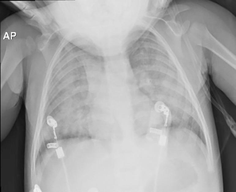
This anteroposterior (AP) chest X-ray of a pediatric patient demonstrates a diagnostic image consistent with Acute Respiratory Distress Syndrome (ARDS). The primary findings include bilateral, diffuse pulmonary opacities and areas of consolidation with ill-defined borders, indicating alveolar-interstitial involvement. The lungs appear hypoexpanded with significant increased density (haziness) throughout both lung fields, suggestive of pulmonary edema. Extrapulmonary findings include two ECG monitoring leads positioned on the lower chest and visible medical tubing. The skeletal structures and heart borders are partially obscured by the extensive pulmonary infiltrates. This image illustrates the typical radiological presentation of progressive respiratory dysfunction following traumatic injury, highlighting the clinical signs of widespread lung injury and fluid accumulation in a pediatric intensive care setting.
Create a PDF guide on respiratory inspection techniques
How to subscribe