
Therapeutic X-ray endovascular interventions 1) coronary angioplasty and stenting of coronary arteries 2) angioplasty of arteries of the lower extremities 3) implantation of a CAV filter in patients with a risk of pulmonary embolism (floating thrombi) or with recurrent pulmonary embolism 4) treatment of brain diseases (aneurysms, arteriovenous fistulas, carotid-cavernous anastomoses)
-
Coronary angioplasty and stenting
- Balloon dilatation of narrowed coronary arteries, usually followed by placement of a drug-eluting stent.
- Used in acute coronary syndromes and selected stable coronary artery disease.
-
Lower-extremity arterial angioplasty
- Endovascular treatment of peripheral arterial disease causing claudication, rest pain, or limb-threatening ischemia.
- May involve balloon angioplasty, drug-coated balloons, stents, atherectomy, or thrombectomy depending on the lesion.
-
IVC-filter implantation
- A filter placed in the inferior vena cava to trap emboli from lower-limb or pelvic venous thrombi before they reach the pulmonary arteries.
- Usually considered when anticoagulation is contraindicated or pulmonary embolism recurs despite appropriate anticoagulation. “Floating” proximal deep-vein thrombus alone is not automatically an indication in all patients.
-
Neuroendovascular treatment
- Catheter-based treatment of cerebral vascular lesions:
- intracranial aneurysm: coil embolization, stent-assisted coiling, flow diversion
- arteriovenous malformation/fistula: embolization with liquid embolic agents or coils
- carotid-cavernous fistula: transarterial or transvenous embolization
- Catheter-based treatment of cerebral vascular lesions:
Explain everything in depth
-
Foundations of therapeutic endovascular radiology
- Angiography, fluoroscopy, vascular access, guidewires, catheters, contrast media
- Balloons, stents, coils, flow diverters, embolic agents, thrombectomy devices, and IVC filters
- Patient preparation, anticoagulant/antiplatelet management, radiation and contrast safety
- General complications and post-procedure monitoring
-
Coronary angioplasty and coronary stenting (PCI)
- Coronary anatomy and pathophysiology of atherosclerotic stenosis
- Indications in acute coronary syndrome and chronic coronary syndrome
- Diagnostic coronary angiography, lesion assessment, balloon angioplasty, and stent deployment
- Drug-eluting versus bare-metal stents, intravascular imaging, antiplatelet therapy
- Complications: dissection, perforation, no-reflow, acute stent thrombosis, restenosis, bleeding, contrast kidney injury
-
Lower-extremity arterial angioplasty
- Peripheral arterial disease, Fontaine/Rutherford clinical staging, limb-threatening ischemia
- Imaging work-up and lesion classification
- Iliac, femoropopliteal, infrapopliteal angioplasty, drug-coated balloons, stenting, atherectomy, and thrombectomy
- Choosing endovascular versus open surgical revascularization
- Technical failures, distal embolization, re-occlusion, access complications, surveillance
-
Venous interventions and IVC filters
- Deep-vein thrombosis and pulmonary embolism pathophysiology
- Correct term: inferior vena cava (IVC) filter
- Indications, contraindications, filter types, implantation steps, retrieval
- Limits of filter use: anticoagulation remains the primary treatment whenever possible
- Complications: filter thrombosis, migration, tilt, penetration, fracture, failure to retrieve
-
Neuroendovascular treatment
- Cerebral aneurysms: coil embolization, balloon remodeling, stent-assisted coiling, flow diversion
- Arteriovenous malformations and dural arteriovenous fistulas: transarterial/transvenous embolization
- Carotid-cavernous fistulas: anatomy, presentation, diagnosis, and endovascular closure
- Periprocedural antiplatelets/anticoagulation and neurological monitoring
- Complications: ischemic stroke, intracranial hemorrhage, aneurysm rupture, cranial nerve deficits, device-related thrombosis
Explain coronary angioplasty and stenting step by step
"percutaneous coronary intervention" AND "systematic review"
2025 ACC AHA guideline percutaneous coronary intervention coronary revascularization patient information
coronary angioplasty stent deployment diagram fluoroscopy
https://www.acc.org/latest-in-cardiology/journal-scans/2025/…
https://www.acc.org/about-acc/press-releases/2025/02/28/17/5…
Coronary angioplasty and stenting: step-by-step
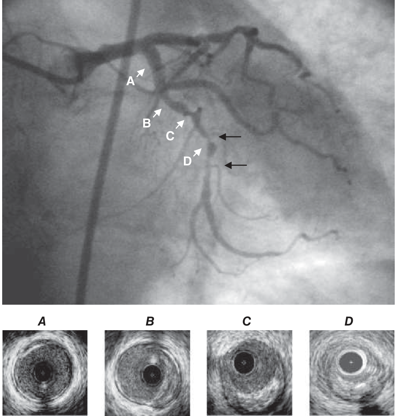
1. Why a coronary artery becomes narrowed
- Lipid accumulates in the coronary artery wall.
- Inflammation, fibrosis, and sometimes calcium form an atherosclerotic plaque.
- The plaque narrows the lumen and reduces blood flow, particularly during exertion.
- If plaque rupture or erosion occurs, a platelet-rich thrombus may rapidly form.
- Acute obstruction produces an acute coronary syndrome, including STEMI or NSTEMI.
2. When PCI is used
A. Acute indications
- NSTEMI or unstable angina with high-risk clinical features
- Ongoing or recurrent ischemic chest pain despite medical therapy
- Cardiogenic shock due to acute coronary occlusion
- Hemodynamically significant coronary occlusion or severe stenosis found during acute coronary angiography
- Acute stent thrombosis or acute closure after a previous intervention
B. Elective or planned PCI
- Angina that limits daily activity despite guideline-directed medical therapy
- Objective evidence of inducible myocardial ischemia
- A suitable focal coronary stenosis that is anatomically amenable to PCI
- Selected cases after prior coronary bypass surgery when a native artery or graft has become narrowed
When bypass surgery may be preferred
3. Pre-procedure assessment and preparation
Clinical assessment
- Symptoms: chest pain, breathlessness, instability
- ECG changes and cardiac troponin in suspected acute coronary syndrome
- Echocardiography when ventricular function or structural complications need assessment
- Past history: prior PCI/CABG, bleeding, stroke, kidney disease, allergy, and peripheral arterial disease
- Review of drugs, especially anticoagulants, antiplatelets, diabetes agents, and allergy history
Laboratory tests
- Hemoglobin and platelet count
- Serum creatinine and estimated glomerular filtration rate
- Electrolytes
- Coagulation status when relevant
- Blood group and screen if bleeding risk is substantial
Antithrombotic preparation
- Aspirin
- A P2Y12 inhibitor as part of dual antiplatelet therapy, or DAPT
- An intraprocedural anticoagulant
Consent
- Expected benefit
- Alternatives: medical therapy, CABG, or no intervention
- Possibility that anatomy may not be suitable for PCI
- Risks, including bleeding, kidney injury, infarction, stroke, emergency surgery, and death
4. Vascular access
Radial access
- Lower bleeding and vascular-complication risk
- Earlier mobility after the procedure
- Easier hemostasis with a wrist compression band
Femoral access
- Radial access is not feasible
- Larger devices are needed
- The radial artery is too small, occluded, or severely diseased
- There is a specific anatomical or procedural reason
5. Introduction of the sheath and diagnostic catheter
- A short plastic tube, the arterial sheath, is inserted into the access artery.
- A diagnostic catheter is advanced through the aorta under fluoroscopy.
- The catheter tip is placed at the opening of the left or right coronary artery.
- Small amounts of contrast are injected.
- Rapid X-ray image sequences, called coronary angiograms, are obtained in several views.
- Which artery is abnormal
- Location and length of narrowing
- Degree of stenosis
- Presence of thrombus, calcium, vessel tortuosity, bifurcation disease, or chronic total occlusion
- Distal flow, usually described using the TIMI flow scale
- Collateral circulation
- Whether there are multiple significant lesions
6. Assessing whether the lesion needs treatment
Physiological assessment
- Fractional flow reserve (FFR)
- Instantaneous wave-free ratio (iFR)
Intravascular imaging
- Intravascular ultrasound (IVUS)
- Optical coherence tomography (OCT)
- True vessel diameter
- Plaque and calcium burden
- Lesion length
- Stent diameter and length
- Stent expansion and wall apposition
- Edge dissection or residual disease
7. Engaging the coronary artery with a guide catheter
- The diagnostic catheter is exchanged for a guide catheter.
- The guide catheter is seated at the coronary ostium.
- It provides a stable route for guidewires, balloons, stents, and imaging devices.
- The operator watches pressure tracing continuously. Deep or unstable engagement can transiently obstruct the coronary ostium and must be corrected immediately.
8. Crossing the narrowing with a coronary guidewire
- Under fluoroscopy, the wire crosses the narrowed or blocked segment.
- Its tip is placed safely in a distal coronary branch.
- The wire now acts as a rail over which treatment devices can be delivered.
9. Lesion preparation
A. Direct stenting
B. Predilatation
- Compresses and redistributes plaque
- Stretches the vessel wall
- Improves the lumen enough for stent delivery
C. Treatment of calcified lesions
- Noncompliant high-pressure balloons
- Scoring or cutting balloons
- Intravascular lithotripsy
- Rotational or orbital atherectomy in selected lesions
10. Stent implantation
What is a coronary stent?
Deployment sequence
- The stent-mounted balloon is advanced over the guidewire.
- Fluoroscopy confirms that the stent covers the full lesion, including appropriate margins at both edges.
- The balloon is inflated to deploy the stent.
- The metallic scaffold expands and presses against the arterial wall.
- The balloon is deflated and withdrawn, while the stent remains permanently in position.
11. Post-dilatation and optimization
- A short, high-pressure noncompliant balloon is placed inside the stent.
- It is inflated to optimize expansion.
- Further imaging or angiography verifies the result.
- Good expansion of the stent
- No major edge dissection
- No significant residual narrowing
- Brisk distal coronary flow
- No evidence of perforation, thrombus, distal embolization, or side-branch compromise
12. Final angiographic check
- Residual stenosis
- TIMI coronary flow
- Distal embolization
- Branch-vessel patency
- Dissection
- Perforation and contrast leak
- New thrombus
- Coronary spasm
13. Removing the equipment and achieving hemostasis
- Guidewire, guide catheter, and sheath are removed after the procedure, depending on anticoagulation status.
- With radial access, a compression device is placed over the wrist artery.
- With femoral access, manual compression, a closure device, or both may be used.
- The access site is monitored for bleeding, hematoma, pain, distal ischemia, and pulse changes.
14. Immediate recovery and monitoring
- Heart rhythm and serial ECGs when indicated
- Blood pressure, pulse, oxygen saturation, and symptoms
- Access-site checks
- Kidney function in patients at risk of contrast-associated kidney injury
- Observation for recurrent chest pain, arrhythmia, heart failure, or bleeding
15. Antiplatelet therapy after a stent
Why DAPT is essential
- Aspirin, usually continued long term
- Plus a P2Y12 inhibitor for a duration selected according to the indication, stent type, ischemic risk, bleeding risk, and concurrent anticoagulation requirement
16. Potential complications
Access-site complications
- Bleeding or hematoma
- Radial artery occlusion
- Femoral pseudoaneurysm or arteriovenous fistula
- Retroperitoneal bleeding after femoral access
- Limb ischemia, rarely
Coronary complications
- Coronary dissection
- Acute vessel closure
- Side-branch occlusion
- Distal embolization
- No-reflow or slow-flow phenomenon
- Coronary perforation with pericardial tamponade
- Acute or subacute stent thrombosis
- In-stent restenosis months to years later
Systemic complications
- Myocardial infarction
- Arrhythmias
- Stroke
- Contrast reaction
- Contrast-associated acute kidney injury
- Bleeding due to antithrombotic therapy
- Radiation skin injury in very long, complex procedures
- Death, though uncommon in elective uncomplicated PCI
17. Long-term care after PCI
- Take prescribed aspirin and P2Y12 therapy exactly as directed.
- Use high-intensity lipid-lowering therapy when indicated.
- Stop tobacco exposure completely.
- Control diabetes, hypertension, body weight, and sleep apnea where present.
- Participate in cardiac rehabilitation after ACS or when recommended.
- Follow an exercise and dietary plan approved by the treating team.
- Attend follow-up for medication review and reassessment of symptoms.
Seek urgent medical help after PCI for
- New or recurrent heavy chest pain
- Severe breathlessness, fainting, or sustained palpitations
- Significant bleeding
- A rapidly enlarging, painful wrist or groin swelling
- A cold, pale, numb, or painful hand or leg beyond the access site
One-line procedural sequence
Angioplasty of arteries of the lower extremies
"peripheral artery disease" AND "endovascular revascularization"
2024 2025 peripheral artery disease guideline lower extremity endovascular revascularization angioplasty ACC AHA
lower extremity peripheral artery disease angioplasty balloon stent angiogram
https://www.ahajournals.org/doi/10.1161/CIR.0000000000001251
Angioplasty of lower-extremity arteries
1. Arterial anatomy relevant to the procedure
| Segment | Typical consequence of disease |
|---|---|
| Aortoiliac arteries | Buttock, hip, or thigh claudication; sometimes erectile dysfunction |
| Common and superficial femoral arteries | Thigh or calf claudication |
| Popliteal artery | Calf claudication or reduced distal flow |
| Tibial and pedal arteries | Rest pain, nonhealing toe/foot ulcers, gangrene, especially in diabetes or chronic kidney disease |
2. Why angioplasty is needed
Clinical presentations
A. Intermittent claudication
- Reproducible aching, cramping, tightness, or fatigue in the buttock, thigh, calf, or foot during walking.
- Improves after resting for a few minutes.
- The site of symptoms often indicates the level of arterial disease.
B. Chronic limb-threatening ischemia, or CLTI
- Ischemic rest pain, usually in the forefoot and often worse at night
- Nonhealing ulcer
- Tissue necrosis or gangrene
C. Acute limb ischemia
- Pain
- Pallor
- Pulselessness
- Paresthesia
- Paralysis
- Poikilothermia, meaning a cold limb
3. Who should undergo lower-limb angioplasty?
- Smoking cessation
- Supervised or structured walking exercise
- Antiplatelet therapy when indicated
- High-intensity statin therapy
- Blood-pressure and diabetes control
- Foot care
- Medication for claudication in selected patients
Main indications for endovascular intervention
-
Lifestyle-limiting claudication
- Symptoms significantly restrict work, daily activity, or quality of life
- Symptoms persist despite exercise and guideline-directed medical therapy
- A hemodynamically significant lesion is present and suitable for treatment
-
Chronic limb-threatening ischemia
- Rest pain, ulceration, or gangrene due to poor arterial supply
- The objective is limb salvage and wound healing
-
Acute limb ischemia
- After restoration of flow by thrombolysis or thrombectomy, angioplasty may treat the underlying stenosis that triggered thrombosis.
4. Evaluation before angioplasty
A. History and physical examination
- Walking distance and functional limitation
- Rest pain, ulcers, necrosis, and prior amputations
- Tobacco use, diabetes, hypertension, dyslipidemia, kidney disease, coronary disease, and stroke
- Femoral, popliteal, dorsalis pedis, and posterior tibial pulses
- Skin temperature, color, hair loss, capillary refill, wounds, and signs of infection
- Motor and sensory function if acute ischemia is suspected
B. Ankle-brachial index, ABI
C. Imaging for procedural planning
- Duplex arterial ultrasound: identifies stenosis, flow velocities, occlusions, and vessel patency.
- CT angiography: maps arterial anatomy and calcification.
- MR angiography: an alternative when appropriate.
- Catheter angiography: often performed immediately before treatment and provides the most detailed live roadmap.
5. Pre-procedure preparation
-
Confirm indication and treatment goal
- Improve walking performance for claudication
- Heal an ulcer or save a limb in CLTI
- Treat an underlying stenosis after thrombectomy or thrombolysis
-
Check kidney function, blood count, and bleeding risk
- Iodinated contrast can worsen kidney function in susceptible patients.
- Anticoagulants and antiplatelets need individualized management.
-
Review imaging and choose access
- Usually common femoral access
- Sometimes radial, brachial, popliteal, pedal, or contralateral femoral access
-
Give antithrombotic treatment
- Antiplatelet therapy is commonly used.
- Intraprocedural heparin is commonly given to reduce catheter-related thrombosis.
- The regimen depends on bleeding risk, lesion treatment, stent placement, and concurrent anticoagulation.
-
Use sterile technique and local anesthesia
- Most patients remain awake with local anesthetic and light sedation if needed.
- General anesthesia is rarely required for routine angioplasty.
6. Step-by-step procedure
Step 1: Arterial access
- Antegrade femoral access: puncture on the same side as the target leg and advance the catheter downward.
- Contralateral retrograde femoral access: puncture the opposite groin, cross the aortic bifurcation, and enter the diseased leg.
Step 2: Diagnostic angiography
- Stenosis or complete occlusion
- Lesion length and location
- Calcification
- Collateral vessels
- Distal runoff into the tibial arteries and foot
- Suitable landing zones if a stent will be used
Step 3: Crossing the lesion
Step 4: Balloon angioplasty
- The balloon is inflated for a short period.
- It compresses plaque and stretches the arterial wall.
- This enlarges the lumen and improves blood flow.
- The balloon is deflated and withdrawn.
- Repeat angiography checks the result.
Types of balloons
- Standard balloon: basic dilation of the stenosis.
- Noncompliant balloon: used when higher pressure is needed, often in rigid or calcified lesions.
- Scoring or cutting balloon: creates controlled plaque disruption in selected resistant lesions.
- Drug-coated balloon, DCB: delivers an antiproliferative drug to reduce recurrent narrowing, especially in selected femoropopliteal disease.
Step 5: Treatment of severe calcification, when necessary
- Atherectomy: removes or modifies plaque using directional, rotational, orbital, or laser devices.
- Intravascular lithotripsy: sonic pressure waves fracture calcium within the arterial wall.
- Specialty balloons: scoring, cutting, or high-pressure balloons.
Step 6: Stent placement, if required
Indications for a stent
- Significant residual narrowing after balloon angioplasty
- Flow-limiting dissection
- Marked elastic recoil
- Occlusion not adequately treated by balloon alone
- Selected long or complex lesions
Types of peripheral stents
| Stent type | Typical use |
|---|---|
| Bare-metal self-expanding stent | Often used in iliac and femoropopliteal arteries |
| Balloon-expandable stent | Useful where precise placement is required, such as selected iliac lesions |
| Drug-eluting stent | Reduces neointimal hyperplasia in selected lesions |
| Covered stent | Used in selected long lesions, aneurysmal disease, perforation, or arterial rupture |
Step 7: Completion angiography
- Adequate luminal expansion
- Improved distal flow
- No significant residual stenosis
- No important dissection
- No distal embolization
- Patency of important branch vessels
- One or more arteries supplying the foot in CLTI, when technically possible
Step 8: Hemostasis and recovery
- The catheter and sheath are removed.
- Hemostasis is achieved using manual compression or a vascular closure device.
- The puncture site is checked for bleeding, hematoma, pain, pseudoaneurysm, or impaired distal pulses.
- The patient is monitored for limb symptoms, circulation, renal function, and bleeding.
7. Segment-specific strategy
Aortoiliac disease
- Often responds well to balloon angioplasty plus stenting.
- Typical symptoms are buttock, hip, or thigh claudication.
- Endovascular treatment is frequently favored because these vessels are relatively large and accessible.
Femoropopliteal disease
- Often treated with plain balloon angioplasty, DCB, stenting, or plaque modification.
- Lesions may be long and calcified, and restenosis can be a major issue.
- Device choice is individualized.
Infrapopliteal or below-knee disease
- Common in diabetes, chronic kidney disease, and CLTI.
- The main aim is often wound healing and limb salvage rather than improved walking distance.
- Balloon angioplasty is commonly used; treatment needs careful coordination with wound and infection management.
8. Complications
Access-site complications
- Bleeding or hematoma
- Pseudoaneurysm
- Arteriovenous fistula
- Arterial thrombosis
- Retroperitoneal bleeding after high femoral puncture
Arterial and device-related complications
- Arterial dissection
- Perforation or rupture
- Acute thrombosis
- Distal embolization to the foot
- Loss of a side branch
- Stent migration, fracture, thrombosis, or occlusion
- Failure to cross or fully dilate the lesion
General complications
- Contrast allergy
- Contrast-associated acute kidney injury
- Radiation exposure
- Infection, uncommon
- Myocardial infarction or stroke in patients with extensive systemic atherosclerosis
Late complication: restenosis
- Neointimal hyperplasia, meaning tissue growth inside the treated segment
- Progression of atherosclerosis
- Stent fracture or mechanical stress
- Inadequate initial expansion
- Persistent smoking, uncontrolled diabetes, or poor lipid management
9. Medical treatment after revascularization
Core measures
- Stop smoking completely.
- Maintain high-intensity lipid-lowering therapy when tolerated and indicated.
- Control diabetes and blood pressure.
- Continue structured walking or supervised exercise when appropriate.
- Inspect feet daily, use protective footwear, and seek early care for wounds.
- Take prescribed antiplatelet and antithrombotic therapy.
Follow-up
- Walking distance and symptom recurrence
- Pulses, skin condition, ulcers, and wound healing
- Access-site healing
- Medication adherence and bleeding
- ABI and duplex ultrasound if new symptoms or signs suggest restenosis
10. Endovascular treatment versus bypass surgery
- Lesions are focal or technically suitable
- Surgical risk is high
- The patient is frail or has major cardiac, pulmonary, or kidney disease
- There is no suitable vein for bypass
- A less invasive approach is preferred
- There is very complex, long-segment occlusive disease
- Endovascular treatment has repeatedly failed
- There is a good autogenous vein conduit
- Durable flow to the foot is needed in selected patients with CLTI
Summary of the procedural sequence
Implantation of a CAV filter in patients with a risk of pulmonary embolism ( floating thrombi ) or with recurrent pulmonary embolism
"vena cava filters" AND "pulmonary embolism"
2025 guideline inferior vena cava filter pulmonary embolism contraindication anticoagulation recurrent PE
inferior vena cava filter implantation fluoroscopy diagram
https://www.acc.org/latest-in-cardiology/ten-points-to-remem…
Inferior vena cava filter implantation
1. Why pulmonary embolism occurs
- Calf veins
- Popliteal vein
- Femoral vein
- Iliac veins
- Inferior vena cava
2. What is a floating thrombus?
- Whether therapeutic anticoagulation is possible
- Size, location, and mobility of the thrombus
- Presence of PE
- Bleeding risk
- Planned surgery or trauma
- Overall expected duration of PE risk
3. Main indications
A. Strongest and most widely accepted indication
- Active major bleeding
- Recent intracranial hemorrhage
- Urgent major surgery with unacceptable bleeding risk
- Severe trauma with ongoing bleeding
- A condition in which anticoagulation is temporarily unsafe
B. Recurrent PE despite adequate anticoagulation
- The recurrent event is truly new PE, not persistent imaging abnormality
- The anticoagulant is being taken correctly
- Drug dose is therapeutic and appropriate for kidney/liver function and body weight
- There is no drug interaction reducing anticoagulant effect
- The diagnosis has been confirmed
- Alternative anticoagulation strategies have been considered
C. Selected high-risk situations
- Large proximal iliofemoral DVT with a free-floating component when anticoagulation cannot be used
- DVT or PE with very limited cardiopulmonary reserve, where another PE could be catastrophic
- Major trauma with acute VTE and a temporary contraindication to anticoagulation
- Perioperative patient with confirmed acute proximal DVT when anticoagulation must be interrupted
- Pregnancy with extensive DVT and special circumstances near delivery, rarely
4. When an IVC filter should generally not be used
- DVT or PE that can be treated safely with anticoagulation
- Primary prevention in most surgical, trauma, or immobile patients without proven DVT/PE
- Mild or unconfirmed PE risk
- A history of previous PE without a current indication
- Recurrent VTE caused by inadequate anticoagulation adherence or incorrect dosing, until these problems have been addressed
5. Types of IVC filters
Retrievable or optional filters
Permanent filters
6. Pre-procedure evaluation
Clinical factors
- Confirmed DVT, PE, or both
- Active bleeding and reason anticoagulation is contraindicated
- Current and planned antithrombotic drugs
- History of previous filter placement
- Infection, sepsis, malignancy, pregnancy, trauma, and planned surgery
Laboratory assessment
- Hemoglobin and platelet count
- Kidney function
- Coagulation studies when relevant
- Contrast allergy history
Imaging
- Compression ultrasound for lower-limb DVT
- CT pulmonary angiography for PE when appropriate
- CT venography, ultrasound, or venography to assess IVC anatomy and thrombus extent
- IVC diameter
- Position of renal veins
- Duplicated IVC or congenital anatomical variants
- IVC thrombus
- Renal-vein thrombosis
- Whether thrombus extends above the renal veins
7. Step-by-step implantation procedure
Step 1: Consent and positioning
- Local anesthetic
- Sterile draping
- Optional light sedation
- Continuous monitoring of heart rate, blood pressure, and oxygen saturation
Step 2: Obtain venous access
- Right internal jugular vein, in the neck
- Common femoral vein, in the groin
Step 3: Inferior vena cavography
- Patency of the IVC
- IVC diameter
- Renal-vein location
- Presence and extent of thrombus
- Anatomic variants, such as duplicated IVC
- Appropriate location for filter deployment
Step 4: Select the deployment site
Standard infrarenal position
- Protects against emboli from lower-limb and pelvic veins
- Avoids obstructing renal venous drainage
- Provides a generally stable, suitable segment of vena cava
Suprarenal position
- Thrombus extends into the infrarenal IVC
- Renal-vein thrombosis is present
- A duplicated IVC or unusual anatomy makes standard placement unsuitable
- Pregnancy-related anatomy or thrombus distribution requires it
- Thrombus is present above a previous filter
Step 5: Advance the filter delivery system
- The sheath is advanced over the guidewire to the chosen IVC level.
- Fluoroscopy confirms correct position.
- The guidewire may be removed while maintaining sheath stability.
- The filter is prepared for controlled release.
Step 6: Deploy the filter
Step 7: Completion imaging
- Filter is fully expanded
- Filter is centered and stable
- No excessive tilt
- Filter lies at the intended level
- No immediate caval injury or extravasation
8. Care after insertion
- Neck or groin bleeding and hematoma
- Hemodynamic stability
- New leg swelling or pain
- Symptoms of PE
- Contrast reaction
- Renal function when risk factors are present
Restart anticoagulation as soon as safe
9. Retrieval of a temporary IVC filter
- The transient contraindication to anticoagulation has resolved.
- The patient can receive therapeutic anticoagulation.
- The acute PE-risk period has passed.
- There is no large trapped thrombus that makes immediate removal unsafe.
Retrieval procedure
- Venous access is usually obtained through the right internal jugular vein.
- A retrieval sheath and snare are advanced into the IVC.
- The snare engages the hook at the filter apex.
- The filter is collapsed into the sheath.
- The device is withdrawn through the sheath.
- Completion venography may be performed.
10. Complications
Early complications
- Bleeding, pain, or hematoma at venous access site
- Infection, uncommon
- Contrast reaction
- Incorrect position
- Filter tilt
- Filter migration
- IVC perforation, uncommon
- Arrhythmia during catheter passage, rarely
Late complications
- Filter thrombosis and IVC occlusion
- New or recurrent lower-limb DVT
- Chronic leg swelling and post-thrombotic syndrome
- Filter migration
- Tilting and embedding in the caval wall
- Strut penetration through the IVC wall
- Fracture of a filter strut and embolization
- Failure of retrieval
- Recurrent PE despite the filter, since small emboli may still pass through or around it
11. Key clinical principles
| Principle | Practical meaning |
|---|---|
| Anticoagulation is first-line treatment | Use anticoagulants for DVT/PE whenever safe and appropriate |
| Filter is selective, not routine | Do not place a filter merely because PE is feared |
| Strongest indication | Acute VTE with an absolute contraindication to anticoagulation |
| Floating proximal DVT | May influence the decision, but is not by itself a universal indication |
| Recurrent PE | Verify therapeutic anticoagulation and adherence before considering a filter |
| Prefer retrievable filter | Use when the risk is temporary |
| Plan retrieval at implantation | Record responsibility and timing for follow-up |
| Resume anticoagulation early | Restart when bleeding risk permits, even if a filter remains in place |
Short procedural sequence
Treatment of brain diseases ( aneurysms, arteriovenous fistula, carotid cavernous anastomoses )
"intracranial aneurysm" AND endovascular
"carotid cavernous fistula" AND endovascular
2025 guideline endovascular treatment intracranial aneurysm arteriovenous malformation carotid cavernous fistula
cerebral aneurysm coil embolization angiogram endovascular
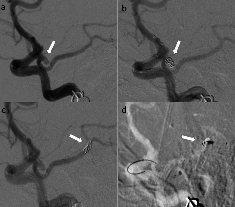
A four-panel digital subtraction angiography (DSA) series (lateral views) documenting a complication and management during endovascular aneurysm coiling. (a) Pre-treatment angiogram of the left internal carotid artery (ICA) reveals an irregular, wide-necked aneurysm at the posterior communicating artery (PCOM) origin. (b) Intra-procedural view demonstrates the initial placement of a radiopaque coil mass within the aneurysm sac. (c) Subsequent angiogram shows a procedural complication where the coil mass has dislodged and migrated distally into the left posterior cerebral artery (PCA), resulting in unintended vessel occlusion. (d) Roadmap image depicts the endovascular rescue maneuver, showing a stent retriever deployed across the migrated coil mass to facilitate mechanical retrieval. The series illustrates the clinical management of thromboembolic or hardware-related complications during neurointerventional procedures, specifically the transition from coil embolization to stent-assisted retrieval.
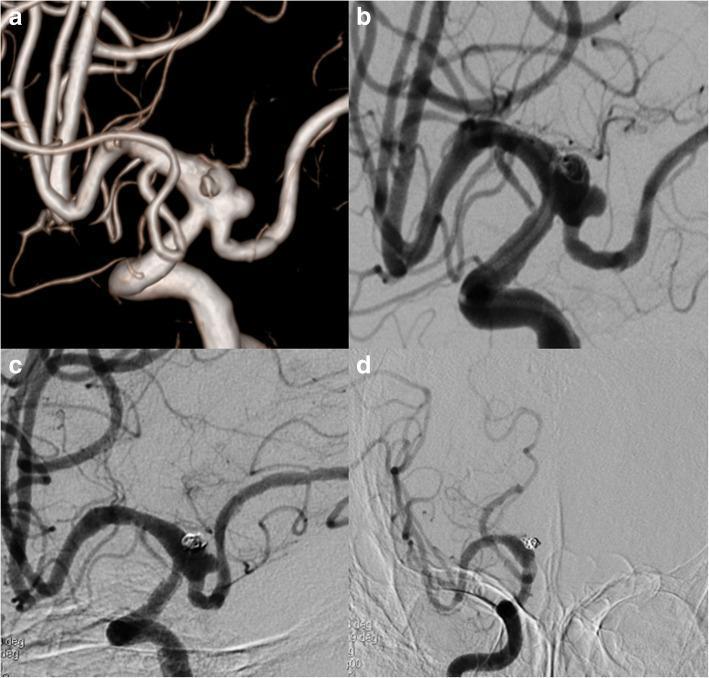
This composite of four cerebral angiographic images illustrates the endovascular treatment and follow-up of a Posterior Communicating Artery (PCoA) aneurysm. Panel (a) shows a pre-treatment 3D Digital Subtraction Angiogram (DSA) highlighting a wide-necked aneurysm with two daughter sacs at the PCoA origin, incorporating a fetal Posterior Cerebral Artery (PCA). Panel (b) is an immediate postoperative angiogram showing the placement of a Pipeline Embolization Device (PED) across the aneurysm neck combined with loose endovascular coiling. Panel (c) displays a four-month follow-up angiogram, demonstrating a reduction in aneurysm size and continued patency of the fetal PCA, though residual contrast filling persists. Panel (d) illustrates a significant hemodynamic change where the right A1 segment of the Anterior Cerebral Artery (ACA) is no longer patent; however, distal ACA flow is maintained via compensatory collateral circulation through the Anterior Communicating Artery (AComA). The series demonstrates flow diversion therapy, coil embolization, and intracranial collateral vascular remodeling.
carotid cavernous fistula angiogram endovascular embolization
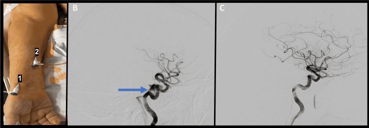
This composite educational image illustrates the diagnosis and treatment of a carotid-cavernous fistula (CCF). Panel A is a clinical photograph of a patient's forearm demonstrating two access points: transradial access (1) and ultrasound-guided transvenous access (2) through the cephalic vein. Panel B is a lateral digital subtraction angiogram (DSA) of the internal carotid artery (ICA) showing a direct carotid-cavernous fistula. A blue arrow highlights the fistula arising from the posterior genu of the cavernous ICA, with contrast drainage into the inferior petrosal sinus. Panel C is a post-procedural lateral angiogram following endovascular coil embolization. The image demonstrates successful obliteration of the fistula, with restoration of normal intracranial arterial flow and absence of premature venous filling. This series demonstrates a neurointerventional approach to vascular malformations using combined arterial and venous access for coil embolization.
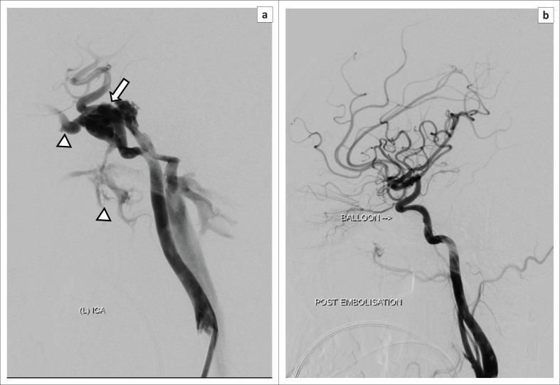
This lateral digital subtraction angiography (DSA) sequence demonstrates the diagnosis and treatment of a carotid-cavernous fistula (CCF). Image (a) is a pre-intervention angiogram of the left internal carotid artery (ICA) showing high-flow arteriovenous shunting. Key findings include rapid, premature opacification of the cavernous sinus (indicated by the white arrow) during the arterial phase, with subsequent retrograde drainage into anterior pathways (superior ophthalmic vein) and inferior pathways (pterygoid plexus), marked by arrowheads. Image (b) shows the post-embolization state, where the fistula has been successfully occluded using a detachable balloon (labeled). There is complete resolution of the abnormal cavernous sinus filling and venous drainage pathways, while the patency of the parent ICA and its distal intracranial branches is preserved. This clinical case illustrates the characteristic neurovascular imaging features of a traumatic CCF and the efficacy of endovascular embolization in restoring normal cerebral hemodynamics.
Endovascular treatment of cerebral vascular diseases
- Intracranial aneurysm
- Cerebral arteriovenous malformation (AVM) and dural arteriovenous fistula (dAVF)
- Carotid-cavernous fistula (CCF), historically called a carotid-cavernous anastomosis
1. General endovascular procedure
Basic sequence
-
Diagnostic imaging and planning
- CT and CT angiography, MRI/MR angiography, and especially catheter digital subtraction angiography (DSA) define the anatomy.
- DSA shows the feeding arteries, lesion, draining veins, collateral circulation, and flow direction.
-
General anesthesia or monitored sedation
- Many intracranial embolization procedures are performed under general anesthesia to prevent movement and permit strict control of blood pressure and ventilation.
-
Arterial access
- A sheath is inserted into the radial or femoral artery.
- A guide catheter is navigated through the aorta into the internal carotid artery or vertebral artery.
-
Microcatheter navigation
- A very small microcatheter is passed through the guide catheter into the aneurysm, arterial feeder, venous sinus, or fistulous point.
- This is performed under high-magnification fluoroscopy.
-
Treatment delivery
- The operator deploys coils, stents, flow diverters, liquid embolic material, plugs, or balloons depending on the lesion.
-
Completion angiography
- DSA confirms occlusion of the lesion, preservation of normal vessels, and absence of dangerous residual shunting.
-
Neurological monitoring and follow-up
- The patient is observed for stroke, bleeding, seizure, cranial-nerve deficit, or access-site complications.
- Follow-up CTA, MRA, or DSA is often needed because some lesions can recur or reopen.
2. Intracranial or cerebral aneurysms
Definition
- Anterior communicating artery
- Posterior communicating artery
- Middle cerebral artery bifurcation
- Internal carotid artery
- Basilar artery tip
- Vertebral and posterior inferior cerebellar arteries
Clinical presentation
Unruptured aneurysm
- Headache
- Oculomotor nerve palsy, especially with a posterior communicating artery aneurysm
- Diplopia
- Facial pain
- Visual symptoms
Ruptured aneurysm
- Sudden severe “thunderclap” headache
- Neck stiffness
- Nausea and vomiting
- Photophobia
- Loss of consciousness
- Seizure
- Focal neurological deficit
Principle of treatment
- Microsurgical clipping
- Endovascular embolization
A. Coil embolization
Principle
Step-by-step coil embolization
- DSA identifies the aneurysm and defines the neck, dome, parent vessel, and adjacent branches.
- A guide catheter is placed in the internal carotid or vertebral artery.
- A microcatheter is advanced through the parent artery into the aneurysm sac.
- A first coil, often called the framing coil, is deployed to form a stable basket.
- Additional coils are inserted to fill the sac densely.
- Each coil can be repositioned before detachment if its position is unsafe.
- The coil is detached electrolytically or mechanically when satisfactory.
- Final angiography confirms absent or minimal aneurysm filling and preserved parent-vessel flow.
Suitable aneurysms
Limitations
- Wide-neck aneurysms may not retain coils safely.
- Coils can compact over time.
- Residual filling or recurrence may require surveillance and retreatment.
B. Balloon-assisted coiling
Why it is used
Procedure
- One microcatheter is placed in the aneurysm for coil insertion.
- A balloon catheter is positioned in the parent artery across the neck.
- The balloon is temporarily inflated during deployment of coils.
- The balloon is deflated after the coil is stable.
- It is removed after adequate aneurysm occlusion is achieved.
Benefit and risk
C. Stent-assisted coiling
Principle
Procedure
- A stent is placed across the aneurysm neck.
- A microcatheter is passed through the stent cells or positioned first by a “jailing” technique.
- Coils are inserted into the aneurysm.
- Angiography confirms stable coil placement and parent-vessel patency.
Important consequence
D. Flow-diverter stent
Principle
Main uses
- Large or giant aneurysms
- Wide-neck aneurysms
- Fusiform aneurysms
- Selected internal carotid artery aneurysms
- Some recurrent aneurysms after coiling
Key points
- The aneurysm is not usually filled directly.
- Aneurysm occlusion occurs gradually over weeks to months.
- Dual antiplatelet therapy is required.
- It may not be suitable if important small branches arise from the treated segment.
E. Parent artery occlusion
Complications of aneurysm embolization
- Aneurysm perforation and intracranial hemorrhage
- Thrombus formation and ischemic stroke
- Coil protrusion or coil migration
- Parent-vessel or branch occlusion
- Vasospasm after aneurysmal SAH
- Recurrence due to coil compaction
- In-stent thrombosis
- Delayed aneurysm rupture, rarely after flow diversion
- Contrast reaction, radiation exposure, and access-site bleeding
3. Cerebral arteriovenous malformation, AVM
Definition
- Veins enlarge and become fragile
- Hemorrhage can occur
- Normal adjacent brain may receive less blood, called “steal”
- Venous hypertension can damage brain tissue
Presentation
- Intracranial hemorrhage
- Seizures
- Headache
- Focal neurological deficit
- Incidental discovery
Diagnosis
- CT or MRI identifies hemorrhage and structural abnormality.
- CTA/MRA may show enlarged vessels.
- DSA is the definitive study for treatment planning.
- Feeding arteries
- Nidus size and compactness
- High-flow shunts
- Deep versus superficial venous drainage
- Associated aneurysm
- Venous stenosis
- Surgical and embolization risk
AVM treatment options
- Observation and medical treatment
- Microsurgical removal
- Stereotactic radiosurgery
- Endovascular embolization
- Multimodal treatment combining two or more methods
Endovascular embolization of an AVM
Goals
- Cure a small, anatomically favorable AVM
- Reduce AVM size before stereotactic radiosurgery
- Reduce flow or remove high-risk features before microsurgery
- Obliterate an associated intranidal or feeding-artery aneurysm
- Treat a source of active or recurrent bleeding
Materials
- n-butyl cyanoacrylate, n-BCA: a tissue adhesive that polymerizes rapidly.
- Onyx: a nonadhesive liquid embolic material injected slowly under fluoroscopy.
- Detachable coils: used selectively, particularly in high-flow fistulous components or aneurysms.
- Particles: less commonly used in modern intracranial AVM treatment because of poor control and risk of non-target embolization.
Step-by-step AVM embolization
- DSA maps all arterial feeders, the nidus, and draining veins.
- A guide catheter is placed in the relevant carotid or vertebral artery.
- A microcatheter is advanced as close as possible to the nidus through a selected arterial feeder.
- The operator performs superselective angiography through the microcatheter to confirm the exact territory supplied.
- Liquid embolic agent is injected slowly into the nidus under fluoroscopy.
- The goal is to fill the nidus and proximal draining vein only as intended, without entering normal branches or excessively occluding the main venous drainage.
- The microcatheter is carefully removed.
- This may be repeated through multiple feeders, sometimes over more than one session.
- Completion DSA assesses residual AVM and normal arterial patency.
Critical technical principle
Important risk
4. Dural arteriovenous fistula, dAVF
Definition
- Dural sinus thrombosis
- Head trauma
- Prior cranial surgery
- Infection
- Occasionally without a clear cause
Why it can be dangerous
Presentation
- Pulsatile tinnitus
- Headache
- Eye symptoms
- Seizure
- Progressive neurological deficit
- Intracranial hemorrhage
Endovascular treatment of dAVF
Goal
Approaches
| Approach | Main use |
|---|---|
| Transarterial embolization | Microcatheter is advanced through a meningeal arterial feeder |
| Transvenous embolization | Microcatheter is placed in the involved venous sinus or venous pouch |
| Combined treatment | Used for complex lesions |
| Surgery or radiosurgery | Considered if endovascular therapy is unsuitable or incomplete |
Embolic agents
- Onyx
- n-BCA
- Coils
- Sometimes plugs or other specialized devices
Step-by-step outline
- Full cerebral DSA identifies every arterial feeder and venous drainage pathway.
- The operator determines whether a transarterial or transvenous route provides safer access to the shunt.
- A microcatheter is placed near the fistulous point or venous pouch.
- Coils, liquid embolic material, or both are deployed.
- The treatment aims to occlude the fistulous point and pathological venous outlet.
- Final DSA confirms elimination of early venous filling and cortical venous reflux.
5. Carotid-cavernous fistula, CCF
Definition
- Internal carotid artery
- Cranial nerves III, IV, V1, V2, and VI
- Ophthalmic veins
- Pituitary-region structures
Types of CCF
A. Direct CCF, Barrow type A
- Head trauma
- Skull-base fracture
- Iatrogenic arterial injury
- Ruptured cavernous internal carotid aneurysm
- Connective-tissue disease, including vascular Ehlers-Danlos syndrome
B. Indirect CCF, Barrow types B, C, and D
| Barrow type | Arterial source |
|---|---|
| B | Meningeal branches of the internal carotid artery |
| C | Meningeal branches of the external carotid artery |
| D | Branches of both internal and external carotid arteries |
Clinical features
- Pulsatile exophthalmos or proptosis
- Red eye and conjunctival chemosis
- Ocular bruit, often described as a pulsating or whooshing sound
- Pulsatile tinnitus
- Diplopia due to cranial-nerve III, IV, or VI palsy
- Eye pain
- Increased intraocular pressure and glaucoma
- Blurred vision or visual loss
- Headache
Diagnosis
CT and MRI findings
- Enlarged superior ophthalmic vein
- Enlarged cavernous sinus
- Proptosis
- Enlarged extraocular muscles
- Orbital congestion
- Flow voids
Definitive test: DSA
- Direct versus indirect fistula
- High-flow versus low-flow shunt
- Exact arterial feeders
- Venous drainage route
- Cortical venous reflux
- Feasibility of transarterial or transvenous treatment
Endovascular treatment of CCF
Treatment objectives
- Close the fistula.
- Preserve the internal carotid artery when possible.
- Relieve orbital venous hypertension.
- Prevent visual loss, intracranial hemorrhage, and cranial-nerve injury.
A. Transvenous embolization
Step-by-step
- Venous access is obtained, usually through the femoral vein.
- A catheter is passed through the internal jugular vein into the inferior petrosal sinus.
- A microcatheter enters the cavernous sinus.
- Detachable coils are deployed in the cavernous sinus or venous pouch.
- Liquid embolic material may be used in selected cases.
- Angiography confirms disappearance of the shunt and restoration of normal arterial circulation.
- Facial vein to superior ophthalmic vein
- Direct superior ophthalmic vein puncture
- Pterygoid plexus
- Superior petrosal sinus
- Other venous routes, depending on the anatomy
B. Transarterial embolization
- Arterial access is obtained.
- A microcatheter enters the cavernous internal carotid artery or crosses the fistulous opening.
- Coils, liquid embolic agent, or a balloon are used to close the communication.
- A temporary balloon may protect the internal carotid artery during coil deployment.
C. Covered stent or flow diverter
D. Parent artery sacrifice
CCF complications and follow-up
- Stroke due to thromboembolism or carotid occlusion
- Cranial-nerve palsy
- Worsening eye congestion or visual impairment
- Intracranial hemorrhage
- Coil migration
- Incomplete occlusion or recurrence
- Parent-vessel injury
- Access-site bleeding
6. Comparison of the lesions
| Feature | Aneurysm | Brain AVM | dAVF | CCF |
|---|---|---|---|---|
| Main abnormality | Weak, dilated artery | Arteries connected to veins through nidus | Meningeal arteries connected to dural sinus/vein | Carotid circulation connected to cavernous sinus |
| Main danger | Rupture causing SAH | Hemorrhage, seizure, neurological deficit | Venous hypertension, hemorrhage, neurological deficit | Visual loss, cranial-nerve palsy, orbital congestion, hemorrhage in high-risk drainage |
| Principal endovascular aim | Exclude aneurysm from circulation | Obliterate nidus or reduce high-risk flow | Disconnect fistula and dangerous venous reflux | Close shunt and preserve ICA if possible |
| Common materials | Coils, stent, flow diverter | Onyx, n-BCA, coils | Onyx, n-BCA, coils | Coils, liquid embolic agent, balloon, covered stent |
| Common route | Transarterial | Mainly transarterial | Transarterial or transvenous | Usually transvenous, sometimes transarterial |
7. General risks of neuroendovascular therapy
- Ischemic stroke from embolus, thrombosis, or vessel occlusion
- Intracranial hemorrhage
- Vessel perforation or dissection
- Brain swelling
- Seizure
- Cranial-nerve deficit
- Contrast-associated kidney injury or contrast allergy
- Radiation exposure
- Groin or wrist bleeding, hematoma, pseudoaneurysm, or arterial occlusion
- Need for emergency open surgery or repeat endovascular treatment