
diabetic foot ulcer detailed examination s das surgery
diabetic foot ulcer Wagner classification examination
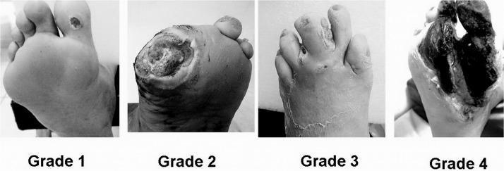
A comparative clinical photograph set illustrating the Wagner Classification System for diabetic foot ulcers across four stages (Grade 1 to Grade 4). Grade 1 (Superficial Ulcer) shows a small, localized skin lesion on the plantar surface near the base of the toes with no deep tissue involvement. Grade 2 (Deep Ulcer) depicts a larger, deeper wound penetrating to the tendon or joint capsule without abscess or osteomyelitis. Grade 3 (Osteitis/Infection) demonstrates a deep ulcer associated with significant inflammation, purulent discharge, and clinical signs of underlying bone infection or deep tissue abscess. Grade 4 (Forefoot Gangrene) shows extensive necrosis and blackening of the tissue localized to the forefoot or toes, indicating severe vascular compromise and tissue death. This series serves as an educational tool for medical professionals to differentiate between stages of diabetic foot pathology, guiding clinical management and prognosis assessment.
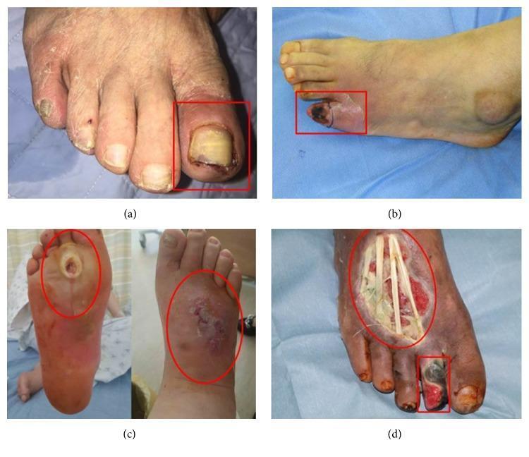
A series of four clinical photographs (a-d) demonstrating the progression of diabetic foot ulcers categorized by the Wagner classification system. (a) Wagner Grade 1: A superficial ulcer on the left hallux (big toe) featuring erythema, periungual inflammation, and a thickened, discolored yellowish toenail, indicative of an ischemic etiology. (b) Wagner Grade 2: A deep ulcer on the fifth digit (pinky toe) showing more extensive tissue involvement and localized cyanosis. (c) Wagner Grade 3: A neuropathic perforating ulcer extending from the plantar surface to the dorsum of the foot, associated with deep infection, abscess, and clinical signs of osteomyelitis. (d) Wagner Grade 4: Advanced diabetic foot pathology exhibiting local gangrene of the second toe and a large, deep dorsal foot ulcer. The necrotic tissue is blackened, and the wound bed is exposed, revealing underlying anatomical structures such as tendons. This set illustrates the clinical manifestations of ischemic, neuropathic, and mixed-type diabetic foot disease for educational use in wound care and endocrinology.
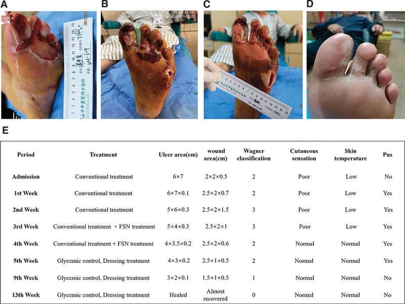
This composite figure presents a clinical progression timeline of a complex diabetic foot ulcer (DFU) on the left foot, categorized under the Wagner classification system. (A) Initial presentation shows a large, 6x7 cm ulcer on the plantar forefoot with surrounding maceration and a lateral abscess. (B) Following initial debridement, the wound exhibits deep tissue involvement (Wagner Grade 3), with necrotic tissue extending to the tendons and musculature of the first through third toes, accompanied by a deep 2x2x0.7 cm abscess cavity. (C) Mid-treatment status demonstrates significant clinical improvement; the wound bed appears ruddy with healthy granulation tissue, reduced exudate, and diminishing necrotic debris. (D) Final follow-up at three months shows complete secondary intention healing with full epithelialization and stable scar tissue formation. (E) An accompanying summary table provides longitudinal data on treatment phases (including Fu's subcutaneous needling), ulcer/wound dimensions, Wagner grade transition (from Grade 3 to 0), and clinical markers such as cutaneous sensation, skin temperature, and presence of pus.
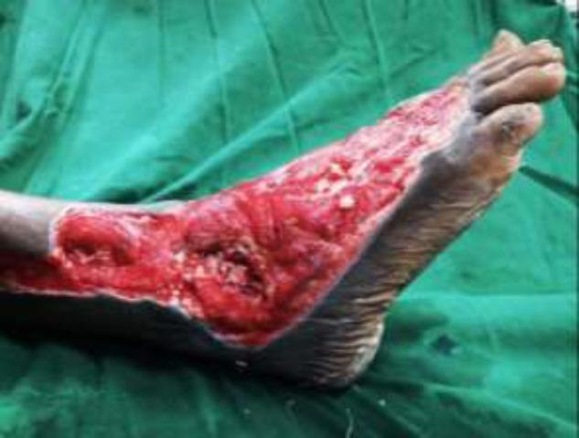
Clinical photograph of a right foot demonstrating a severe, extensive Wagner Grade III or IV diabetic foot ulcer. The wound spans the lateral and dorsal aspects of the foot, extending from the distal lower leg across the ankle joint to the midfoot region. The ulcer bed is characterized by beefy red granulation tissue and deep cavitation, with visible exposure of underlying musculoskeletal structures including the ankle joint. There are focal areas of yellow/white slough and fibrin, indicative of potential infection or chronic inflammatory processes. The wound margins are irregular but well-demarcated against the surrounding hyperpigmented, intact skin. This image serves as a clinical example of complex diabetic wound progression requiring advanced surgical intervention, such as debridement and microvascular flap reconstruction. Key educational concepts include diabetic foot classification, wound bed assessment, and the management of deep tissue infections with joint involvement.
Diabetic Foot Ulcer - Detailed Examination
I. History Taking
- Duration of ulcer, previous episodes of ulceration
- Prior amputations or surgeries on the foot
- Neuropathic symptoms: paresthesias, numbness, burning pain (note: many patients deny trauma due to impaired sensation)
- Vascular symptoms: claudication, rest pain
- Glycemic control: current medications, last HbA1c
- Renal history, impaired vision (retinopathy)
- Footwear history: type of footwear used, any foreign bodies noticed inside shoes
- Known Charcot neuroarthropathy
- Systemic symptoms (fever, chills) - suggest ascending infection requiring hospital admission
II. General Inspection - Start at the Shoes
| Finding | Significance |
|---|---|
| Undersized shoes | Increased pressure over bony prominences -> ulceration |
| Inadequate toe box | Cannot accommodate forefoot deformities |
| Abnormal wear pattern | Structural or dynamic foot deformity |
| Prominent seams / foreign bodies not noticed by patient | Evidence of neuropathy |
III. Inspection of the Foot (Without Socks/Shoes)
A. Deformities
- Claw toes / hammertoes / foot drop - indicate motor neuropathy
- Atrophy of extensor digitorum brevis - motor neuropathy
- Fixed flexion deformities of IP joints - shift plantar pressure, cause ulceration at tip of toes
- Arch and hindfoot alignment (weight-bearing) - altered plantar pressure distribution
- Charcot foot (rocker-bottom deformity) - flattened arch, bony prominence on plantar surface
B. Skin Changes
| Finding | Significance |
|---|---|
| Shiny, taut skin without hair growth | Peripheral arterial disease (PAD) |
| Dry, scaly skin | Autonomic dysfunction (loss of sweat glands) |
| Erythema, swelling, calor | Infection OR Charcot neuroarthropathy |
| Corns, calluses | High-pressure areas - precursors to ulceration |
| Maceration between toes | Fungal infection, interdigital ulceration |
Tip to differentiate Charcot from infection: Elevate the limb. Erythema/warmth from Charcot subsides with elevation; infection does not.
IV. Wound Assessment (The Ulcer Itself)
A. Site
B. Size
- Measure the longest dimension x widest perpendicular dimension to get cross-sectional area
- Serial measurements at each visit track healing (a 50% reduction in size at 4 weeks predicts healing at 12 weeks)
C. Depth and Probing
- Use a sterile cotton swab/blunt probe to assess depth
- Note involvement of: skin only, subcutaneous tissue, tendon, joint capsule, bone
- Probe-to-bone test: if bone is palpable with the back of a cotton swab, assume osteomyelitis (PPV 57%, NPV 96% - Lavery et al.)
D. Wound Bed
- Granulation tissue (healthy, pink/red)
- Fibrous/sloughy tissue (pale, yellow)
- Necrotic tissue (black, dry eschar or wet gangrene)
E. Wound Margins and Surrounding Skin
- Raised, thickened callus around the margin increases plantar pressure and blocks inward epithelialization
- Spreading erythema beyond 2 cm from wound margin - suggests active infection
- Purulent drainage, odor, maceration - signs of infection
- Penetration of fascial layers - requires surgical evaluation
V. Vascular Assessment
- Medial calcinosis ("lead pipeline" vessels) - palpable but non-compressible
- Pedal edema obscures pulse palpation
- Small vessel disease with poor collateralization (angiosomal ischemia despite palpable pulse)
A. Pulse Palpation
- Dorsalis pedis (between 1st and 2nd metatarsals, dorsum of foot)
- Posterior tibial (behind medial malleolus)
- Absent or diminished pulses -> refer for Doppler
B. Capillary Refill Time
- Prolonged (>2 seconds) suggests poor perfusion
C. Buerger's Test
- Elevate legs to 45 degrees for 1-2 min - pallor of the sole indicates ischemia
- Then hang the legs down - reactive hyperemia (rubor) of dependency suggests ischemic limb
D. Ankle-Brachial Index (ABI)
- Noninvasive, rapid perfusion screen
- Interpretation: ABI ≥0.9 normal | 0.6-0.79 mild ischemia | 0.4-0.59 moderate | <0.4 severe
- Limitation in diabetes: arteriosclerosis -> non-compressible vessels -> falsely elevated ABI
- An ABI difference >0.15 between limbs is significant
E. Toe-Brachial Index (TBI) / Toe Pressures
- More reliable in diabetics (digital arteries often spared from calcification)
- Normal TBI >0.7; Toe pressure >40 mmHg = adequate perfusion for wound healing
- Toe pressure >60-70 mmHg needed for healing in diabetics
- TBI <0.4 or toe pressure <30 mmHg = severe ischemia -> vascular referral
WIFI Ischemia Grading (Current Surgical Therapy)
| Grade | ABI | Toe Pressure |
|---|---|---|
| 0 | ≥0.80 | ≥60 mmHg |
| 1 | 0.60-0.79 | 40-59 mmHg |
| 2 | 0.40-0.59 | 30-39 mmHg |
| 3 | <0.40 | <30 mmHg |
Important: Do NOT perform aggressive debridement until adequate perfusion is confirmed, unless severe infection demands immediate action.
VI. Neurological Examination
A. Semmes-Weinstein Monofilament Test (10-g / 5.07 monofilament)
- Inability to perceive 10 g of pressure = loss of protective sensation = one of the strongest predictors of foot morbidity
- Test 10 sites on the plantar surface (metatarsal heads, heel, arch, toes)
- Equivalent screening: 4.5-g monofilament beneath both first metatarsal heads
- Failure to perceive the 5.07 monofilament = 30% risk of ulcer development
B. Vibration Sensation
- 128 Hz tuning fork applied at great toe
- Reduced/absent = large fiber neuropathy
C. Pain and Temperature Sensation
- Pin-prick and temperature testing for small fiber neuropathy
D. Deep Tendon Reflexes
- Absent ankle jerk (Achilles reflex) = peripheral neuropathy
E. Proprioception
- Romberg's test for balance
VII. Musculoskeletal / Biomechanical Examination
Silfverskiold Test (Ankle Dorsiflexion)
- Record ankle ROM with knee flexed then knee extended
- Gastrocnemius contracture: dorsiflexion limited with knee extended but improved with knee flexed
- Achilles tendon contracture: equally reduced in both positions
- Gastrocnemius/Achilles tightness increases forefoot pressure and predisposes to plantar ulceration
Range of Motion
- Assess subtalar, midfoot, and MTP joint mobility
Gait Assessment
- Observe patient walking - balance, offloading behavior, antalgic gait
VIII. Ulcer Classification
Wagner-Meggitt Classification (Most Commonly Used in S Das Surgery)
| Grade | Description |
|---|---|
| 0 | Intact skin; pre-ulcerative lesion (callus, corn); healed ulcer |
| 1 | Superficial ulcer, skin only |
| 2 | Deep ulcer to tendon, joint capsule, or bone (no abscess/osteomyelitis) |
| 3 | Deep ulcer with abscess, osteomyelitis, or septic arthritis |
| 4 | Localized gangrene (forefoot/heel) |
| 5 | Extensive gangrene involving the whole foot |
IWGDF/IDSA Infection Classification
| Grade | Description |
|---|---|
| 1 (Uninfected) | No local/systemic signs |
| 2 (Mild) | ≥2 local signs (erythema 0.5-2 cm, induration, warmth, purulent discharge), no systemic involvement |
| 3 (Moderate) | Erythema ≥2 cm, involves deeper tissue (tendon, muscle, joint, bone), no SIRS |
| 4 (Severe) | Any foot infection with systemic inflammatory response (SIRS criteria) |
IX. Wound Images (Wagner Classification - Clinical Examples)
X. Laboratory and Imaging Investigations
| Investigation | Purpose |
|---|---|
| HbA1c | Glycemic control - >7% associated with higher complications |
| Fasting blood glucose | Pre-operative glucose >200 mg/dL = higher infection/wound healing risk |
| CBC | Leukocytosis suggests infection |
| ESR, CRP | Elevated supports osteomyelitis (non-specific) |
| X-ray (3 views) | Detect gas in soft tissue, foreign bodies, late osteomyelitis changes |
| MRI foot | Gold standard for early osteomyelitis; use if equivocal exam or non-healing at 4-6 weeks |
| Bone scan / tagged WBC scan | If MRI contraindicated (metal hardware, pacemaker) |
| Bone biopsy / culture | Gold standard diagnosis AND microbiological speciation for osteomyelitis |
| Doppler / ABI / toe pressures | Vascular assessment |
XI. Key Surgical Decision Points from Examination
- Adequate perfusion? - Do not debride aggressively until vascular status confirmed (unless life/limb threatened by severe infection)
- Probe-to-bone positive? - Assume osteomyelitis; plan MRI/bone biopsy
- Spreading erythema >2 cm or SIRS? - Hospital admission, IV antibiotics, urgent surgical review
- Wagner Grade 3 or higher / WIFI Infection Grade 2-3? - Surgical debridement, possible amputation planning
- Poor glycemic control (HbA1c >7%)? - Involve endocrinology before elective surgery
lymphnode palpation in lower limb with picture
inguinal lymph node palpation technique lower limb
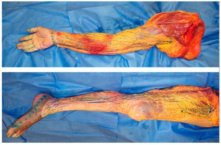
This composite clinical photograph displays two dissection specimens of the upper (top) and lower (bottom) human limbs, illustrating the superficial lymphatic system through a microinjection technique. The specimens have had the skin and superficial adipose tissue removed to reveal the superficial lymph-collecting vessels, which have been injected with blue acrylic dye for visualization. In the upper limb, the blue-stained lymphatic vessels are seen originating from the hand and extending proximally toward the axillary region, where a white arrow identifies a sentinel lymph node. In the lower limb, vessels traverse from the foot and lower leg toward the groin, with a black arrow marking a sentinel node in the superficial inguinal region. The vessels appear as fine, independent, dark blue branching pathways within the subcutaneous layer, demonstrating a lack of extensive interconnections or network formation. These images serve as an anatomical reference for understanding normal lymphatic drainage, lymphosome territories, and the baseline pathways involved in conditions like lymphedema or cancer metastasis.
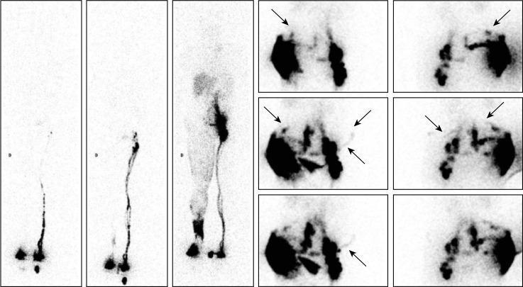
Diagnostic lymphoscintigraphic images of a 65-year-old male with secondary right lower limb lymphedema (LLLE) following inguinal node dissection for Merkel cell carcinoma. The panel includes three consecutive whole-body scans (WBS) and static pelvic images. Phase 1 (rest) shows normal tracer drainage to the left inguinal lymph node, while the right side displays delayed outflow. Phase 2 (active movement) demonstrates increased activity in the left inguinal region and limited distal drainage on the right. Phase 3 (post-walking) reveals significant pathological findings on the right, including dermal backflow at the foot and ankle, and a superficial collateral network extending proximally toward visible inguinal and iliac nodes. Detailed pelvic views (anterior and posterior) show collateral lymphatic drainage vessels (LVs), specifically median and oblique pathways. These include LVs crossing the midline and drainage toward the contralateral (left) inguinal lymph nodes. Arrows highlight specific collateral channels and areas of midline crossover, demonstrating physiological adaptations to lymphatic obstruction.
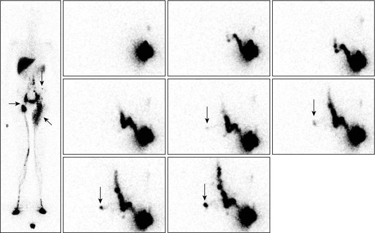
This composite diagnostic image features Phase 3 and Phase 4 99mTc-nanocolloid lymphoscintigraphy of a 13-year-old patient with primary lower limb lymphedema. The left panel shows an anterior whole-body scan (WBS) obtained after walking. It illustrates tracer progression from the feet. The right limb displays a horizontal arrow marking an inguinal nodal gap and decreased uptake in the common iliac and lumbo-aortic chains. The left limb shows lymphatic reflux toward the superficial dermal network of the thigh (oblique arrow) and an isolated lymph node in the left flank (vertical arrow). The right panel comprises a sequence of eight static images centered on the pelvis/abdomen following a supplemental intradermal injection in the left external buttock. This sequence demonstrates abnormal collateral drainage pathways: a lymphatic vessel (marked by vertical arrows) is seen crossing the anterior midline from the left inguinocrural region toward the right inguinal area, eventually reaching a contralateral inguinal lymph node. These findings characterize collateral lymphatic compensation in the setting of primary lymphatic insufficiency.
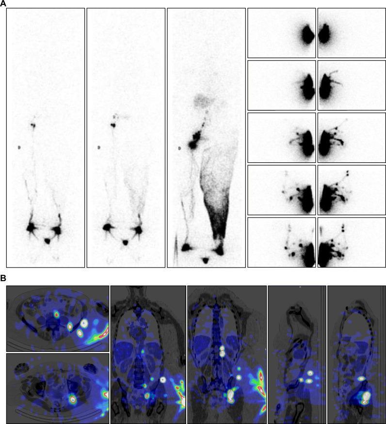
This composite figure displays diagnostic lymphoscintigraphic and SPECT-CT imaging of a 74-year-old female with secondary lower limb lymphedema following inguinal and iliac lymph node dissection. Panel A (Top): Anterior Whole-Body Scans (WBS) across three phases. Phase 1 (resting) shows normal drainage on the right and limited distal tracer progression on the left. Phase 2 (post-exercise) reveals increased right inguinal nodal activity and collateral vessels above the left ankle. Phase 3 (post-walking) demonstrates extensive dermal backflow on the left extending from the ankle to the groin, without visualizing distinct lymph nodes. Subsequent intradermal gluteal injections (right sub-panels) reveal alternate lymphatic pathways. Panel B (Bottom): SPECT-CT fusion images in transverse, coronal, and sagittal planes. These images localize radiocolloid uptake to deep collateral pathways, identifying intergluteal lymphatic vessels and intercalated nodes near the iliac crest and paravertebral regions. The imaging highlights the clinical significance of identifying deep lymphatic drainage when superficial inguinal nodes are absent, aiding in the surgical planning for lymph node-to-vein anastomosis or targeted physical therapy.
superficial inguinal lymph nodes anatomy diagram groin
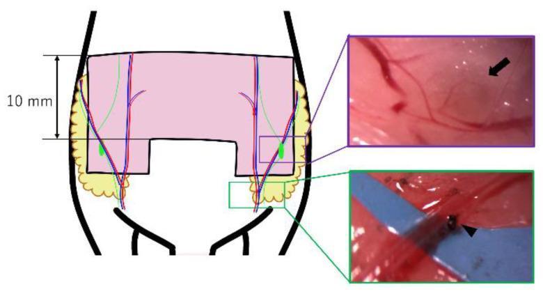
This medical illustration and surgical photograph composite details a murine model flap design for researching vascularized lymph node transfer (VLNT). The primary diagram shows a 10 mm wide bilateral pedicled abdominal flap (pink U-shaped skin paddle) that incorporates subcutaneous fat (yellow) and inguinal lymph nodes (green ovals). The vascular anatomy is depicted with abdominal arteries (red) and veins (blue). A purple inset provides a high-magnification view identifying a left groin lymph node (black arrow) and its associated feeding vessels. A green inset demonstrates the surgical technique for creating a unilateral congestion model through venous ligation; a black arrowhead indicates the specific ligation point on the vein, performed with a 10-0 nylon suture. This model is used to simulate clinical scenarios of impaired blood flow in transferred lymph nodes, allowing for the comparative study of ischemic, congestive, and control flap survival rates and pathological changes.
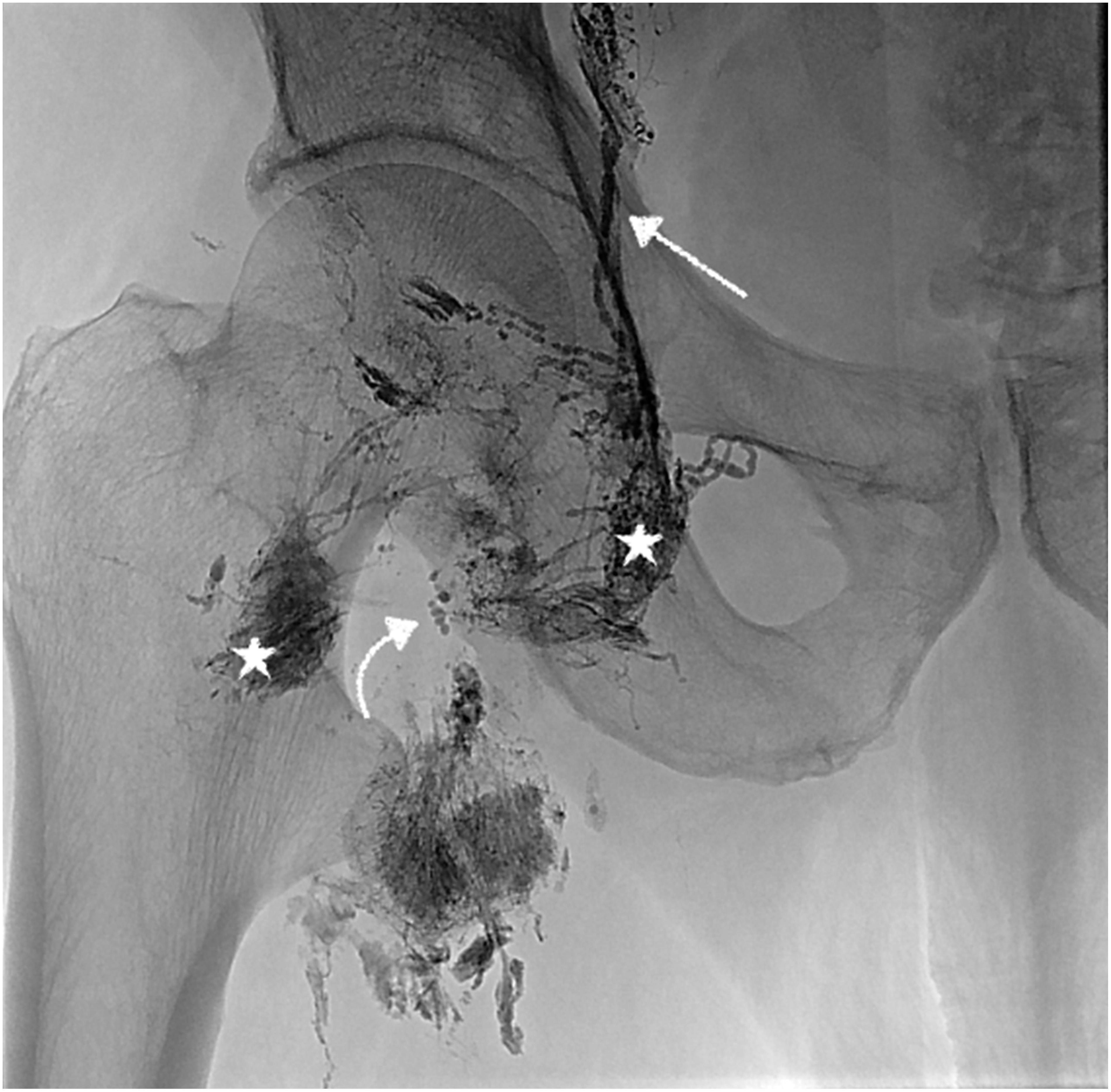
This diagnostic image is an intranodal lymphangiogram of the right groin region, demonstrating contrast-enhanced lymphatic anatomy and pathology. The image reveals multiple inguinal lymph nodes injected with contrast material (marked with stars), appearing as dense, opaque aggregates. Connecting these nodes are normal lymphatic vessels, visualized as thin, well-defined, linear branching structures (indicated by a straight arrow). A significant finding is the presence of lymphatic extravasation, characterized by an amorphous, cloud-like distribution of contrast material leaking from a medially located lymph node (indicated by a curved arrow). This extravasation represents a lymphatic leak or lymphorrhea, contrasting with the discrete margins of the healthy vessels. The imaging provides essential clinical context for identifying specific leakage sites in patients with post-surgical wound complications, such as those following vascular interventions, and serves as a roadmap for subsequent interventional procedures like N-butyl cyanoacrylate (NBCA) glue embolization.
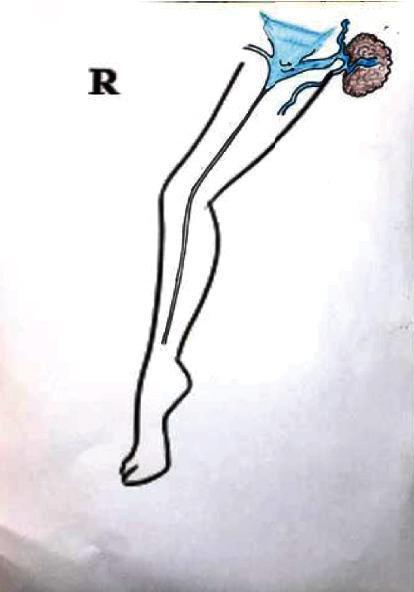
This medical schematic illustrates the vascular anatomy of the right lower extremity, focusing on mechanisms for the recurrence of varicose veins in the groin region. The diagram depicts the outline of a leg with a primary vertical vessel, representing the great saphenous vein (GSV), extending from the foot to the thigh. In the inguinal (groin) area, specifically within Scarpa's triangle, the illustration highlights a complex vascular network in blue. Key components include a triangular-shaped structure representing the saphenofemoral junction or a venous confluence, and a brown, textured cluster representing a lymph node. Small blue vessels are shown interconnecting the lymph node with the main venous system, demonstrating lymphatic-venous communication. The diagram serves an educational purpose by visualizing how accessory lymph node vessels can serve as pathways for venous reflux, leading to recurrent varicosities after surgical or endovenous treatments like radiofrequency ablation (RFA). This clinical illustration is designed for vascular surgery and phlebology education.
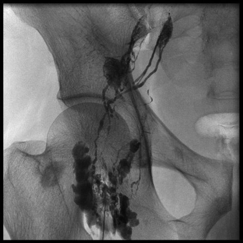
This diagnostic fluoroscopic image is a groin lymphangiogram utilizing Lipiodol contrast. The radiograph displays the anatomical distribution of the inguinal lymphatic system in relation to the pelvic skeletal structures, including the iliac bone, acetabulum, and proximal femur. Opacified lymphatic vessels appear as thin, branching linear channels that ascend from the femoral region toward the pelvis. Multiple dense, lobulated clusters represent contrast-filled superficial and deep inguinal lymph nodes. The nodes vary in morphology and contrast density, with some showing intense opacification indicating successful uptake. The vessels track along the expected medial aspect of the thigh and course superiorly toward the external iliac chain. This imaging modality is typically used for investigating lymphatic leaks, such as chylothorax or chylous ascites, and for guiding interventions like thoracic duct embolization. The image demonstrates normal and variant branching patterns of the lymphatic network essential for procedural planning in interventional radiology.
lymph node examination palpation technique clinical
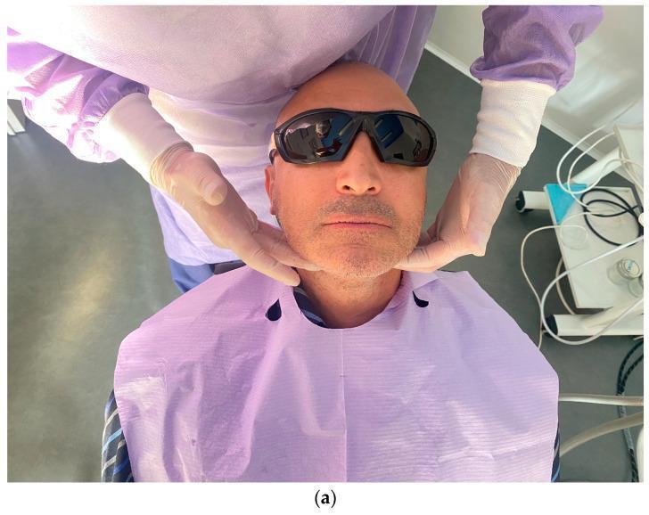
This clinical photograph demonstrates an extraoral examination of the submandibular and cervical lymph nodes. The patient is positioned seated and reclined, wearing protective eyewear and a lavender dental bib. A clinician, donned in a lavender surgical gown and light-colored gloves, is shown performing manual palpation by placing their hands bilaterally along the patient's jawline and upper neck area. This procedure is a critical component of a comprehensive head and neck examination in dental and oral surgery contexts, used to detect lymphadenopathy or signs of inflammatory processes related to dental implants or oral infections. The image highlights the correct hand positioning for systematic palpation of the facial and neck soft tissues.
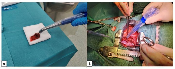
Clinical photographs demonstrating sentinel lymph node (SLN) detection techniques using a handheld gamma probe. Panel A shows an ex vivo examination of an excised lymph node specimen, which appears as a dark red, fleshy tissue on a sterile white gauze pad over a surgical drape. A gamma probe with a blue handle, encased in a sterile transparent sheath, is pointed at the specimen to measure radioactive uptake. Panel B displays the in vivo intraoperative application of the same technique. An open surgical field is maintained by a metallic self-retaining retractor, revealing deep reddish tissues and anatomical structures. The surgeon is utilizing the sterile gamma probe to scan the wound bed for any remaining radioactive 'hot spots' to identify additional sentinel lymph nodes. The image illustrates the workflow of radioguided surgery for oncological staging, focusing on the identification and removal of SLNs following the injection of a radioactive tracer.
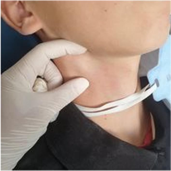
This clinical photograph displays a lateral view of a pediatric patient's right neck, highlighting the submandibular region. A medical professional, wearing a white nitrile glove, is performing physical palpation of a visible anatomical abnormality. The key finding is a localized, rounded swelling or bulge measuring approximately 3 cm in the right submandibular area, consistent with lymphadenopathy. The overlying skin exhibits mild erythema compared to the adjacent cervical tissue but remains intact without ulceration or drainage. The image demonstrates the clinical examination technique for assessing cervical lymph nodes, specifically focused on size, mobility, and contour in the context of oral or systemic infection. The presence of a surgical face mask around the patient's neck and a dark shirt collar provide additional orientation.
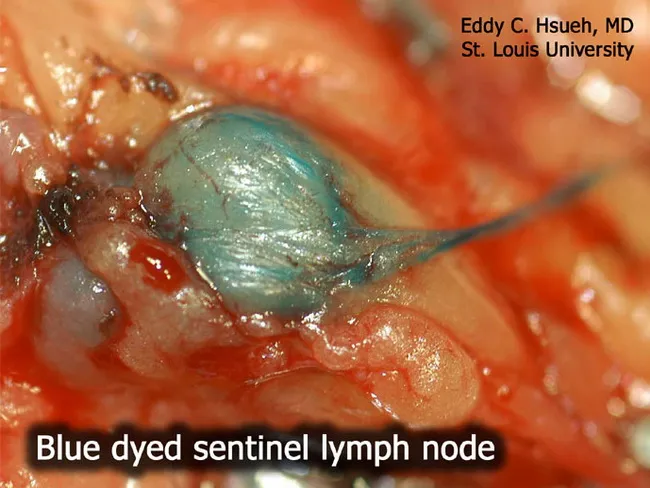
Imaging modality: Intraoperative surgical photography of a sentinel lymph node biopsy for malignant melanoma. Technique: blue dye mapping using Lymphazurin blue (isosulfan blue) injected near the primary lesion to trace afferent lymphatics to the sentinel node, which is visually stained a blue-green hue. Anatomical context: axillary regional lymphatics draining the tumor bed are highlighted by dye transport, with the sentinel lymph node identified by conspicuous color uptake. Visual features: a single lymph node is enlarged and engorged, with surrounding adipose tissue and operating field in clear view; the node exhibits uniform blue coloration within the cortex and hilum, with surrounding soft tissues appearing erythematous from operative exposure. Notable findings: blue-stained sentinel node ready for targeted excision; no gross metastatic nodal disease is discernible on this view, though microscopic metastasis may be present. Clinical significance: sentinel lymph node biopsy is a standard staging procedure in melanoma, enabling nodal assessment while minimizing extensive lymphadenectomy. Differential considerations: accidental intradermal staining, dye leakage, or allergic reaction to isosulfan blue. Clinical correlation: histopathologic examination of the excised node is required to determine nodal metastasis status, guiding adjuvant therapy and prognosis. This image serves educational and surgical planning purposes for clinicians, trainees, and researchers studying lymphatic mapping in cutaneous melanoma.
popliteal lymph node anatomy location posterior knee
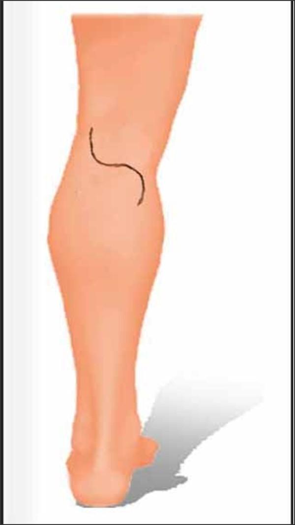
This medical illustration depicts the surgical planning for a posterior approach to the knee, specifically for popliteal lymph node dissection. The image shows a posterior view of the left lower limb with a planned S-shaped (Lazy-S) incision line. The central transverse segment of the incision is positioned directly over the popliteal crease. The cranial (superior) portion of the incision curves laterally toward the thigh, while the caudal (inferior) portion curves medially toward the calf. This specific curvilinear geometry is designed to provide optimal exposure of the popliteal fossa contents—including the neurovascular bundle and lymph nodes—while preventing vertical scar contracture across the joint flexion line. The anatomical landmarks shown include the posterior thigh, the popliteal region, and the gastrocnemius muscle bulk of the calf. This type of incision is standard in surgical oncology and vascular surgery to minimize functional impairment of the knee joint postoperatively.
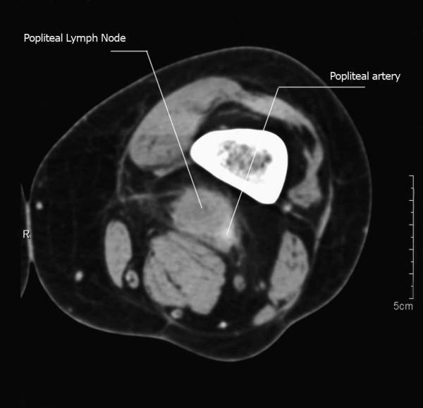
Axial MRI (T1-weighted image) of the popliteal region showing a metastatic mass originating from squamous cell carcinoma. The visual identifies a primary soft-tissue mass, labeled as a popliteal lymph node, measuring approximately 3 cm in size within the popliteal fossa. The mass is hyperintense relative to adjacent muscle and demonstrates a close anatomical relationship with the vascular bundle, specifically encasing the popliteal artery, which is labeled for reference. The image illustrates the complex anatomy of the posterior knee, including the femur and surrounding musculature. This diagnostic image is intended for medical education regarding oncological staging, regional lymphatic spread, and the surgical considerations of popliteal lymphadenectomy when tumors approximate major neurovascular structures.
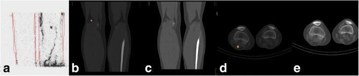
A multi-panel diagnostic imaging series demonstrating sentinel lymph node (SLN) localization in a patient with malignant melanoma of the right foot. (a) Planar lymphoscintigraphy of the right popliteal region showing diffuse radiotracer uptake and faint lymphatic channels. (b) Sagittal SPECT/CT fusion image of the popliteal region, providing high-contrast localization of a single focal SLN within the posterior leg anatomy. (c) Corresponding sagittal low-dose CT alone, displaying the anatomical structures of the knee and lower limb without tracer data. (d) Axial SPECT/CT fusion image clearly identifying the SLN located posteriorly to the popliteal fossa. (e) Axial low-dose CT at the same level showing cross-sectional musculoskeletal anatomy of both knees. The comparison highlights the clinical advantage of hybrid SPECT/CT over planar lymphoscintigraphy for precise 3D anatomical mapping and surgical planning during sentinel node excision (SLNE) for cutaneous melanoma.
Lymph Node Palpation in the Lower Limb
Lymph Nodes of the Lower Limb - Overview
- Inguinal lymph nodes (in the groin) - primary group
- Popliteal lymph nodes (behind the knee) - secondary/satellite group
I. INGUINAL LYMPH NODES
Anatomy
| Group | Location | Drains |
|---|---|---|
| Horizontal group (superolateral + superomedial) | Parallel to inguinal ligament, just inferior to it | Lower trunk (below umbilicus), perineum, external genitalia, perianal region, lower anterior abdominal wall |
| Vertical group (inferior superficial) | Along the terminal great saphenous vein in the femoral triangle | Lower extremity - entire leg and thigh via superficial lymphatics |
- Located along the medial aspect of the femoral vein
- Most superior = Node of Cloquet (in femoral ring) - sentinel node indicating spread beyond inguinal ligament to pelvic nodes
- Drain: deep lymphatics of lower limb + efferents from superficial nodes -> external iliac nodes
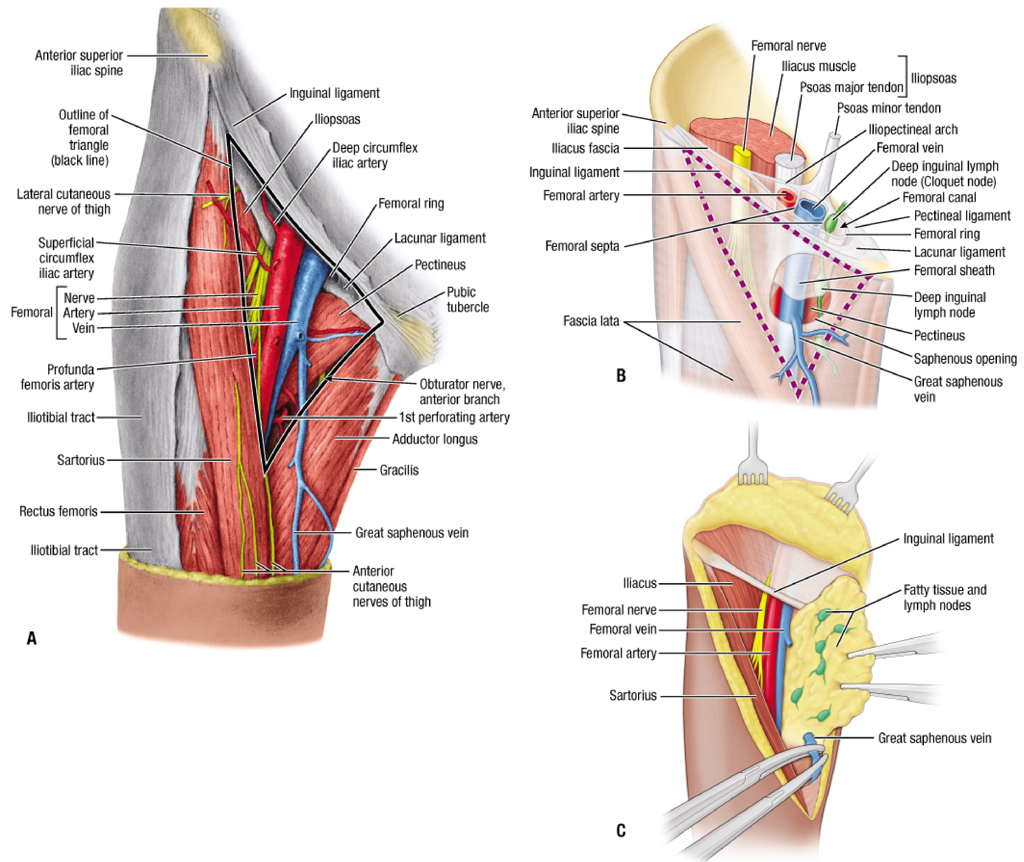
Anatomical Diagram (Fischer's Mastery of Surgery)
Palpation Technique - Inguinal Nodes
-
Identify landmarks: Palpate the anterior superior iliac spine (ASIS) and the pubic tubercle. The inguinal ligament runs between them.
-
Horizontal group: Place fingers just below and parallel to the inguinal ligament, sweeping from ASIS medially toward the pubic tubercle. These nodes are felt along the inguinal crease in the subcutaneous tissue.
-
Vertical group: Move fingers inferiorly along the line of the great saphenous vein (medial thigh, just below the saphenous opening/fossa ovalis). These nodes drain the leg and foot.
-
Palpate using the pulp of 2-4 fingers in a gentle rolling motion against the underlying femoral vessels.
-
Deep inguinal nodes: Not usually palpable unless enlarged. They lie deep to fascia lata, medial to the femoral pulse. Press firmly medial to the femoral artery to attempt palpation.
Horizontal = Hip/trunk/perineum (above the waist line of drainage) Vertical = Vein (along great saphenous vein) = Vertebral column of the leg
II. POPLITEAL LYMPH NODES
Anatomy
- Located in the popliteal fossa (diamond-shaped space behind the knee)
- Small cluster of 4-6 nodes embedded in the fat around the popliteal vessels
- Drain: dorsum of the foot, lateral leg (via short saphenous vein territories), deep structures of leg (via anterior/posterior tibial and peroneal vessels)
- Efferents pass upward with the femoral vessels to deep inguinal nodes
Popliteal Fossa Boundaries (for orientation)
- Superomedially: Semimembranosus and semitendinosus
- Superolaterally: Biceps femoris
- Inferomedially: Medial head of gastrocnemius
- Inferolaterally: Lateral head of gastrocnemius
Palpation Technique - Popliteal Nodes
-
Prone position (best): Patient lies face down, examiner stands at foot end. Cup both hands around the back of the knee, thumbs on the patella anteriorly.
-
Flex the knee to about 30-40 degrees - this slackens the popliteal fascia and makes nodes accessible.
-
Press finger pads deeply into the popliteal fossa, between the two heads of gastrocnemius inferiorly. The nodes are deep and small - significant force is required.
-
Supine alternative: Support the knee with one hand under the popliteal fossa, flex to 30 degrees; use the other hand's fingers to press deeply.
Note: Popliteal nodes are not palpable normally. They enlarge with infections of the heel/lateral foot/lower leg.
Superficial Lymphatic Drainage of Lower Limb - Visual
III. WHAT TO ASSESS ON PALPATION (Characters of a Lymph Node)
| Feature | Normal | Abnormal findings + meaning |
|---|---|---|
| Size | <1 cm | >1 cm = lymphadenopathy; >2 cm = significant |
| Number | 1-2 small nodes | Multiple enlarged = systemic disease |
| Consistency | Soft/rubbery | Firm/rubbery = lymphoma; Hard/stony = metastasis; Soft = reactive/infective |
| Tenderness | Non-tender | Tender = acute infection/inflammation |
| Surface | Smooth | Irregular = malignant |
| Mobility | Freely mobile | Fixed/matted = malignant infiltration or TB (periadenitis) |
| Skin over node | Normal | Warm, red = acute infection; Sinus = TB (collar stud abscess) |
| Matting | None | Matted = TB or metastatic carcinoma |
IV. DRAINAGE TERRITORIES (Surgical Relevance)
| Primary site of pathology | Enlarged node group |
|---|---|
| Foot and leg (lateral border, heel) | Popliteal nodes |
| Entire lower limb (superficial) | Vertical inguinal nodes |
| Scrotum/vulva/perineum/perianal skin | Horizontal inguinal nodes |
| Penis/clitoris/lower vagina | Horizontal inguinal nodes |
| Anal canal below dentate line | Horizontal inguinal nodes |
| Melanoma of lower leg | Popliteal -> inguinal |
| Melanoma of thigh/buttock | Inguinal nodes directly |
| Deep structures of leg | Deep inguinal nodes (via popliteal) |
Clinical pearl: In a patient with a diabetic foot ulcer with proximal spread, ALWAYS palpate inguinal (especially the vertical group) and popliteal nodes to assess for ascending lymphangitis and regional lymphadenopathy.
V. COMMON CAUSES OF INGUINAL LYMPHADENOPATHY
| Cause | Type |
|---|---|
| Cellulitis / infected ulcer of lower limb | Tender, soft, reactive |
| STI (syphilis, herpes, LGV, chancroid) | Variable - may be matted |
| Metastatic carcinoma (penis, vulva, anal, melanoma) | Hard, fixed, painless |
| Lymphoma | Rubbery, non-tender, mobile |
| Filariasis | Chronic, fibrotic |
| Cat scratch disease | Tender, fluctuant |
tell me landmarks to palpate
Landmarks for Lymph Node Palpation - Lower Limb
1. INGUINAL LYMPH NODES
| Landmark | How to find |
|---|---|
| Anterior Superior Iliac Spine (ASIS) | Follow the iliac crest anteriorly to its tip |
| Pubic tubercle | Palpate suprapubically, 2.5 cm lateral to the midline |
| Inguinal ligament | Runs between ASIS and pubic tubercle |
| Femoral artery pulsation | Mid-inguinal point (midpoint between ASIS and pubic symphysis) |
| Saphenous opening (fossa ovalis) | ~3.5 cm below and lateral to the pubic tubercle, where great saphenous vein drains into femoral vein |
ASIS ----[Horizontal group nodes]---- Pubic tubercle
              (just below inguinal ligament)

              ↓
         Femoral pulse
              ↓
         [Vertical group nodes]
      (along great saphenous vein,
       medial thigh below fossa ovalis)
- Horizontal group: Finger pads just below and parallel to the inguinal ligament, from ASIS to pubic tubercle
- Vertical group: Move 4-5 cm inferiorly down the medial thigh along the saphenous vein - these drain the leg and foot
- Deep inguinal nodes (Node of Cloquet): Press medial to the femoral pulse, deep to fascia lata - not palpable unless enlarged
2. POPLITEAL LYMPH NODES
| Landmark | How to find |
|---|---|
| Popliteal crease | Transverse crease at the back of the knee |
| Popliteal artery pulsation | Deep midline of popliteal fossa - press firmly with knee slightly flexed |
| Medial and lateral hamstring tendons | Semimembranosus/semitendinosus (medial) and biceps femoris (lateral) - form the upper boundaries of the fossa |
| Gastrocnemius heads | Form the lower boundaries of the fossa |
- Centre of the popliteal fossa, between the two heads of gastrocnemius
- Knee flexed 30-40 degrees to relax the popliteal fascia
- Press deeply with all four finger pads - nodes are buried in fat alongside the popliteal vessels
Quick Memory Map
         ASIS
          |
          |  <-- Inguinal ligament
          |
      Pubic tubercle
          |
    [Horizontal nodes] -- along inguinal ligament (drain trunk/perineum)
          |
    Femoral pulse (mid-inguinal point)
          |
    [Vertical nodes] -- medial thigh below fossa ovalis (drain LOWER LIMB)
          |
    Great saphenous vein (medial thigh)
          |
          ↓
    [Popliteal nodes] -- posterior knee, centre of popliteal fossa
                         (drain heel, lateral foot, lower leg)