
Teach me like I am a Gastro reg in nhs, about upper gi bleed,
"Gastrointestinal Hemorrhage"[MeSH Terms] AND "upper gastrointestinal bleeding"
upper gastrointestinal bleeding guideline ESGE 2021 BSG NICE NG245 recommendations
BSG multisociety care bundle acute upper GI bleeding Siau 2020 key points; Baveno VII acute variceal bleeding terlipressin 2-5 days early TIPS; HALT-IT tranexamic acid
Forrest classification peptic ulcer bleeding endoscopic stigmata
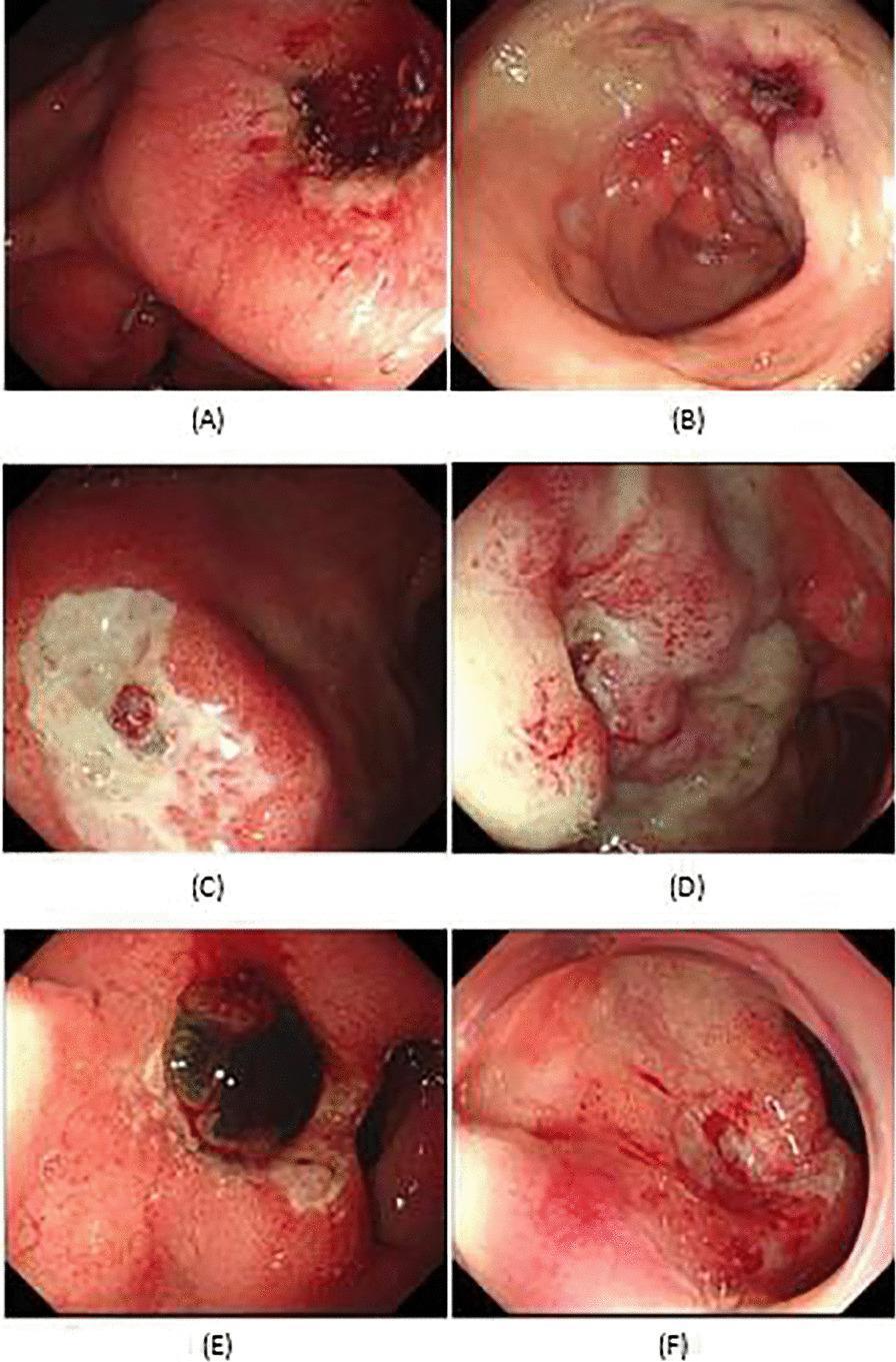
This set of six upper gastrointestinal endoscopic images (A-F) illustrates the pre- and post-therapeutic appearance of peptic ulcer bleeding (PUB) categorized by the modified Forrest classification. The images demonstrate standard epinephrine injection (EI) monotherapy outcomes. Panels A and B show a Forrest Ib ulcer (oozing hemorrhage) before and after therapy, with B exhibiting cessation of active blood flow. Panels C and D depict a Forrest IIa lesion (visible non-bleeding vessel), where the ‘after’ image shows a reduced prominence of the vascular stump. Panels E and F represent a Forrest IIb ulcer (adherent clot), with E displaying a dark, protruding clot and F showing the ulcer base after clot management and hemostasis. Key visual features across the series include varying degrees of mucosal erythema, edema, and the presence of fibrin or hematin. The collection serves as an educational reference for endoscopic stigmata of recent hemorrhage and the immediate visual impact of endoscopic hemostatic techniques.
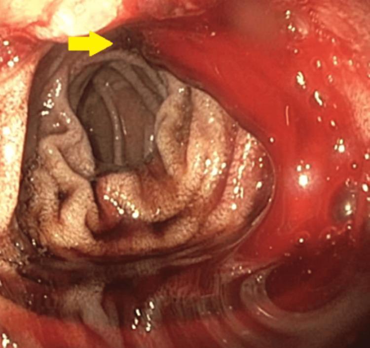
This endoscopic image (Esophagogastroduodenoscopy) provides a view of the duodenal bulb and proximal duodenum. A yellow arrow points to a cratered duodenal ulcer located on the mucosal wall. The ulcer exhibits stigmata of active hemorrhage, specifically a slow, consistent oozing of liquid, dark red blood into the duodenal lumen, characteristic of a Forrest Class Ib ulcer. The surrounding duodenal mucosa appears erythematous and edematous. This visual serves as a clinical example of an upper gastrointestinal bleed (UGIB) originating from peptic ulcer disease. The image is intended for medical training in gastroenterology, focusing on the endoscopic identification, classification, and assessment of bleeding risk in peptic ulcers.
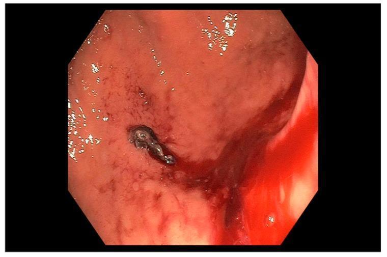
This endoscopic clinical photograph depicts a gastric ulcer located within the gastric body. The image demonstrates a prominent adherent clot, categorized as a Forrest IIb lesion, which appears as a dark, irregular, fibrinous mass firmly attached to the ulcer base. Significant active oozing of fresh, bright red blood is visible at the margins of the ulcer and extending into the surrounding lumen. The surrounding gastric mucosa exhibits marked erythema and vascular congestion, indicative of acute inflammation. This finding represents a high-risk stigmata of recent hemorrhage (SRH), highlighting the clinical need for endoscopic intervention to prevent recurrent bleeding. The image serves as a pedagogical tool for classifying peptic ulcer disease based on the Forrest classification system, emphasizing the visual identification of non-bleeding adherent clots versus active oozing hemorrhage.
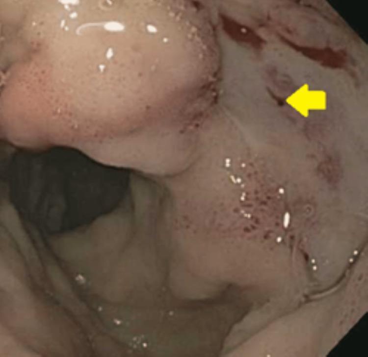
This endoscopic clinical photograph displays the internal mucosal surface of the duodenal bulb during an esophagogastroduodenoscopy (EGD). The primary finding is a large, cratered duodenal ulcer characterized by mucosal architectural disruption and erythema. A yellow arrow points to a prominent, non-bleeding visible vessel (Forrest Class IIa) located within the ulcer base, which represents a high-risk stigmata of recent hemorrhage. The surrounding duodenal mucosa is diffusely erythematous and edematous, showing scattered petechiae and small spots of oozing blood, indicative of active inflammation and recent bleeding. Bubbles of gastric/duodenal secretions are visible on the mucosal surface, and the distal lumen is partially visualized in the background. This image is an essential educational resource for identifying endoscopic risk factors for recurrent peptic ulcer bleeding and for training in Forrest classification for gastrointestinal pathology.
Quick answer