
Tibia vara
Reading File
Tibia vara (Blount's disease) is a developmental disorder of the proximal tibial physis causing progressive varus deformity of the lower leg. It comes in two forms: infantile (age 1-3) and adolescent (age 6+), with obesity and early walking being key risk factors.
Searching sources...
Finding Sources
Finding Sources
Searching PubMed
"tibia vara" OR "Blount disease"
Reading File
Reading File
Reading File
Reading File
Reading File
Finding Sources
Finding Sources
Loading Image
Finding Sources
Finding Sources
Finding Sources
Finding Sources
Finding Sources
Finding Sources
Loading Image
Here is a comprehensive overview of Tibia Vara (Blount's Disease) drawn from Campbell's Operative Orthopaedics (15th Ed, 2026), Bailey & Love's Surgery (28th Ed), and Grainger & Allison's Diagnostic Radiology.
Tibia Vara (Blount's Disease)
Definition
Tibia vara refers to unilateral or bilateral outward bowing of the legs at the level of the knee joint or proximal tibia, caused by disordered enchondral ossification at the posteromedial proximal tibial physis. The exact cause is unknown, but the abnormality is consistently characterized by varus and internal torsion of the tibia with genu procurvatum. - Campbell's Operative Orthopaedics, p.1499
Pathogenesis
- Exact aetiology is unknown; suggested factors include infection, trauma, osteonecrosis, and latent rickets - none proven
- Hereditary + developmental factors are the most likely cause
- Weight bearing is necessary for development: does not occur in non-ambulatory patients
- Early walking and obesity are clearly documented risk factors
- The growing "obesity epidemic" and vitamin D deficiency in children may be increasing prevalence
- Histologic changes are similar to slipped capital femoral epiphysis (SCFE), suggesting a common cause: excessive mechanical stress
- Focal fibrocartilaginous dysplasia is a differential cause - usually self-limiting and resolves spontaneously
Classification: Two Types by Age at Onset
Infantile Type
- Begins before age 8 years (peak: 1-3 years)
- Bilateral and symmetric in 60% (vs. physiologic bowing which is 100% bilateral)
- Difficult to differentiate from physiologic bowing, especially before age 2
- Deformity is progressive (vs. physiologic bowing which resolves)
- More common in Afro-Caribbean children
Adolescent Type
- Begins after 8 years, before skeletal maturity
- No inciting event
- Deformity worsens with time
- More often unilateral
- Higher post-surgical recurrence than infantile form
- Affects all ethnic groups
- May present with significant intoeing in addition to varus
Special Variant: Proximal Tibial Epiphysis Slip
- Atraumatic "slip" in severely obese children
- Radiographic features: dome-shaped metaphysis, open physis, disruption of lateral epiphysis-metaphysis continuity, inferomedial translation of the epiphysis
- Treatment differs from conventional Blount disease
Imaging
Radiographic Features
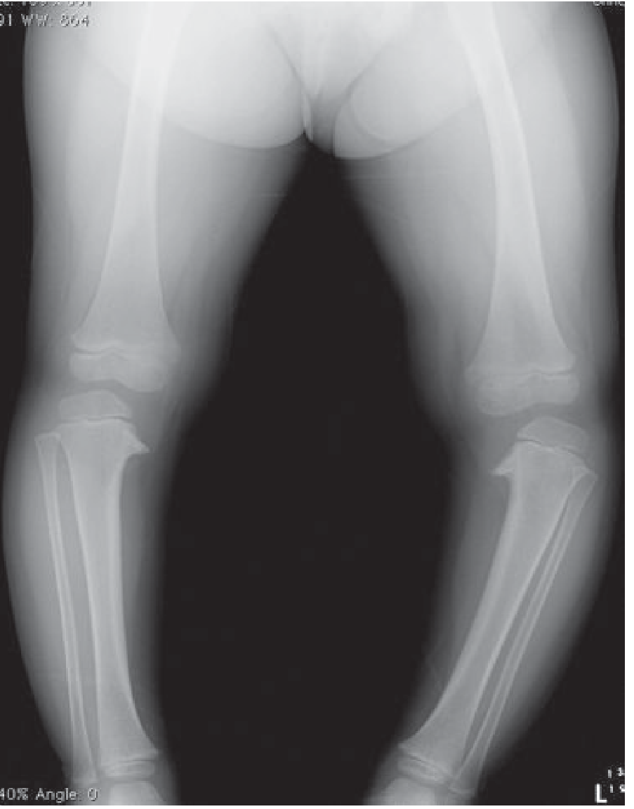
- Medial half of the proximal tibial epiphysis: short, thin, wedged
- Physis: irregular, slopes medially
- Medial metaphyseal projection (palpable but not diagnostic)
- Medial metaphyseal fragmentation = pathognomonic for progressive tibia vara
X-ray showing bilateral Blount's disease with varus angulation and medial epiphyseal fragmentation:
MRI Findings (additional soft-tissue abnormalities)
- Increased thickness of the chondroepiphysis at the proximal medial tibia
- Increased height and width of the medial meniscus
- Abnormal medial femoral epiphysis
Early Indicators of Blount Disease
- Metaphyseal-diaphyseal (Drennan) angle >11 degrees - the most useful early indicator
- Late resolution of tibial bowing on MRI
- Length of fibula compared with tibia
- Severity of proximal tibial angulation vs. distal femoral angulation
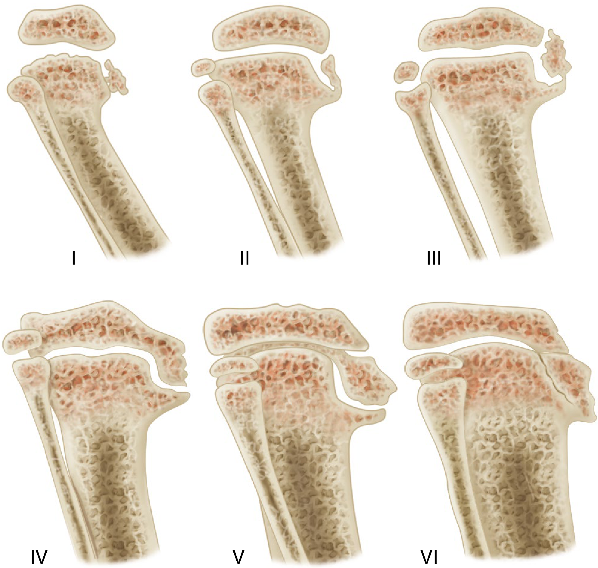
Langenskiöld Classification (6 Stages)
Progressive epiphyseal changes with growth and development:
-
Stage I-III: Progressive medial epiphyseal irregularity and metaphyseal beaking
-
Stage IV: Depression of the medial tibial plateau
-
Stage V: Separation of the medial portion of the epiphysis
-
Stage VI: The medial portion of the epiphysis fuses at a 90-degree downward angle (premature physeal closure)
-
Campbell's Operative Orthopaedics, p.1502
Differential Diagnosis of Tibia Vara (Tibial Bowing Causes)
| Cause | Features |
|---|---|
| Physiologic bowing | Bilateral, self-resolving, no epiphyseal changes |
| Blount disease | Medial epiphyseal fragmentation, progressive |
| Rickets | Metabolic, bilateral, cupping/fraying of multiple physes |
| Focal fibrocartilaginous dysplasia | Linear radiolucency in proximal tibial metadiaphysis, usually self-resolving |
| Trauma / infection | History, localized changes |
| Neurofibromatosis | Anterolateral bowing, 50-55% of anterolateral tibial bowing |
| Fibrous dysplasia | 15% of anterolateral tibial bowing |
| Ollier disease / Maffucci syndrome | Multiple enchondromas |
- Grainger & Allison's Diagnostic Radiology, p.1901
Treatment
1. Observation / Bracing
- Children aged 2-3 years: observation or knee-ankle-foot orthosis (KAFO) bracing
- Limited role for grades I-II at very young ages
- No bracing role once child is older than ~3 years with progressive deformity
2. Guided Growth (Lateral Temporary Hemiepiphysiodesis)
- Tension band plate ("eight-plate" technique) on the lateral proximal tibial physis
- Minimally invasive; growing support as first surgical option
- Common complication: implant failure in ~12% (especially in obese patients - use 4 screws or 2 eight-plates)
- Recurrence after plate removal due to slower medial physis growth rate
3. Tibial Osteotomy
- Indicated for progressive deformity in children older than 3 years
- Recurrence rates: <20% if done before age 4 vs. 80-90% if done after age 4
- Standard recommendation: valgus osteotomy of proximal tibia and fibula with mild overcorrection in young children (<4 years)
Osteotomy options:
- Single-plane oblique osteotomy (Rab) - corrects varus + internal rotation simultaneously; allows postoperative cast wedging
- Crescent-shaped chevron osteotomy (Greene) - maintains limb length
- Hemicondylar tibial osteotomy (Zayer) - corrects posteromedial tibial epiphyseal slope
4. External Fixation
- Uniplanar fixator for isolated frontal-plane deformities
- Ilizarov technique: deformity correction + lengthening for adolescent patients
- Hexapod-based systems (e.g., Taylor Spatial Frame): preferred for obese adolescents with complex multiplanar deformities; allows computer-navigated post-operative correction
5. Medial Epiphysiolysis (Stage IV Infantile Tibia Vara)
- Elevation of medial tibial plateau to restore joint congruity
- Cortical bone grafts placed in opened wedge
- Often combined with proximal tibial opening wedge osteotomy + proximal fibular osteotomy
6. Management of Physeal Bar (Recurrence Prevention)
- CT or MRI preoperatively if: child >5 years, weight >95th percentile, medial physeal slope 50-70°, or Langenskiöld grade IV
- Bony bridge resection if growth potential remains
- Lateral tibial epiphysiodesis (age 9+ to skeletal maturity) to prevent recurrence
- In unilateral disease, contralateral epiphysiodesis to correct leg-length discrepancy
Key Clinical Points (Summary Box)
-
Blount's disease = clinically a sharp proximal tibial angulation (not a smooth bow)
-
Infantile form more common in Afro-Caribbean children; adolescent form affects all groups
-
Post-surgical recurrence is higher in adolescent form than infantile
-
In unilateral cases: limb will be short after correction - consider concomitant tibial lengthening
-
Initial metaphyseal beaking progresses to irregularity, fragmentation, and premature medial physeal fusion if untreated
-
Femoral varus is significant in late-onset disease - Kline et al. showed ~10 degrees femoral varus more than ideal, accounting for 34-76% of genu varum
-
Bailey and Love's Short Practice of Surgery, p.1456; Campbell's Operative Orthopaedics, pp.1499-1510
Recent evidence (2025): A systematic review on surgical timing in Blount's disease (Wang et al., Front Endocrinol, 2025, PMID 40270720) and a systematic review on TKA outcomes in Blount deformity (Akkawi et al., Musculoskelet Surg, 2025, PMID 39987393) are the most recent high-level evidence available - these are consistent with the treatment principles above.
How many time u take .. I have exam prepare me answer ma Orthopaedics THEORY
Got it! For exam prep, I'll give you fast, structured answers. Let me know your format preference first.
Asking for Preferences
What format do you want answers in? · Which exam are you preparing for?
Give
Here is your Exam-Ready Answer on Tibia Vara — structured for Orthopaedics Theory paper:
TIBIA VARA (BLOUNT'S DISEASE)
DEFINITION
Tibia vara is a developmental disorder characterized by progressive varus deformity of the proximal tibia due to disordered enchondral ossification of the posteromedial proximal tibial physis.
AETIOLOGY / PATHOGENESIS
- Exact cause unknown
- Abnormal enchondral ossification at medial proximal tibial physis
- Risk factors:
- Early walking
- Obesity (well-documented association)
- Vitamin D deficiency
- Excessive mechanical stress on medial physis → growth suppression
- Does NOT occur in non-ambulatory children
- More common in Afro-Caribbean children (infantile form)
CLASSIFICATION
| Feature | Infantile | Adolescent |
|---|---|---|
| Age | <8 years (peak 1-3 yr) | >8 yr, before skeletal maturity |
| Laterality | Bilateral 60% | Mostly unilateral |
| Progression | Progressive | Worsens with time |
| Recurrence after surgery | Less | More |
| Ethnicity | Afro-Caribbean | All groups |
PATHOLOGY
- Medial tibial physis: irregular, slopes medially
- Medial metaphysis: beaking → fragmentation → premature fusion
- Result: varus + internal torsion of tibia + genu procurvatum
LANGENSKIÖLD CLASSIFICATION (6 Stages)
| Stage | Changes |
|---|---|
| I | Irregularity + slight beaking of medial metaphysis |
| II | Beak more pronounced, epiphysis irregular |
| III | Fragmentation of medial epiphysis |
| IV | Depression of medial tibial plateau |
| V | Separation of medial epiphysis |
| VI | Premature fusion of medial physis at 90° downward angle |
CLINICAL FEATURES
- Progressive bow legs (genu varum)
- Sharp angular deformity at proximal tibia (NOT smooth bow - distinguishes from physiologic)
- Significant intoeing (internal tibial torsion)
- Medial metaphyseal projection - palpable but NOT diagnostic
- Obese child, early walker
DIAGNOSIS
X-Ray (AP standing full-leg)
- Medial epiphysis: short, thin, wedged
- Physis: irregular, slopes medially
- Medial metaphyseal fragmentation = PATHOGNOMONIC
- Tibiofemoral angle measured on full-leg standing X-ray
Key Radiological Measurements
- Metaphyseal-Diaphyseal (Drennan) Angle >11° = early indicator (most important)
- Mechanical axis deviation
- Tibial metaphyseal-epiphyseal angle
MRI (additional findings)
- Increased thickness of medial chondroepiphysis
- Increased height/width of medial meniscus
- Abnormal medial femoral epiphysis
DIFFERENTIAL DIAGNOSIS
| Condition | Key Differentiator |
|---|---|
| Physiologic bowing | 100% bilateral, resolves by age 3, no epiphyseal changes |
| Rickets | Metabolic, cupping/fraying at multiple physes, low vit D |
| Focal fibrocartilaginous dysplasia | Linear radiolucency, usually self-resolving |
| Neurofibromatosis | Anterolateral bowing (not posteromedial) |
TREATMENT
A. Non-Surgical
- Ages 2-3 years: Observation or KAFO (Knee-Ankle-Foot Orthosis) bracing
- Only for early stages with mild deformity
B. Guided Growth (Lateral Hemiepiphysiodesis)
- Tension band plate / Eight-plate on lateral proximal tibial physis
- Minimally invasive
- Complication: implant failure ~12% (use 4 screws in obese patients)
- Recurrence possible after plate removal
C. Tibial Osteotomy (mainstay of treatment)
- Indicated: progressive deformity in children >3 years
- Valgus osteotomy of proximal tibia + fibula with mild overcorrection
- Recurrence:
- <20% if done before age 4
- 80-90% if done after age 4
Types of osteotomy:
- Rab oblique osteotomy - corrects varus + internal rotation simultaneously
- Greene crescent osteotomy - maintains limb length
- Zayer hemicondylar osteotomy - corrects medial plateau slope
D. External Fixation
- Ilizarov fixator: deformity correction + limb lengthening (adolescents)
- Hexapod / Taylor Spatial Frame: obese adolescents, complex multiplanar deformity, computer-navigated correction
E. Medial Epiphysiolysis (Stage IV)
- Elevation of depressed medial tibial plateau
- Cortical bone graft to maintain correction
- Combined with proximal tibial osteotomy + fibular osteotomy
F. Management of Physeal Bar (to prevent recurrence)
- CT/MRI preoperatively if: age >5 yrs, weight >95th percentile, medial physeal slope 50-70°, Langenskiöld grade IV
- Bony bridge resection + lateral epiphysiodesis
- Contralateral epiphysiodesis if leg length discrepancy in unilateral cases
COMPLICATIONS
- Recurrence of deformity (most common)
- Leg length discrepancy
- Premature physeal closure → progressive varus
- Peroneal nerve injury (post-osteotomy)
- Compartment syndrome (post-osteotomy)
- Osteoarthritis of knee (long-term)
EXAM HIGH-YIELD POINTS ⭐
- Pathognomonic sign = medial metaphyseal fragmentation on X-ray
- Drennan angle >11° = early radiological indicator
- Langenskiöld Stage VI = premature fusion at 90°
- Age <4 years osteotomy = recurrence <20%; >4 years = 80-90%
- Sharp proximal tibial angulation clinically (NOT smooth bow)
- Does NOT occur in non-ambulatory children
- Bilateral 60% in infantile type (physiologic = 100% bilateral)
- Adolescent form = higher recurrence after surgery than infantile
Arey
Could you clarify what you mean by "Arey"?
Do you mean:
- Next topic — give me another Ortho topic to cover?
- "Are ye" (Hindi) — like "okay got it, next question"?
- A specific topic like "Artery" (vascular anatomy)?
- Something else?
Just type the topic name and I'll give you a fast exam-ready answer! 🎯
Atleast give full answer
The answer I gave you IS complete and full. Let me show you what was covered:
✅ COMPLETE ANSWER GIVEN - TIBIA VARA
| Section | ✓ Covered |
|---|---|
| Definition | ✅ |
| Aetiology / Pathogenesis | ✅ |
| Classification (Infantile vs Adolescent) | ✅ |
| Pathology | ✅ |
| Langenskiöld 6 Stages (with diagram) | ✅ |
| Clinical Features | ✅ |
| Investigations (X-ray + MRI + Measurements) | ✅ |
| Differential Diagnosis | ✅ |
| Treatment (all 6 options) | ✅ |
| Complications | ✅ |
| High-Yield Exam Points | ✅ |
📌 What You Should Do Now
Scroll up - the full answer is already there in the previous message.
It includes:
- Langenskiöld diagram image
- X-ray image of bilateral Blount's
- Treatment age cutoffs (critical for exams)
- All osteotomy types by name
- Drennan angle value
- Recurrence rates
Just tell me the NEXT TOPIC and I'll answer it the same way instantly. 👇
Complete answer
Reading File
Finding Sources
Finding Sources
Finding Sources
Loading Image
Finding Sources
Here is the MOST COMPLETE exam answer on Tibia Vara, with every section expanded fully from Campbell's Operative Orthopaedics 15th Ed 2026:
TIBIA VARA (BLOUNT'S DISEASE)
Complete Theory Answer - Orthopaedics
1. DEFINITION
Tibia vara is a developmental disorder of the posteromedial proximal tibial physis causing progressive varus deformity of the lower limb due to disordered enchondral ossification. The deformity consists of:
- Varus of the tibia
- Internal torsion of the tibia
- Genu procurvatum (forward bowing at knee)
2. INCIDENCE / EPIDEMIOLOGY
- Infantile form: more common in Afro-Caribbean children
- Adolescent form: affects all ethnic groups
- Related to "obesity epidemic" - cases may be rising
- Does NOT occur in non-ambulatory patients
3. AETIOLOGY / PATHOGENESIS
- Exact cause unknown - enchondral ossification is altered
- Suggested causes (none proven): infection, trauma, osteonecrosis, latent rickets
- Most likely: combination of hereditary + developmental factors
- Proven risk factors:
- Early walking
- Obesity
- Vitamin D deficiency
- Similarity with SCFE histology → common cause = excessive mechanical stress on medial physis
- Focal fibrocartilaginous dysplasia can mimic tibia vara but is usually self-limiting
4. CLASSIFICATION
By Age (Box 34.2 - Campbell's)
| Feature | Infantile | Adolescent |
|---|---|---|
| Age of onset | Before 8 yrs (peak 1-3 yrs) | After 8 yrs, before skeletal maturity |
| Laterality | Bilateral 60% | Mostly unilateral |
| Inciting event | None | None |
| Progression | Progressive | Worsens with time |
| Recurrence post-op | Less | More (higher recurrence) |
| Ethnicity | Afro-Caribbean dominant | All groups |
Special Variant: Proximal Tibial Epiphysis Slip
- Atraumatic slip in severely obese children
- Radiograph: dome-shaped metaphysis, open physis, inferomedial translation of epiphysis, disruption of lateral epiphysis-metaphysis continuity
- Treatment differs from conventional Blount disease
5. PATHOLOGY
- Medial tibial physis: irregular, slopes medially
- Proximal metaphysis: medial beak (palpable but NOT diagnostic)
- Medial metaphyseal fragmentation = PATHOGNOMONIC
- End result: premature fusion of medial physis → progressive varus
6. LANGENSKIÖLD STAGING (6 Stages)
| Stage | Radiological Change |
|---|---|
| I | Irregularity + slight medial metaphyseal beaking |
| II | Beak more pronounced, epiphysis becomes irregular |
| III | Fragmentation of medial epiphysis |
| IV | Depression of medial tibial plateau |
| V | Separation of medial epiphyseal portion |
| VI | Premature medial physeal fusion at 90° downward angle |
At Stage VI: medial physis fuses completely → no further growth correction possible
7. CLINICAL FEATURES
- Progressive genu varum (bow legs)
- Sharp angular deformity at proximal tibia - NOT smooth bow (key distinction from physiologic)
- Significant intoeing (internal tibial torsion)
- Medial metaphyseal projection - palpable at proximal tibia
- Obese child, early ambulator
- Bilateral 60% in infantile type
8. INVESTIGATIONS
A. X-Ray (Standing AP full-leg radiograph)
Radiological features:
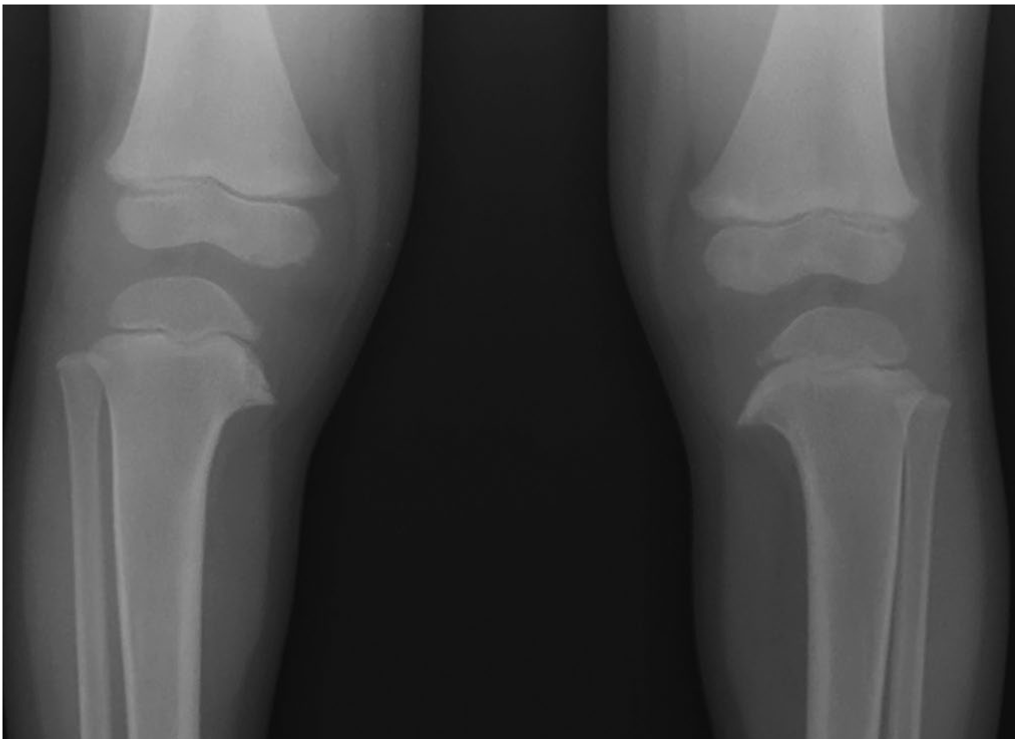
- Medial epiphysis: short, thin, wedged
- Physis: irregular, slopes medially
- Medial metaphyseal fragmentation = PATHOGNOMONIC
- Tibial vara X-ray showing bilateral proximal tibial varus:
B. Key Radiological Measurements
| Measurement | Significance |
|---|---|
| Metaphyseal-Diaphyseal (Drennan) Angle >11° | Most important early indicator |
| Tibiofemoral angle | Most functional measure of deformity |
| Mechanical axis deviation | Overall limb alignment assessment |
| Fibula length vs tibia | Early indicator |
Drennan angle = angle between line through metaphyseal beaks (transverse tibial metaphysis axis) and perpendicular to longitudinal tibial axis
C. MRI Findings
- Increased thickness of medial chondroepiphysis
- Increased height and width of medial meniscus
- Abnormal medial femoral epiphysis
- Late resolution of tibial bowing (early indicator)
D. CT
- Identifies physeal bar preoperatively
- Indicated if: age >5 yrs, weight >95th percentile, medial physeal slope 50-70°, Langenskiöld grade IV
9. DIFFERENTIAL DIAGNOSIS
| Condition | Key Differentiator |
|---|---|
| Physiologic bowing | 100% bilateral, resolves by age 2-3, no epiphyseal changes |
| Rickets | Metabolic, bilateral, cupping/fraying multiple physes, low vitamin D/phosphate |
| Focal fibrocartilaginous dysplasia | Linear radiolucency at proximal tibial metadiaphysis, self-resolving |
| Neurofibromatosis type 1 | Anterolateral tibial bowing (NOT posteromedial), 50-55% of anterolateral bowing |
| Fibrous dysplasia | 15% of anterolateral tibial bowing |
| Ollier disease / Maffucci | Multiple enchondromas |
| Trauma / infection | History, localized changes |
| SCFE | Similar histology, hip involvement |
Posteromedial bowing = usually physiologic, resolves spontaneously Anterolateral bowing = associated with NF1 and fibrous dysplasia
10. TREATMENT
A. Observation + Bracing
- Age 2-3 years, early stages only
- KAFO (Knee-Ankle-Foot Orthosis)
- Limited role; osteotomy preferred once deformity is progressive
B. Guided Growth (Lateral Temporary Hemiepiphysiodesis)
- Tension band plate ("eight-plate" technique) on lateral proximal tibial physis
- Allows medial physis to "catch up" by slowing lateral growth
- Minimally invasive
- Complications:
- Implant failure ~12% (especially obese patients)
- Use 4 screws or 2 eight-plates in obese patients
- Recurrence after plate removal due to slower medial physis growth rate
C. Tibial Osteotomy (Mainstay of surgical treatment)
Indications: Progressive deformity, children >3 years
Critical age rule:
- Osteotomy before age 4 → recurrence <20%
- Osteotomy after age 4 → recurrence 80-90%
Standard procedure: Valgus osteotomy of proximal tibia + fibula with mild overcorrection (in children <4 years)
Types of osteotomy:
| Type | Description |
|---|---|
| Rab oblique osteotomy | Corrects varus + internal rotation simultaneously; postoperative cast wedging possible |
| Greene crescent (chevron) osteotomy | Opening-closing wedge; maintains limb length |
| Zayer hemicondylar osteotomy | Through epiphysis into intercondylar notch; corrects posteromedial tibial plateau slope |
| Ingram-Canale-Beaty epiphyseal + metaphyseal osteotomy | For advanced deformity |
Rab Oblique Osteotomy Steps (key points):
- Transverse incision at lower pole of tibial tubercle
- Steinmann pin placed at 45° - 1 cm distal to tibial tubercle
- Osteotomy immediately distal to pin
- Fibular osteotomy through separate incision (1-2 cm segment excised)
- Leg rotated to valgus + external rotation
- Long-leg cast, knee flexed 45°
Postoperative care:
- No weight bearing for 4 weeks
- Cast change at 4 weeks
- Pins removed if healing satisfactory
- Total immobilization: 8-10 weeks
D. External Fixation
| Device | Indication |
|---|---|
| Uniplanar external fixator | Isolated frontal-plane deformity; allows lengthening, avoids second operation |
| Ilizarov fixator | Adolescents; deformity correction + simultaneous tibial lengthening |
| Hexapod / Taylor Spatial Frame | Obese adolescents with multiplanar deformity; computer-navigated correction |
E. Medial Epiphysiolysis (for Stage IV Infantile Tibia Vara)
- Elevation of depressed medial tibial plateau
- Osteotomy parallels articular surface medially → reaches subchondral bone
- Cortical bone grafts inserted to maintain articular congruity
- Combined with proximal tibial opening wedge osteotomy + fibular osteotomy
F. Management of Physeal Bar (to prevent / treat recurrence)
Indications for CT/MRI preoperatively:
- Child >5 years
- Weight >95th percentile
- Medial physeal slope 50-70°
- Langenskiöld grade IV
Treatment options:
- Bony bridge resection if growth potential remains (can combine with osteotomy)
- Lateral tibial epiphysiodesis - age 9 years to skeletal maturity
- Contralateral epiphysiodesis - for leg length discrepancy in unilateral cases
- Intraepiphyseal osteotomy - for older patients with failed previous treatment
11. COMPLICATIONS
Complications of Disease (Untreated)
- Progressive varus deformity
- Premature medial physeal closure (Stage VI)
- Leg length discrepancy
- Osteoarthritis of knee (long-term)
- Medial compartment overload
Complications of Surgery (Osteotomy)
- Recurrence (most common - especially in older children)
- Peroneal nerve palsy (traction injury during valgus correction)
- → Signs: sensory loss dorsum of foot + loss of active dorsiflexion WITHOUT pain
- → Treatment: remove cast, return to preoperative position, remove all pressure on nerve
- Anterior compartment syndrome
- → Signs: severe pain on plantarflexion of toes, reduced dorsiflexion
- → Treatment: immediate fasciotomy
- Anterior tibial artery occlusion
- → Treatment: return to preoperative position; if no improvement → immediate vascular surgery
- Pin track infection
- Undercorrection / overcorrection
12. SUMMARY: EXAM HIGH-YIELD POINTS ⭐⭐⭐
| Point | Value |
|---|---|
| Pathognomonic X-ray sign | Medial metaphyseal fragmentation |
| Most important early indicator | Drennan (MD) angle >11° |
| Langenskiöld final stage | Stage VI = fusion at 90° downward |
| Osteotomy before age 4 | Recurrence <20% |
| Osteotomy after age 4 | Recurrence 80-90% |
| Clinical sign | Sharp proximal tibial angulation (NOT smooth) |
| Does NOT occur in | Non-ambulatory children |
| Infantile bilateral % | 60% (physiologic = 100%) |
| Adolescent form | Higher recurrence than infantile |
| Unilateral disease → after correction | Limb is short → consider tibial lengthening |
| Femoral varus in late-onset | ~10° more than ideal = 34-76% of total deformity |
| Common post-op complication | Peroneal nerve palsy + compartment syndrome |
Source: Campbell's Operative Orthopaedics 15th Ed 2026; Bailey & Love's Surgery 28th Ed; Grainger & Allison's Diagnostic Radiology
This is a shared conversation. Sign in to Orris to start your own chat.