
Pectoralis muscles name and position image
Reading File
Finding Sources
Searching the Web
pectoralis major minor muscle anatomy diagram labeled
Reading File
Reading File
Loading Image
Loading Image
Reading File
Loading Image
Here is a complete, image-rich overview of the pectoralis muscles from Gray's Anatomy for Students and the THIEME Atlas of Anatomy:
Pectoralis Muscles - Name, Position & Anatomy
1. Pectoralis Major
The largest and most superficial muscle of the anterior chest wall. It forms the anterior wall of the axilla and creates the anterior axillary fold.
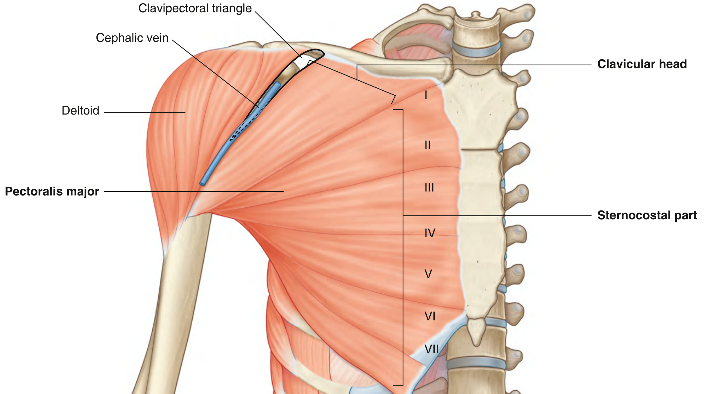
Parts / Origin:
| Part | Origin |
|---|---|
| Clavicular head | Medial half of the clavicle |
| Sternocostal part | Sternum + costal cartilages of ribs 1-6 |
| Abdominal part | Anterior layer of the rectus sheath |
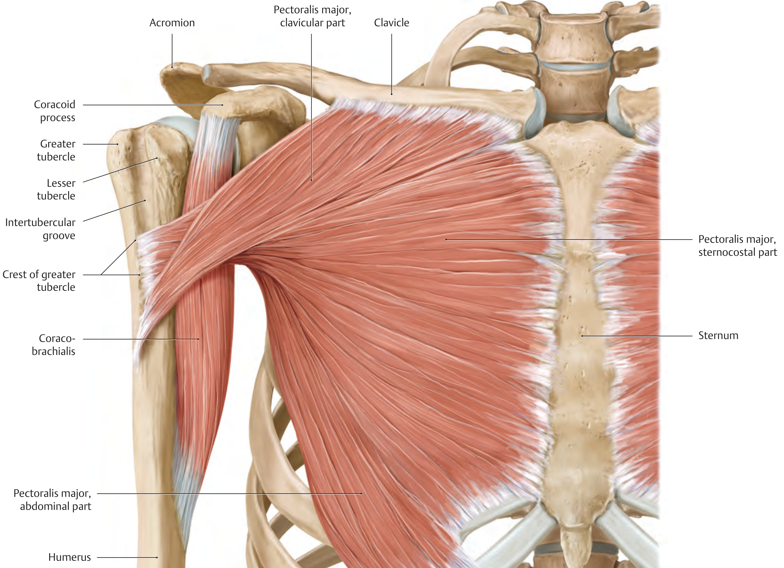
Insertion: Crest of the greater tubercle (lateral lip of the intertubercular sulcus) of the humerus - the tendon twists on itself so the clavicular fibers insert lower than the sternocostal fibers.
Actions: Adduction + internal rotation of the arm (whole muscle); flexion (clavicular and sternocostal parts); assists respiration when the shoulder girdle is fixed.
Innervation: Medial and lateral pectoral nerves (C5-T1)
2. Pectoralis Minor
A small, triangular muscle lying deep to pectoralis major, against the anterior thoracic wall. It is an important surgical landmark because the brachial plexus and axillary vessels pass around it.
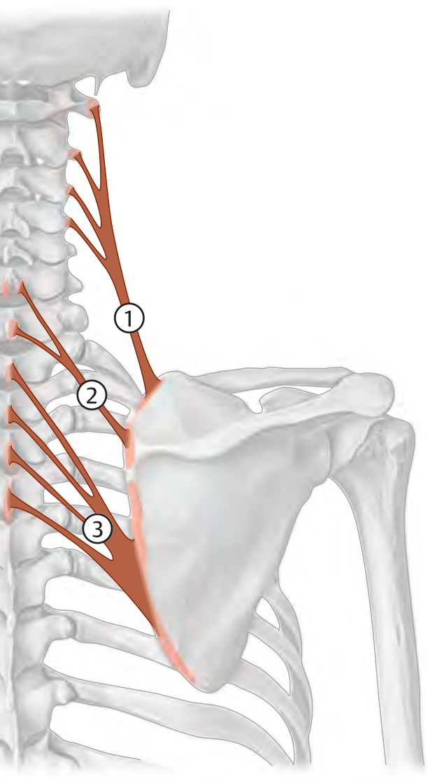
Origin: Anterior surfaces and upper margins of ribs III, IV, and V (as three muscular slips - numbered 1, 2, 3 in the diagram above)
Insertion: Medial and upper surface of the coracoid process of the scapula
Actions: Protracts the scapula (pulls it anteriorly); depresses the lateral angle of the scapula; assists in respiration
Innervation: Medial and lateral pectoral nerves (C8, T1)
Quick Position Summary
| Muscle | Layer | Location | Key Landmark |
|---|---|---|---|
| Pectoralis major | Superficial | Entire anterior chest wall | Forms anterior axillary fold |
| Pectoralis minor | Deep (under major) | Ribs 3-5 to coracoid process | Divides the axilla into parts |
- Gray's Anatomy for Students, p. 844
- THIEME Atlas of Anatomy - General Anatomy and Musculoskeletal System, p. 329-330
This is a shared conversation. Sign in to Orris to start your own chat.