
Cardiovascular disorders
Reading File
Finding Sources
Asking for Preferences
Which topic do you want in detail?
Cardiovascular disorders explain
Finding Sources
Finding Sources
Searching PubMed
"cardiovascular diseases"[MeSH Terms] AND "global burden"
Searching Images
atherosclerosis plaque progression diagram coronary artery
Cardiovascular disorders: an overview
Cardiovascular disorders are diseases of the heart and blood vessels. They are the leading cause of death worldwide. In the US they account for about 1 in 3 deaths, and they stayed the leading cause even during the COVID-19 pandemic (Goldman-Cecil Medicine International Edition, "Burden in the United States").
1. Main groups
| Group | Examples | Core problem |
|---|---|---|
| Coronary artery disease | Stable angina, unstable angina, NSTEMI, STEMI | Atherosclerotic plaque narrows or blocks coronary arteries, causing myocardial ischemia or infarction |
| Heart failure | HFrEF (reduced ejection fraction), HFpEF (preserved ejection fraction) | The heart cannot meet the body's demand without high filling pressures |
| Arrhythmias | Atrial fibrillation, SVT, ventricular tachycardia, heart block | Abnormal impulse formation or conduction |
| Valvular disease | Aortic stenosis, mitral regurgitation, rheumatic disease, endocarditis | Stenosis or regurgitation causing pressure or volume overload |
| Myocardial and pericardial disease | Cardiomyopathies, myocarditis, pericarditis, tamponade | Primary muscle or pericardial pathology |
| Hypertension | Primary and secondary hypertension | Sustained high pressure that damages the heart, brain, kidney, and vessels |
| Vascular disease | Aortic aneurysm and dissection, peripheral artery disease, venous thromboembolism, pulmonary hypertension | Arterial, venous, or pulmonary circulation pathology |
| Congenital heart disease | ASD, VSD, tetralogy of Fallot, coarctation | Structural defects present from birth |
2. How most of it develops
Atherosclerosis is the common substrate.
- Endothelial injury and LDL retention in the vessel wall begin the process.
- Macrophages take up the lipid and become foam cells, and inflammation follows.
- A fibrous cap forms over a lipid core.
- A stable plaque causes fixed narrowing and exertional angina.
- Plaque rupture or erosion triggers thrombosis, which causes acute coronary syndrome, stroke, or limb ischemia.
Hemodynamic stress is the second pathway.
- Pressure overload (hypertension, aortic stenosis) leads to hypertrophy and diastolic dysfunction.
- Volume overload (regurgitant valves) leads to dilation.
- Over time both end in heart failure.
Electrical and structural abnormalities are the third pathway.
- They include scar, fibrosis, atrial stretch, ion channel defects, and congenital malformations.
- They cause arrhythmias, sudden cardiac death, and congenital heart disease.
3. Risk factors
- Modifiable: hypertension, high LDL cholesterol, smoking, diabetes, obesity, physical inactivity, poor diet, and air pollution.
- Non-modifiable: age, male sex (risk in women rises after menopause), family history, and genetics.
- Burden data: a 2026 JAMA analysis of the Global Burden of Disease Study covers elevated LDL-C, and a 2024 systematic review covers air pollution. Both support these as major contributors. I did not find recent reviews that contradict the textbook picture.
4. Typical presentations
- Chest pain: pressure-like and exertional suggests ischemia. Sharp and positional suggests pericarditis. Tearing pain radiating to the back suggests aortic dissection.
- Dyspnea, orthopnea, edema, fatigue: suggest heart failure.
- Palpitations or syncope: suggest arrhythmia, or severe aortic stenosis or hypertrophic cardiomyopathy.
- Claudication: suggests peripheral artery disease.
- Asymptomatic: many patients have no symptoms (Goldman-Cecil), for example those with hypertension or early valve disease.
5. Evaluation
- History and examination: blood pressure, JVP, heart sounds and murmurs, edema, pulses.
- 12-lead ECG, which is also the first test for suspected acute coronary syndrome.
- Blood tests: troponin, BNP or NT-proBNP, lipids, glucose or HbA1c, renal function, thyroid function, and a blood count.
- Imaging:
- Chest X-ray and echocardiography, which is the key test for structure, function, and valves.
- Stress testing, coronary CT angiography, or cardiac MRI.
- Invasive coronary angiography.
- Rhythm monitoring (Holter or event monitor) when arrhythmia is suspected.
6. Principles of management
- Risk-factor control: blood pressure, statins and other lipid lowering, glucose control, smoking cessation, weight loss, exercise, and diet.
- Drugs by condition:
- Antiplatelets, beta-blockers, and statins for coronary disease.
- ACE inhibitors or ARNI, beta-blockers, MRAs, and SGLT2 inhibitors for HFrEF.
- Anticoagulants and rate or rhythm control for atrial fibrillation.
- Procedures and devices: PCI, CABG, valve repair or replacement (surgical or transcatheter), ablation, pacemakers, ICDs, and CRT.
- Acute care: time-critical pathways for STEMI (reperfusion), acute decompensated heart failure, and aortic dissection.
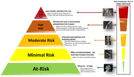
This figure links plaque and anatomical features to the risk of ischemia, acute coronary syndrome, or major coronary events.
Sources
The library holds Braunwald's Heart Disease, Fuster and Hurst's The Heart (15th ed.), and Textbook of Clinical Echocardiography. Goldman-Cecil Medicine is cited above for the epidemiology.
This is a broad overview. I can go deeper on any one area: acute coronary syndromes, heart failure, arrhythmias, valve disease, cardiomyopathies, hypertension, vascular disease, or congenital heart disease. Tell me which one.
Only disease name explain
Cardiovascular disorders: disease names with a one-line explanation
Coronary artery disease
- Stable angina: exertional chest pain from a fixed coronary narrowing.
- Unstable angina: chest pain at rest or with a crescendo pattern, caused by a ruptured plaque, with no myocardial necrosis.
- NSTEMI: partial coronary occlusion with troponin rise and no persistent ST elevation.
- STEMI: complete coronary occlusion with ST elevation and transmural infarction.
Heart failure
- HFrEF: heart failure with reduced ejection fraction (40% or below), a pumping problem.
- HFpEF: heart failure with preserved ejection fraction (50% or above), a stiff, poorly relaxing ventricle.
- Acute decompensated heart failure: sudden worsening with pulmonary congestion.
- Cardiogenic shock: pump failure causing hypotension and organ hypoperfusion.
Arrhythmias
- Atrial fibrillation: chaotic atrial activity with an irregularly irregular pulse and a risk of stroke.
- Atrial flutter: a regular atrial macro-reentry circuit, with a sawtooth pattern on ECG.
- SVT (AVNRT, AVRT): fast regular narrow-complex tachycardia.
- Ventricular tachycardia and fibrillation: life-threatening ventricular rhythms that cause sudden cardiac arrest.
- Heart block: delayed or failed conduction from the atria to the ventricles (first, second, or third degree).
- Long QT syndrome: a repolarization defect that predisposes to torsades de pointes.
Valvular heart disease
- Aortic stenosis: a narrowed aortic valve, causing angina, syncope, and dyspnea.
- Aortic regurgitation: a leaking aortic valve with a wide pulse pressure.
- Mitral stenosis: usually rheumatic, causing left atrial enlargement and atrial fibrillation.
- Mitral regurgitation: backflow into the left atrium.
- Mitral valve prolapse: billowing of the mitral leaflets into the left atrium.
- Infective endocarditis: infection of the valves or endocardium.
- Rheumatic heart disease: valve damage after group A streptococcal infection.
Myocardial and pericardial disease
- Dilated cardiomyopathy: a dilated ventricle with poor contraction.
- Hypertrophic cardiomyopathy: an inherited thickened septum or ventricle, a cause of sudden death in athletes.
- Restrictive cardiomyopathy: a stiff ventricle, as in amyloidosis.
- Myocarditis: inflammation of the heart muscle, usually viral.
- Acute pericarditis: pleuritic, positional chest pain with a friction rub and diffuse ST elevation.
- Pericardial effusion and tamponade: fluid compressing the heart, with Beck's triad (hypotension, raised JVP, muffled heart sounds).
- Constrictive pericarditis: a rigid, scarred pericardium that restricts filling.
Hypertension
- Primary (essential) hypertension: high blood pressure with no single cause.
- Secondary hypertension: caused by renal artery stenosis, primary aldosteronism, pheochromocytoma, and similar.
- Hypertensive emergency: severe hypertension with acute end-organ damage.
- Pulmonary hypertension: raised pulmonary artery pressure, leading to right heart failure.
Vascular disease
- Atherosclerosis: lipid-rich plaque in the arterial wall, the base of most cardiovascular disease.
- Peripheral artery disease: leg artery narrowing, causing claudication.
- Aortic aneurysm: abnormal aortic dilation, with a risk of rupture.
- Aortic dissection: a tear in the intima, with tearing chest or back pain.
- Deep vein thrombosis: clot in the deep veins, usually of the leg.
- Pulmonary embolism: a clot that travels to the pulmonary arteries.
- Vasculitis (for example giant cell arteritis, Takayasu arteritis): inflammation of vessel walls.
Congenital heart disease
- Atrial septal defect (ASD): a hole between the atria.
- Ventricular septal defect (VSD): a hole between the ventricles, the most common defect.
- Patent ductus arteriosus (PDA): the ductus arteriosus fails to close after birth.
- Tetralogy of Fallot: VSD, pulmonary stenosis, overriding aorta, and right ventricular hypertrophy.
- Coarctation of the aorta: aortic narrowing, with a blood pressure difference between arms and legs.
- Transposition of the great arteries: the aorta and pulmonary artery arise from the wrong ventricles.
- Eisenmenger syndrome: a shunt reverses to right-to-left because of pulmonary hypertension.
I can give a detailed explanation of any one disease: causes, features, diagnosis, and treatment. Tell me which one.
This is a shared conversation. Sign in to Orris to start your own chat.