
Heamburgers phenomenon short note with diagram in easy words
chloride shift Hamburger phenomenon red blood cell CO2 transport diagram
carbon dioxide transport in blood bicarbonate chloride exchange erythrocyte
Hamburger Phenomenon (Chloride Shift) - Short Note
Why it happens (easy explanation)
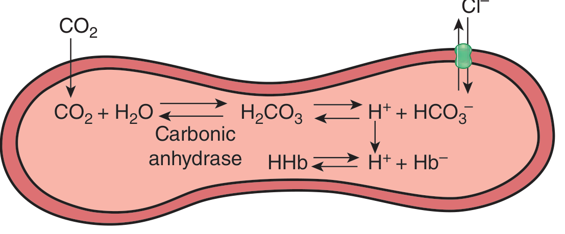
- CO2 produced by tissues diffuses into the RBC.
- Inside the RBC, the enzyme carbonic anhydrase rapidly converts CO2 + H2O into carbonic acid (H2CO3).
- H2CO3 immediately splits into H+ and HCO3-.
- The H+ is buffered by binding to deoxyhemoglobin (this also drives the Haldane effect).
- HCO3- builds up much faster inside the RBC than in the plasma, so about 70% of it moves out of the cell into the plasma through a membrane transporter called anion exchanger 1 (AE1 / Band 3).
- Since the cell cannot lose a negative charge without gaining one back (electroneutrality), Cl- moves into the RBC from the plasma to replace the HCO3- that left. This inward Cl- movement is the "chloride shift."
Simple diagram (tissue capillary side)
        TISSUE CAPILLARY (CO2 loading)

  Plasma                         RBC (inside)
  ------                         ------------
                CO2  ---------->  CO2 + H2O
                                     |  (carbonic anhydrase)
                                     v
                                   H2CO3
                                     |
                              H+  <--+-->  HCO3-
                              |             |
                     binds Hb (Hb.H+)       | (~70% leaves cell)
                                             v
                    <-------------------  HCO3- (out to plasma)

                    Cl-  ------------->  Cl- (in, via AE1/Band3)
                       (to balance charge lost by HCO3- leaving)
- Book Title: Ganong's Review of Medical Physiology, 26th Edition, p. 644
Key points to remember
- Mediator protein: AE1 (Band 3), present in high amounts on the RBC membrane.
- About 70% of the HCO3- formed in RBCs enters plasma; Cl- moves in to replace it.
- Process is fast - essentially complete within 1 second.
- Because an extra osmotically active particle (Cl- or HCO3-) enters the cell for every CO2 that comes in, RBCs swell slightly in venous blood and shrink again in the lungs. This is why venous hematocrit is about 3% higher than arterial hematocrit.
- Works together with the Haldane effect (deoxygenated Hb carries more CO2 and H+) to make CO2 transport efficient.
Top 20 topics of upper that will must come in university mbbs exam
Upper Limb - Top 20 High-Yield Topics for MBBS Anatomy Exam
Topic Breakdown
- Pectoral region & Breast - muscles, nerve supply, breast quadrants/lymphatic drainage (clinical: breast cancer spread)
- Axilla - boundaries, contents, axillary lymph nodes (levels I-III)
- Brachial plexus - formation, cords, branches, and injuries (Erb's palsy, Klumpke's palsy)
- Shoulder joint - type, ligaments, rotator cuff muscles, dislocation, stability
- Scapular region - quadrangular space, triangular space, muscles acting on scapula
- Arm (front) - flexor compartment, biceps brachii, musculocutaneous nerve
- Arm (back) - triceps, radial nerve in radial groove (wrist drop)
- Cubital fossa - boundaries, contents, clinical importance (venepuncture, BP measurement)
- Elbow joint - type, ligaments, movements, dislocation
- Forearm (flexor compartment) - muscle layers, median and ulnar nerve course
- Forearm (extensor compartment) - muscles, radial nerve/posterior interosseous nerve
- Wrist joint and carpal tunnel - boundaries, contents, carpal tunnel syndrome
- Hand - palm - palmar aponeurosis, thenar/hypothenar muscles, palmar spaces
- Hand - intrinsic muscles - lumbricals, interossei (median vs ulnar claw hand)
- Peripheral nerve injuries - median, ulnar, radial, axillary nerve lesions (clinical signs)
- Arterial supply - axillary artery, brachial artery, arterial anastomosis around elbow
- Venous drainage - cephalic vein, basilic vein, median cubital vein (clinical: IV access)
- Bones - clavicle, scapula, humerus, radius, ulna - ossification and common fractures
- Joints of upper limb summary - sternoclavicular, acromioclavicular, radioulnar joints
- Cutaneous innervation & dermatomes - clinical correlation with nerve injuries
Quick overview
Which upper limb topic should I cover in detail first?
Hamburger Phenomenon (Chloride Shift) - Short Note
Why it happens (easy explanation)
- CO2 produced by tissues diffuses into the RBC.
- Inside the RBC, the enzyme carbonic anhydrase rapidly converts CO2 + H2O into carbonic acid (H2CO3).
- H2CO3 immediately splits into H+ and HCO3-.
- The H+ is buffered by binding to deoxyhemoglobin (this also drives the Haldane effect).
- HCO3- builds up much faster inside the RBC than in the plasma, so about 70% of it moves out of the cell into the plasma through a membrane transporter called anion exchanger 1 (AE1 / Band 3).
- Since the cell cannot lose a negative charge without gaining one back (electroneutrality), Cl- moves into the RBC from the plasma to replace the HCO3- that left. This inward Cl- movement is the "chloride shift."
Simple diagram (tissue capillary side)
        TISSUE CAPILLARY (CO2 loading)

  Plasma                         RBC (inside)
  ------                         ------------
                CO2  ---------->  CO2 + H2O
                                     |  (carbonic anhydrase)
                                     v
                                   H2CO3
                                     |
                              H+  <--+-->  HCO3-
                              |             |
                     binds Hb (Hb.H+)       | (~70% leaves cell)
                                             v
                    <-------------------  HCO3- (out to plasma)

                    Cl-  ------------->  Cl- (in, via AE1/Band3)
                       (to balance charge lost by HCO3- leaving)
- Book Title: Ganong's Review of Medical Physiology, 26th Edition, p. 644
Key points to remember
- Mediator protein: AE1 (Band 3), present in high amounts on the RBC membrane.
- About 70% of the HCO3- formed in RBCs enters plasma; Cl- moves in to replace it.
- Process is fast - essentially complete within 1 second.
- Because an extra osmotically active particle (Cl- or HCO3-) enters the cell for every CO2 that comes in, RBCs swell slightly in venous blood and shrink again in the lungs. This is why venous hematocrit is about 3% higher than arterial hematocrit.
- Works together with the Haldane effect (deoxygenated Hb carries more CO2 and H+) to make CO2 transport efficient.
Topic Breakdown
- Pectoral region & Breast - muscles, nerve supply, breast quadrants/lymphatic drainage (clinical: breast cancer spread)
- Axilla - boundaries, contents, axillary lymph nodes (levels I-III)
- Brachial plexus - formation, cords, branches, and injuries (Erb's palsy, Klumpke's palsy)
- Shoulder joint - type, ligaments, rotator cuff muscles, dislocation, stability
- Scapular region - quadrangular space, triangular space, muscles acting on scapula
- Arm (front) - flexor compartment, biceps brachii, musculocutaneous nerve
- Arm (back) - triceps, radial nerve in radial groove (wrist drop)
- Cubital fossa - boundaries, contents, clinical importance (venepuncture, BP measurement)
- Elbow joint - type, ligaments, movements, dislocation
- Forearm (flexor compartment) - muscle layers, median and ulnar nerve course
- Forearm (extensor compartment) - muscles, radial nerve/posterior interosseous nerve
- Wrist joint and carpal tunnel - boundaries, contents, carpal tunnel syndrome
- Hand - palm - palmar aponeurosis, thenar/hypothenar muscles, palmar spaces
- Hand - intrinsic muscles - lumbricals, interossei (median vs ulnar claw hand)
- Peripheral nerve injuries - median, ulnar, radial, axillary nerve lesions (clinical signs)
- Arterial supply - axillary artery, brachial artery, arterial anastomosis around elbow
- Venous drainage - cephalic vein, basilic vein, median cubital vein (clinical: IV access)
- Bones - clavicle, scapula, humerus, radius, ulna - ossification and common fractures
- Joints of upper limb summary - sternoclavicular, acromioclavicular, radioulnar joints
- Cutaneous innervation & dermatomes - clinical correlation with nerve injuries
Quick overview
Top 20 topics of upper that will must come in ouhs university mbbs exam and the sequence which i follow so that i understand these topics easily
OUHS university MBBS anatomy exam upper limb important topics
Upper Limb - Top 20 MBBS Exam Topics + Recommended Study Sequence
Recommended Study Sequence (with reasoning)
- Clavicle - osteology, attachments, fractures
- Scapula - osteology, attachments, winging of scapula
- Humerus - osteology, surgical neck/shaft fractures and nerve involvement
- Radius & Ulna - osteology, Colles' fracture, Monteggia/Galeazzi fractures
- Joints overview - sternoclavicular, acromioclavicular, shoulder, elbow, wrist joints
Why this order works
- You cannot understand nerve injuries (topic 19) until you know the brachial plexus (18) and the regional anatomy each nerve passes through (topics 6-17).
- Cubital fossa (12) makes more sense once you already know the arm's front compartment (10) and elbow joint (13).
- Saving brachial plexus and nerve injuries for the end lets you revisit everything you learned as a "clinical correlation pass" - this is usually the most effective revision strategy right before exams, since university papers heavily favor applied/clinical questions on nerve lesions.
Top 20 topics of upper limb according to weighagethat will must come in ouhs university mbbs exam and the sequence which i follow so that i understand these topics easily
"Odisha University of Health Sciences" OR OUHS MBBS anatomy previous year question papers upper limb
most repeated important anatomy questions upper limb MBBS exam weightage frequently asked
Top 20 Upper Limb Topics Ranked by Weightage
- Brachial plexus - formation, branches, Erb's palsy, Klumpke's palsy
- Axilla - boundaries and contents
- Cubital fossa - boundaries and contents
- Nerve injuries - median, ulnar, radial, axillary (claw hand, wrist drop, winging of scapula)
- Pectoral region and lymphatic drainage of the breast
Easiest Sequence to Study (builds understanding step by step)
- Bones first: Clavicle -> Scapula -> Humerus -> Radius & Ulna (topics 17, 18 and fracture correlations from 16)
- Joints: Shoulder joint (6) -> Elbow joint (11) -> Wrist joint (19)
- Regions front to back: Pectoral region & breast (5) -> Axilla (2) -> Scapular region (9) -> Arm front/back -> Cubital fossa (3)
- Forearm and hand: Forearm compartments (20) -> Carpal tunnel (10) -> Palm and intrinsic hand muscles (13, 14)
- Tie it together with vessels and nerves: Axillary artery (7) -> Anastomosis around elbow (8) -> Cephalic/basilic veins (15) -> Brachial plexus (1) -> Nerve injuries (4) -> Dermatomes (12)

Predict a paper 1 question paper according to this pattern for ouhs anatomy paper 1 which covers head neak face upper limb neuroanatomy general embryology thorax systematic embryology of above topics and general anatomy
Predicted OUHS Anatomy Paper 1 (100 Marks)
SECTION A (50 Marks) - Head & Neck, Neuroanatomy, General Topics
SECTION B (50 Marks) - Upper Limb (Superior Extremity), Thorax, Embryology
Why this prediction fits the pattern
- Long questions (15 marks each) always target a "big" applied topic that combines a structure's anatomy with a classic clinical correlation - facial nerve/Bell's palsy and brachial plexus/Erb's palsy are the single most repeated long questions in Head & Neck and Upper Limb across Indian universities.
- Short notes are picked to spread marks across sub-syllabus items: gross anatomy (axilla, cubital fossa, parotid), neuroanatomy (Circle of Willis, cranial nerve nuclei), thorax (mediastinum), and embryology (heart/thyroid development) - matching the syllabus bullet "Relevant systemic Histology and Embryology of the above parts."
- Give reason questions test the applied/clinical linkage between structure and function, which OUHS explicitly lists as "Applied anatomy of the above parts."
- Case-based questions are new-curriculum (CBME) additions that test nerve lesions with a clinical vignette - almost always drawn from the same high-yield nerve injury topics (facial, radial, axillary, median, ulnar).

Predict a paper 1 question paper according to this pattern for ouhs anatomy paper 1 which covers head neak face upper limb neuroanatomy general embryology thorax systematic embryology of above topics and general anatomy
Predicted OUHS Anatomy Paper 1 (New CBME Format - 100 Marks, 3 Hours)
Q1. Scenario Based MCQ (10 x 2 = 20 marks)
Q2. Long Answer Question (1 x 10 marks)
Q3. Reasoning Questions (5 x 3 = 15 marks)
Q4. Short Notes - Applied Aspects (4 x 5 = 20 marks)
Q5. Short Notes (3 x 5 = 15 marks)
Q6. Short Notes - including 5 marks from AETCOM (4 x 5 = 20 marks)
Why this prediction fits the pattern
- Q1 (MCQs) deliberately spans all listed topics (upper limb, head & neck, embryology, thorax, neuroanatomy, general histology) since scenario-based MCQs in CBME papers sample broadly across the whole syllabus rather than one region.
- Q2 (10-mark long answer) uses the single most repeated high-yield topic (facial nerve/Bell's palsy) since only one long question is asked now, universities pick topics with maximum applied value.
- Q3 (Reasoning, 3 marks each) targets structure-function/clinical links, exactly the "give reason" style seen in your first pattern, just now worth 3 marks instead of 5.
- Q4 (Applied short notes) matches the syllabus line "Applied anatomy of the above parts" directly.
- Q6 AETCOM item is mandatory per the new NMC curriculum integration, universities routinely attach it to a Head & Neck or Embryology topic since those have strong patient-communication scenarios (congenital anomalies, cancer, nerve palsies).
# Predicted OUHS Anatomy Paper 1 (New CBME Format - 100 Marks, 3 Hours) This new format image shows the actual current OUHS answer-script structure (MCQ + Long Answer + Reasoning + Short Notes + AETCOM), which is different and more updated than the older Section A/B format. Here is a full predicted paper built strictly on this template, covering Head & Neck, Face, Upper Limb, Neuroanatomy, General/Systemic Embryology, Thorax, and General Anatomy. *This is a predicted/practice paper for preparation purposes, not a leaked official paper.* --- **Course: MBBS | Subject: Anatomy | Paper: 1 | Max Marks: 100 | Time: 3 Hours** *All questions are compulsory. Illustrate with diagrams wherever relevant.* --- ### Q1. Scenario Based MCQ (10 x 2 = 20 marks) a) A 30-year-old man sustains a fracture of the mid-shaft of the humerus and now cannot extend his wrist or fingers. Which nerve is injured? i) Median ii) Ulnar iii) Radial iv) Axillary b) A patient has drooping of the right angle of the mouth and cannot close the right eye, but forehead wrinkling is normal. What is this most consistent with? i) UMN facial palsy ii) LMN facial (Bell's) palsy iii) Trigeminal neuralgia iv) Horner's syndrome c) Failure of fusion of the maxillary process with the medial nasal process leads to: i) Cleft palate ii) Cleft lip iii) Macrostomia iv) Choanal atresia d) Which structure passes through the carotid canal? i) Internal jugular vein ii) Internal carotid artery iii) Vagus nerve iv) Glossopharyngeal nerve e) A child has a midline neck swelling that rises on protrusion of the tongue. Diagnosis? i) Branchial cyst ii) Thyroglossal cyst iii) Cystic hygroma iv) Dermoid cyst f) The artery most commonly injured in a supracondylar fracture of the humerus is: i) Radial ii) Ulnar iii) Brachial iv) Profunda brachii g) The bulbus cordis of the primitive heart tube mainly gives rise to: i) Right atrium ii) Left atrium iii) Right ventricle iv) Left ventricle h) Loss of pain/temperature on the same side of the face and opposite side of the body after a brainstem stroke suggests: i) Weber syndrome ii) Wallenberg (lateral medullary) syndrome iii) Millard-Gubler syndrome iv) Benedikt syndrome i) The esophagus is lined by: i) Simple columnar epithelium ii) Stratified squamous non-keratinized epithelium iii) Pseudostratified columnar epithelium iv) Transitional epithelium j) Winging of the scapula results from injury to which nerve? i) Axillary ii) Suprascapular iii) Long thoracic iv) Dorsal scapular --- ### Q2. Long Answer Question (1 x 10 marks) Describe the extracranial course, branches, and distribution of the facial nerve. Add a note on the anatomical basis of Bell's palsy. --- ### Q3. Reasoning Questions (5 x 3 = 15 marks) Give reasons for the following: a) Fracture of the surgical neck of the humerus is associated with paralysis of the deltoid muscle. b) A thyroglossal cyst moves upward on protrusion of the tongue. c) Mitral stenosis leads to enlargement of the left atrium. d) Injury to the recurrent laryngeal nerve causes hoarseness of voice. e) Klumpke's palsy is often associated with Horner's syndrome. --- ### Q4. Short Notes - Applied Aspects (4 x 5 = 20 marks) a) Applied anatomy of the cubital fossa b) Applied anatomy of the carotid triangle c) Applied anatomy of the mediastinum d) Applied anatomy of the cavernous sinus --- ### Q5. Short Notes (3 x 5 = 15 marks) a) Development of the interventricular septum b) Cranial nerve nuclei in the brainstem c) Axillary lymph nodes --- ### Q6. Short Notes - including 5 marks from AETCOM (4 x 5 = 20 marks) a) Boundaries and contents of the posterior triangle of the neck b) Development of the palate and cleft palate c) Dermatomes of the upper limb d) **(AETCOM)** As a first-year medical student observing in the pediatric ward, discuss the ethical and communication principles you would apply while a senior doctor breaks the news of a cleft lip and palate diagnosis to anxious new parents. --- ## Why this prediction fits the pattern - **Q1 (MCQs)** deliberately spans all listed topics (upper limb, head & neck, embryology, thorax, neuroanatomy, general histology) since scenario-based MCQs in CBME papers sample broadly across the whole syllabus rather than one region. - **Q2 (10-mark long answer)** uses the single most repeated high-yield topic (facial nerve/Bell's palsy) since only one long question is asked now, universities pick topics with maximum applied value. - **Q3 (Reasoning, 3 marks each)** targets structure-function/clinical links, exactly the "give reason" style seen in your first pattern, just now worth 3 marks instead of 5. - **Q4 (Applied short notes)** matches the syllabus line "Applied anatomy of the above parts" directly. - **Q6 AETCOM item** is mandatory per the new NMC curriculum integration, universities routinely attach it to a Head & Neck or Embryology topic since those have strong patient-communication scenarios (congenital anomalies, cancer, nerve palsies). If you'd like, I can generate a second variant paper with different long-answer and short-note topics (e.g., brachial plexus as the long question, or thorax-heavy short notes) so you have more than one predicted set to practice with.
Topic Map for OUHS Anatomy Paper 1
- General Anatomy / Gross Anatomy - terminology, joints, muscles, fascia classifications
- General Histology - basic tissue types (epithelium, connective tissue, muscle, nerve)
- General Embryology - gametogenesis, fertilization, gastrulation, folding, germ layer derivatives
- Neuroanatomy - brain (cerebrum, cerebellum, brainstem, ventricles) and 12 cranial nerves
- Head and Neck - skull, face, triangles of neck, salivary glands, thyroid, cranial nerve applied anatomy
- Face - muscles of facial expression, blood supply, sensory/motor nerve supply
- Superior Extremity (Upper Limb) - shoulder, arm, forearm, hand, brachial plexus, nerve injuries
- Thorax - heart, lungs, pleura, mediastinum, great vessels
- Structural anatomy with bones - osteology across all above regions (skull, vertebrae, ribs, clavicle, scapula, humerus, radius/ulna)
- Applied anatomy - clinical correlations across all regions above
- Systemic Histology and Embryology - development of heart, face, palate, pharyngeal apparatus, CNS
Overview
Which syllabus area should I build the next detailed predicted question set for?
Hamburger Phenomenon (Chloride Shift) - Short Note
Why it happens (easy explanation)
- CO2 produced by tissues diffuses into the RBC.
- Inside the RBC, the enzyme carbonic anhydrase rapidly converts CO2 + H2O into carbonic acid (H2CO3).
- H2CO3 immediately splits into H+ and HCO3-.
- The H+ is buffered by binding to deoxyhemoglobin (this also drives the Haldane effect).
- HCO3- builds up much faster inside the RBC than in the plasma, so about 70% of it moves out of the cell into the plasma through a membrane transporter called anion exchanger 1 (AE1 / Band 3).
- Since the cell cannot lose a negative charge without gaining one back (electroneutrality), Cl- moves into the RBC from the plasma to replace the HCO3- that left. This inward Cl- movement is the "chloride shift."
Simple diagram (tissue capillary side)
        TISSUE CAPILLARY (CO2 loading)

  Plasma                         RBC (inside)
  ------                         ------------
                CO2  ---------->  CO2 + H2O
                                     |  (carbonic anhydrase)
                                     v
                                   H2CO3
                                     |
                              H+  <--+-->  HCO3-
                              |             |
                     binds Hb (Hb.H+)       | (~70% leaves cell)
                                             v
                    <-------------------  HCO3- (out to plasma)

                    Cl-  ------------->  Cl- (in, via AE1/Band3)
                       (to balance charge lost by HCO3- leaving)
- Book Title: Ganong's Review of Medical Physiology, 26th Edition, p. 644
Key points to remember
- Mediator protein: AE1 (Band 3), present in high amounts on the RBC membrane.
- About 70% of the HCO3- formed in RBCs enters plasma; Cl- moves in to replace it.
- Process is fast - essentially complete within 1 second.
- Because an extra osmotically active particle (Cl- or HCO3-) enters the cell for every CO2 that comes in, RBCs swell slightly in venous blood and shrink again in the lungs. This is why venous hematocrit is about 3% higher than arterial hematocrit.
- Works together with the Haldane effect (deoxygenated Hb carries more CO2 and H+) to make CO2 transport efficient.
Topic Breakdown
- Pectoral region & Breast - muscles, nerve supply, breast quadrants/lymphatic drainage (clinical: breast cancer spread)
- Axilla - boundaries, contents, axillary lymph nodes (levels I-III)
- Brachial plexus - formation, cords, branches, and injuries (Erb's palsy, Klumpke's palsy)
- Shoulder joint - type, ligaments, rotator cuff muscles, dislocation, stability
- Scapular region - quadrangular space, triangular space, muscles acting on scapula
- Arm (front) - flexor compartment, biceps brachii, musculocutaneous nerve
- Arm (back) - triceps, radial nerve in radial groove (wrist drop)
- Cubital fossa - boundaries, contents, clinical importance (venepuncture, BP measurement)
- Elbow joint - type, ligaments, movements, dislocation
- Forearm (flexor compartment) - muscle layers, median and ulnar nerve course
- Forearm (extensor compartment) - muscles, radial nerve/posterior interosseous nerve
- Wrist joint and carpal tunnel - boundaries, contents, carpal tunnel syndrome
- Hand - palm - palmar aponeurosis, thenar/hypothenar muscles, palmar spaces
- Hand - intrinsic muscles - lumbricals, interossei (median vs ulnar claw hand)
- Peripheral nerve injuries - median, ulnar, radial, axillary nerve lesions (clinical signs)
- Arterial supply - axillary artery, brachial artery, arterial anastomosis around elbow
- Venous drainage - cephalic vein, basilic vein, median cubital vein (clinical: IV access)
- Bones - clavicle, scapula, humerus, radius, ulna - ossification and common fractures
- Joints of upper limb summary - sternoclavicular, acromioclavicular, radioulnar joints
- Cutaneous innervation & dermatomes - clinical correlation with nerve injuries
Quick overview
Recommended Study Sequence (with reasoning)
- Clavicle - osteology, attachments, fractures
- Scapula - osteology, attachments, winging of scapula
- Humerus - osteology, surgical neck/shaft fractures and nerve involvement
- Radius & Ulna - osteology, Colles' fracture, Monteggia/Galeazzi fractures
- Joints overview - sternoclavicular, acromioclavicular, shoulder, elbow, wrist joints
Why this order works
- You cannot understand nerve injuries (topic 19) until you know the brachial plexus (18) and the regional anatomy each nerve passes through (topics 6-17).
- Cubital fossa (12) makes more sense once you already know the arm's front compartment (10) and elbow joint (13).
- Saving brachial plexus and nerve injuries for the end lets you revisit everything you learned as a "clinical correlation pass" - this is usually the most effective revision strategy right before exams, since university papers heavily favor applied/clinical questions on nerve lesions.
Top 20 Upper Limb Topics Ranked by Weightage
- Brachial plexus - formation, branches, Erb's palsy, Klumpke's palsy
- Axilla - boundaries and contents
- Cubital fossa - boundaries and contents
- Nerve injuries - median, ulnar, radial, axillary (claw hand, wrist drop, winging of scapula)
- Pectoral region and lymphatic drainage of the breast
Easiest Sequence to Study (builds understanding step by step)
- Bones first: Clavicle -> Scapula -> Humerus -> Radius & Ulna (topics 17, 18 and fracture correlations from 16)
- Joints: Shoulder joint (6) -> Elbow joint (11) -> Wrist joint (19)
- Regions front to back: Pectoral region & breast (5) -> Axilla (2) -> Scapular region (9) -> Arm front/back -> Cubital fossa (3)
- Forearm and hand: Forearm compartments (20) -> Carpal tunnel (10) -> Palm and intrinsic hand muscles (13, 14)
- Tie it together with vessels and nerves: Axillary artery (7) -> Anastomosis around elbow (8) -> Cephalic/basilic veins (15) -> Brachial plexus (1) -> Nerve injuries (4) -> Dermatomes (12)
SECTION A (50 Marks) - Head & Neck, Neuroanatomy, General Topics
SECTION B (50 Marks) - Upper Limb (Superior Extremity), Thorax, Embryology
Why this prediction fits the pattern
- Long questions (15 marks each) always target a "big" applied topic that combines a structure's anatomy with a classic clinical correlation - facial nerve/Bell's palsy and brachial plexus/Erb's palsy are the single most repeated long questions in Head & Neck and Upper Limb across Indian universities.
- Short notes are picked to spread marks across sub-syllabus items: gross anatomy (axilla, cubital fossa, parotid), neuroanatomy (Circle of Willis, cranial nerve nuclei), thorax (mediastinum), and embryology (heart/thyroid development) - matching the syllabus bullet "Relevant systemic Histology and Embryology of the above parts."
- Give reason questions test the applied/clinical linkage between structure and function, which OUHS explicitly lists as "Applied anatomy of the above parts."
- Case-based questions are new-curriculum (CBME) additions that test nerve lesions with a clinical vignette - almost always drawn from the same high-yield nerve injury topics (facial, radial, axillary, median, ulnar).
Q1. Scenario Based MCQ (10 x 2 = 20 marks)
Q2. Long Answer Question (1 x 10 marks)
Q3. Reasoning Questions (5 x 3 = 15 marks)
Q4. Short Notes - Applied Aspects (4 x 5 = 20 marks)
Q5. Short Notes (3 x 5 = 15 marks)
Q6. Short Notes - including 5 marks from AETCOM (4 x 5 = 20 marks)
Why this prediction fits the pattern
- Q1 (MCQs) deliberately spans all listed topics (upper limb, head & neck, embryology, thorax, neuroanatomy, general histology) since scenario-based MCQs in CBME papers sample broadly across the whole syllabus rather than one region.
- Q2 (10-mark long answer) uses the single most repeated high-yield topic (facial nerve/Bell's palsy) since only one long question is asked now, universities pick topics with maximum applied value.
- Q3 (Reasoning, 3 marks each) targets structure-function/clinical links, exactly the "give reason" style seen in your first pattern, just now worth 3 marks instead of 5.
- Q4 (Applied short notes) matches the syllabus line "Applied anatomy of the above parts" directly.
- Q6 AETCOM item is mandatory per the new NMC curriculum integration, universities routinely attach it to a Head & Neck or Embryology topic since those have strong patient-communication scenarios (congenital anomalies, cancer, nerve palsies).
Topic Map for OUHS Anatomy Paper 1
- General Anatomy / Gross Anatomy - terminology, joints, muscles, fascia classifications
- General Histology - basic tissue types (epithelium, connective tissue, muscle, nerve)
- General Embryology - gametogenesis, fertilization, gastrulation, folding, germ layer derivatives
- Neuroanatomy - brain (cerebrum, cerebellum, brainstem, ventricles) and 12 cranial nerves
- Head and Neck - skull, face, triangles of neck, salivary glands, thyroid, cranial nerve applied anatomy
- Face - muscles of facial expression, blood supply, sensory/motor nerve supply
- Superior Extremity (Upper Limb) - shoulder, arm, forearm, hand, brachial plexus, nerve injuries
- Thorax - heart, lungs, pleura, mediastinum, great vessels
- Structural anatomy with bones - osteology across all above regions (skull, vertebrae, ribs, clavicle, scapula, humerus, radius/ulna)
- Applied anatomy - clinical correlations across all regions above
- Systemic Histology and Embryology - development of heart, face, palate, pharyngeal apparatus, CNS
Overview
Predict a paper 1 question paper according to this pattern for ouhs anatomy paper 1 which covers head neak face upper limb neuroanatomy general embryology thorax systematic embryology of above topics and general anatomy make this as a pdf
mkdir -p /tmp/workspace/ouhs-anatomy-paper1 && ls -la /tmp/workspace/
~/ouhs-anatomy-paper1/generate_paper.py
# -*- coding: utf-8 -*-
from reportlab.lib.pagesizes import A4
from reportlab.lib.units import cm
from reportlab.lib import colors
from reportlab.platypus import (SimpleDocTemplate, Table, TableStyle, Paragraph,
Spacer, ListFlowable, ListItem)
from reportlab.lib.styles import getSampleStyleSheet, ParagraphStyle
from reportlab.lib.enums import TA_CENTER, TA_LEFT
OUT = "/tmp/workspace/ouhs-anatomy-paper1/OUHS_Anatomy_Paper1_Predicted.pdf"
styles = getSampleStyleSheet()
normal = ParagraphStyle('normal', parent=styles['Normal'], fontSize=10, leading=14)
bold = ParagraphStyle('bold', parent=normal, fontName='Helvetica-Bold')
italic = ParagraphStyle('italic', parent=normal, fontName='Helvetica-Oblique', fontSize=9)
qhead = ParagraphStyle('qhead', parent=normal, fontName='Helvetica-Bold', fontSize=10.5,
spaceBefore=10, spaceAfter=4)
subitem = ParagraphStyle('subitem', parent=normal, leftIndent=14, spaceAfter=3)
title_style = ParagraphStyle('title', parent=normal, fontName='Helvetica-Bold', fontSize=13,
alignment=TA_CENTER, spaceAfter=6)
note_style = ParagraphStyle('note', parent=normal, fontName='Helvetica-Oblique', fontSize=9,
alignment=TA_CENTER, textColor=colors.grey, spaceAfter=10)
doc = SimpleDocTemplate(OUT, pagesize=A4,
topMargin=1.3*cm, bottomMargin=1.3*cm,
leftMargin=1.6*cm, rightMargin=1.6*cm)
elems = []
# ---------- Header block (mimics official answer script cover) ----------
reg_cells = [Paragraph("Registration no:", bold)] + [Paragraph("", normal) for _ in range(9)]
reg_table = Table([reg_cells], colWidths=[3.2*cm] + [1.2*cm]*9)
reg_table.setStyle(TableStyle([
('BOX', (1,0), (-1,-1), 0.8, colors.black),
('INNERGRID', (1,0), (-1,-1), 0.8, colors.black),
('VALIGN', (0,0), (-1,-1), 'MIDDLE'),
]))
elems.append(reg_table)
elems.append(Spacer(1, 10))
info_data = [
[Paragraph("Total Number of Pages: (XX)", normal), Paragraph("Course: MBBS", normal)],
["", Paragraph("SUB_CODE:", normal)],
]
info_table = Table(info_data, colWidths=[10*cm, 7*cm])
info_table.setStyle(TableStyle([
('BOX', (0,0), (-1,-1), 0.8, colors.black),
('LINEAFTER', (0,0), (0,-1), 0.8, colors.black),
('VALIGN', (0,0), (-1,-1), 'MIDDLE'),
]))
elems.append(info_table)
meta_lines = [
"( X ) Professional Regular / Supplementary / Attempt / Detained Examination: 2025-26",
"<b>SUBJECT NAME:</b> ANATOMY",
"<b>BRANCH:</b> MBBS - First Phase",
"<b>Max Time:</b> 3 Hours <b>Sitting:</b> 1st 10.00 AM to 1.00 PM",
"<b>Max Marks:</b> 100",
"<b>Q. CODE:</b> <i>(To be allotted by the University)</i>",
]
meta_rows = [[Paragraph(l, normal)] for l in meta_lines]
meta_table = Table(meta_rows, colWidths=[17*cm])
meta_table.setStyle(TableStyle([
('BOX', (0,0), (-1,-1), 0.8, colors.black),
('LINEBELOW', (0,0), (-1,-2), 0.4, colors.grey),
('TOPPADDING', (0,0), (-1,-1), 4),
('BOTTOMPADDING', (0,0), (-1,-1), 4),
]))
elems.append(meta_table)
instr_data = [
[Paragraph("All Questions are compulsory. Illustrate answer with diagrams wherever relevant / required", italic)],
[Paragraph("The figures in the right-hand margin indicate marks.", italic)],
]
instr_table = Table(instr_data, colWidths=[17*cm])
instr_table.setStyle(TableStyle([
('BOX', (0,0), (-1,-1), 0.8, colors.black),
('ALIGN', (0,0), (-1,-1), 'CENTER'),
]))
elems.append(instr_table)
elems.append(Spacer(1, 6))
elems.append(Paragraph("SUBJECT: ANATOMY | PAPER 1", title_style))
elems.append(Paragraph("Predicted / Practice Question Paper - Not an official or leaked paper", note_style))
def q_row(qno, label, marks, sub_items=None):
"""Build a table row mimicking the official grid: Q no | content | marks"""
rows = []
header_cell = [Paragraph(f"<b>{qno}</b>", normal), Paragraph(f"<b>{label}</b>", normal),
Paragraph(f"<b>{marks}</b>", normal)]
rows.append(header_cell)
if sub_items:
for tag, text in sub_items:
rows.append(["", Paragraph(f"<b>{tag})</b> {text}", subitem), ""])
t = Table(rows, colWidths=[1.3*cm, 13.7*cm, 2*cm])
t.setStyle(TableStyle([
('BOX', (0,0), (-1,-1), 0.8, colors.black),
('INNERGRID', (0,0), (-1,-1), 0.4, colors.grey),
('VALIGN', (0,0), (-1,-1), 'TOP'),
('TOPPADDING', (0,0), (-1,-1), 4),
('BOTTOMPADDING', (0,0), (-1,-1), 4),
]))
return t
elems.append(Spacer(1, 4))
# ---------------- Q1 MCQ ----------------
mcqs = [
("a", "A 30-year-old man sustains a fracture of the mid-shaft of the humerus and now cannot extend his wrist or fingers. Which nerve is injured? i) Median ii) Ulnar iii) Radial iv) Axillary"),
("b", "A patient has drooping of the right angle of the mouth and cannot close the right eye, but forehead wrinkling is preserved. This is most consistent with: i) UMN facial palsy ii) LMN facial (Bell's) palsy iii) Trigeminal neuralgia iv) Horner's syndrome"),
("c", "Failure of fusion of the maxillary process with the medial nasal process leads to: i) Cleft palate ii) Cleft lip iii) Macrostomia iv) Choanal atresia"),
("d", "Which structure passes through the carotid canal? i) Internal jugular vein ii) Internal carotid artery iii) Vagus nerve iv) Glossopharyngeal nerve"),
("e", "A child has a midline neck swelling that rises on protrusion of the tongue. Diagnosis? i) Branchial cyst ii) Thyroglossal cyst iii) Cystic hygroma iv) Dermoid cyst"),
("f", "The artery most commonly injured in a supracondylar fracture of the humerus is: i) Radial ii) Ulnar iii) Brachial iv) Profunda brachii"),
("g", "The bulbus cordis of the primitive heart tube mainly gives rise to: i) Right atrium ii) Left atrium iii) Right ventricle iv) Left ventricle"),
("h", "Loss of pain/temperature on the same side of the face and opposite side of the body after a brainstem stroke suggests: i) Weber syndrome ii) Wallenberg (lateral medullary) syndrome iii) Millard-Gubler syndrome iv) Benedikt syndrome"),
("i", "The esophagus is lined by: i) Simple columnar epithelium ii) Stratified squamous non-keratinized epithelium iii) Pseudostratified columnar epithelium iv) Transitional epithelium"),
("j", "Winging of the scapula results from injury to which nerve? i) Axillary ii) Suprascapular iii) Long thoracic iv) Dorsal scapular"),
]
elems.append(q_row("Q1", "Scenario Based MCQ", "(10 x 2 = 20 Marks)", mcqs))
elems.append(Spacer(1, 6))
# ---------------- Q2 Long Answer ----------------
long_q = [("", "Describe the extracranial course, branches, and distribution of the facial nerve. "
"Add a note on the anatomical basis of Bell's palsy.")]
elems.append(q_row("Q2", "Long Answer Question", "(1 x 10 = 10 Marks)", long_q))
elems.append(Spacer(1, 6))
# ---------------- Q3 Reasoning ----------------
reasoning = [
("a", "Fracture of the surgical neck of the humerus is associated with paralysis of the deltoid muscle."),
("b", "A thyroglossal cyst moves upward on protrusion of the tongue."),
("c", "Mitral stenosis leads to enlargement of the left atrium."),
("d", "Injury to the recurrent laryngeal nerve causes hoarseness of voice."),
("e", "Klumpke's palsy is often associated with Horner's syndrome."),
]
elems.append(q_row("Q3", "Reasoning Questions - Give reasons for the following", "(5 x 3 = 15 Marks)", reasoning))
elems.append(Spacer(1, 6))
# ---------------- Q4 Short Notes Applied ----------------
q4 = [
("a", "Applied anatomy of the cubital fossa"),
("b", "Applied anatomy of the carotid triangle"),
("c", "Applied anatomy of the mediastinum"),
("d", "Applied anatomy of the cavernous sinus"),
]
elems.append(q_row("Q4", "Short Notes (Applied Aspects)", "(4 x 5 = 20 Marks)", q4))
elems.append(Spacer(1, 6))
# ---------------- Q5 Short Notes ----------------
q5 = [
("a", "Development of the interventricular septum"),
("b", "Cranial nerve nuclei in the brainstem"),
("c", "Axillary lymph nodes"),
]
elems.append(q_row("Q5", "Short Notes", "(3 x 5 = 15 Marks)", q5))
elems.append(Spacer(1, 6))
# ---------------- Q6 Short Notes incl AETCOM ----------------
q6 = [
("a", "Boundaries and contents of the posterior triangle of the neck"),
("b", "Development of the palate and cleft palate"),
("c", "Dermatomes of the upper limb"),
("d", "(AETCOM) As a first-year medical student observing in the pediatric ward, discuss the "
"ethical and communication principles you would apply while a senior doctor breaks the news "
"of a cleft lip and palate diagnosis to anxious new parents."),
]
elems.append(q_row("Q6", "Short Notes (5 marks from AETCOM included)", "(4 x 5 = 20 Marks)", q6))
elems.append(Spacer(1, 10))
elems.append(Paragraph("*" * 25 + " END OF PAPER " + "*" * 25, ParagraphStyle(
'end', parent=normal, alignment=TA_CENTER, fontName='Helvetica-Bold')))
elems.append(Spacer(1, 14))
elems.append(Paragraph(
"Marks Break-up Check: Q1=20 + Q2=10 + Q3=15 + Q4=20 + Q5=15 + Q6=20 = <b>100 Marks</b>",
ParagraphStyle('chk', parent=normal, fontSize=9, alignment=TA_CENTER)))
doc.build(elems)
print("PDF generated at:", OUT)
cd /tmp/workspace/ouhs-anatomy-paper1 && python3 generate_paper.py
ls -la /tmp/workspace/ouhs-anatomy-paper1/
OUHS Anatomy Paper1 Predicted
PDF Document · PDF
What this paper covers (topic breakdown)
- Q1 - Scenario Based MCQs (20 marks) - spans Upper Limb, Head & Neck, Embryology, Thorax, and Neuroanatomy
- Q2 - Long Answer (10 marks) - Facial nerve course/branches + Bell's palsy
- Q3 - Reasoning Questions (15 marks) - clinical reasoning across Upper Limb, Head & Neck, Thorax
- Q4 - Applied Short Notes (20 marks) - cubital fossa, carotid triangle, mediastinum, cavernous sinus
- Q5 - Short Notes (15 marks) - interventricular septum development, cranial nerve nuclei, axillary lymph nodes
- Q6 - Short Notes + AETCOM (20 marks) - posterior triangle, cleft palate development, upper limb dermatomes, and an AETCOM ethics/communication scenario
Overview
- Head, Neck & Face (deeper variant)
- Upper Limb (Superior Extremity)
- Thorax
- Neuroanatomy (Brain & Cranial Nerves)
- General Anatomy / Histology / Embryology