
Leukemias
leukemia blood smear peripheral blood film blast cells
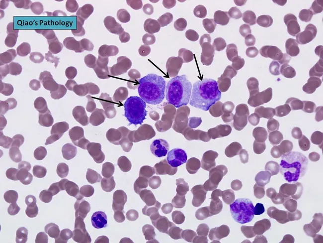
Peripheral blood smear prepared from a patient with suspected plasma cell leukemia (PCL) was examined by bright‑field light microscopy after Wright‑Giemsa staining. The smear shows several circulating plasma‑cell–like elements, including four conspicuous proplasmacytes with dispersed nuclear chromatin and prominent nucleoli, as indicated by arrows in the original figure. In addition, background erythrocytes exhibit rouleaux formation, a common feature in paraproteinemic states. The cells display eccentrically placed nuclei with pronounced basophilic cytoplasm and a coarse chromatin pattern; occasional cells show a perinuclear clearing (Golgi rim) suggestive of plasmacytic differentiation. Immunoglobulin light‑chain restriction is not determined on this smear alone but flow cytometry or immunohistochemistry would typically confirm clonal plasma cells. Relative proportions indicate that neoplastic plasma cells constitute a substantial fraction of leukocytes, consistent with plasma cell leukemia rather than reactive plasmacytosis. PCL is an aggressive myelomatous process often presenting with cytopenias, organomegaly, and sometimes lymphadenopathy; prognosis is poor, and treatment responsiveness is limited. This image illustrates characteristic morphologic features used for differential diagnosis against multiple myeloma, Waldenström macroglobulinemia, and other causes of plasmacytosis. Clinically relevant keywords include plasma cells, rouleaux, Wright‑Giemsa stain, 100X oil, peripheral blood, leukemia, myeloma, and hematologic malignancy.
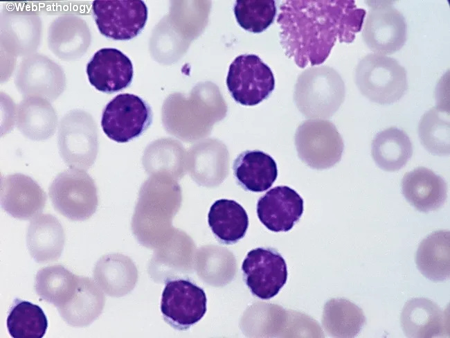
Peripheral blood smear from a patient with suspected chronic lymphocytic leukemia (CLL) shows a predominance of small, mature-appearing lymphocytes with scant cytoplasm and clumped nuclear chromatin, accompanied by fragile cells that readily disrupt during smear preparation, producing numerous smudge or basket cells visible on the slide. The image depicts a single smudge cell in the upper right, with numerous small darkly staining lymphocytes occupying the field. The smear was prepared with Wright-Giemsa stain and examined under light microscopy at high magnification (approximately 1000x total magnification with oil immersion). Clinically, CLL is defined by absolute mature lymphocytosis of ≥5.0 × 10^9/L sustained for ≥3 months, often asymptomatic and detected incidentally on complete blood count. Morphology alone is insufficient for diagnosis; immunophenotyping showing co-expression of CD5, CD19, CD23 with weak surface immunoglobulin and light chain restriction supports CLL diagnosis. Smudge cells result from fragile leukemic cells and reflect underlying cytoskeletal fragility, though they are not specific. The image illustrates typical features: monomorphic lymphocytes, scant cytoplasm, condensed chromatin, and absent prominent nucleoli. This finding has diagnostic significance, guiding further workup, including flow cytometry, cytogenetics, and molecular studies, to distinguish CLL from other chronic lymphoproliferative disorders such as small lymphocytic lymphoma (SLL), mantle cell lymphoma, prolymphocytic leukemia, and reactive lymphocytosis.
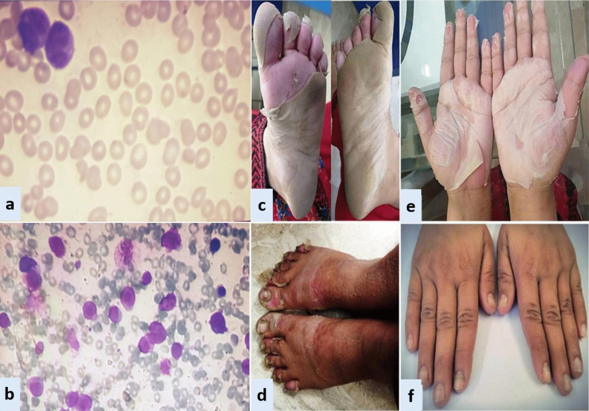
A composite medical image featuring microscopic and clinical findings associated with Acute Promyelocytic Leukemia (APL) and its treatment complications. Panels (a) and (b) show Wright-Giemsa stained light microscopy images: (a) a peripheral blood smear with scattered red blood cells and rare large atypical promyelocytes, and (b) a hypercellular bone marrow aspirate demonstrating a dense infiltration of blast cells with high nuclear-to-cytoplasmic ratios and prominent purple staining. Panels (c) through (f) illustrate dermatological manifestations consistent with exfoliative dermatitis and onychomadesis likely secondary to All-Trans Retinoic Acid (ATRA) therapy. (c) and (d) show the plantar surfaces of the feet with extensive, sheet-like desquamation and underlying erythema. (e) displays bilateral palmar exfoliation with large flaps of peeling epidermis. (f) shows the dorsal aspect of both hands with nail bed changes including yellow discoloration and signs of proximal nail plate separation (onychomadesis). The educational focus is the correlation between hematological malignancy and rare cutaneous drug reactions during induction chemotherapy.
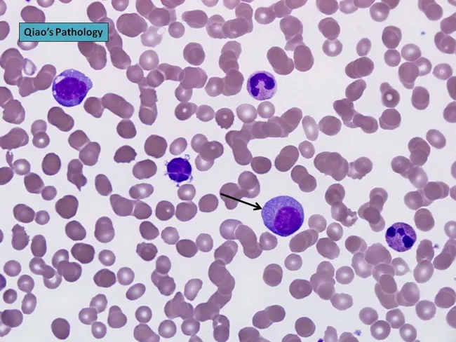
Peripheral blood smear prepared with Wright-Giemsa stain and examined under brightfield illumination using a 100× oil immersion objective reveals a single large plasma cell among a field of mature erythrocytes. The plasma cell displays features typical of clonal plasma cells: basophilic cytoplasm, an eccentrically placed round nucleus, and a conspicuous perinuclear hof; background shows rouleaux formation of red cells. This image exemplifies hematologic involvement by a plasma cell dyscrasia. In plasma cell leukemia, circulating plasma cells constitute more than 20% of leukocytes, a hallmark distinguishing it from conventional multiple myeloma, although both conditions share monoclonal plasma cell proliferation. The presence of circulating plasma cells is associated with younger patient age, higher incidence of anemia, thrombocytopenia, organomegaly, and possible lymphadenopathy; however lytic bone lesions may be less common. Immunophenotypic patterns often include lack of CD56 expression relative to myeloma. Clinically, this morphological finding warrants confirmation with complete blood count, peripheral smear review, quantitative plasma cell percentage, serum protein electrophoresis, and immunofixation to identify monoclonal immunoglobulin isotype (IgD/IgE or light-chain predominance). Early detection of circulating plasma cells supports prognosis assessment and guides therapy decisions in plasma cell dyscrasia management and monitoring, including risk stratification and treatment response evaluation for practice in clinics.
acute myeloid leukemia AML bone marrow biopsy Auer rods
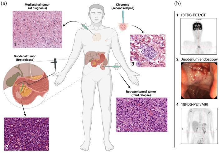
This composite educational infographic illustrates the diagnostic workup and progression of extramedullary manifestations (EM) of Acute Myeloid Leukemia (AML) in a 25-year-old patient. Panel (a) contains a central anatomical diagram showing the chronicity and location of four EM sites: (1) mediastinum, (2) duodenum, (3) skin (chloroma), and (4) retroperitoneum. Arrows point to corresponding hematoxylin and eosin (H&E) stained histopathology images for each site, demonstrating myeloid blasts and varying cellular density. The mediastinal and retroperitoneal tumors are shown being sampled via CT-guided biopsy, the duodenal tumor via endoscopy, and the chloroma via punch biopsy. Panel (b) provides diagnostic imaging correlation: (b1) 18FDG-PET/CT showing a hypermetabolic mediastinal mass, (b2) a duodenoscopy photograph revealing a friable, obstructive mass, and (b4) an 18FDG-PET/MRI depicting retroperitoneal disease. The visual summarizes the complex clinical course of multi-site extramedullary relapse despite bone marrow remission, emphasizing the role of integrated imaging and histological confirmation in AML management.
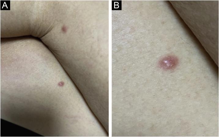
Two-part clinical photograph showing cutaneous manifestations of leukemia cutis in a patient with Acute Myeloid Leukemia (AML). Panel A presents a wide-angle view of the lower extremities, specifically the thighs, showing two isolated, discrete erythematous papules. Panel B provides a high-magnification close-up of a single lesion. The lesions are characterized as smooth-surfaced, rounded, and infiltrated (hardened) papules with a reddish, erythematous hue. They are well-demarcated from the surrounding healthy skin. These findings represent a recurrence of systemic leukemia following an allogeneic bone marrow transplant (BMT). The imagery illustrates the typical clinical presentation of extramedullary leukemic infiltration, which can be polymorphic and non-pathognomonic, requiring clinical correlation and biopsy for definitive diagnosis. The educational focus is on identifying skin-colored to erythematous papulonodular lesions as a sign of systemic hematologic malignancy progression or relapse.
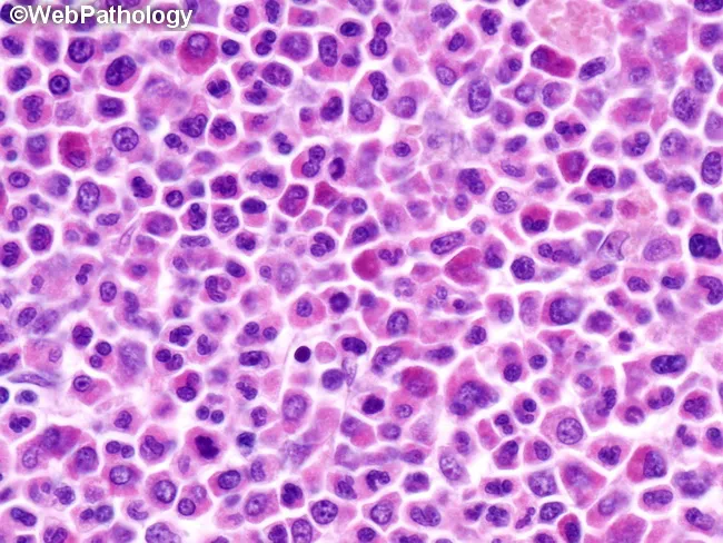
This high-magnification light microscopy image shows an H&E stained tissue section documenting an extramedullary myeloid tumor (myeloid sarcoma) composed of immature myeloid precursors arranged in diffuse sheets. The cellular population includes myeloblasts with high nuclear-to-cytoplasmic ratio, fine nuclear chromatin, prominent nucleoli, and frequent mitotic figures, accompanied by promyelocytes and maturation to myelocytes in variable proportions. Cytoplasm is scant to moderate and can contain occasional cytoplasmic granules; rod-shaped inclusions (Auer rods) may be seen in scattered blasts. The architectural pattern lacks normal tissue stratification, with cohesive clusters and dispersed cells infiltrating the stroma, consistent with an extramedullary granulocytic tumor. The image corresponds to a histopathology specimen most often obtained from soft tissue, lymph node, skin, or other extramedullary sites in patients with or without overt bone marrow involvement. Clinically, myeloid sarcoma is diagnostic of acute myeloid leukemia (AML) and often precedes or heralds AML relapse; immunohistochemical profiling (MPO, CD34, CD117, CD43, lysozyme) and cytogenetic/molecular testing are essential for classification and prognosis. Potential diagnostic significance includes associations with AML subtypes and recurrent translocations (e.g., t(8;21), inv(16), 11q23). This image is valuable for educational illustration of AML-related solid tumors, hematopathology education, differential diagnosis with lymphoma or metastatic carcinoma, and correlation with systemic hematologic disease and treatment planning.
chronic myeloid leukemia CML Philadelphia chromosome BCR-ABL peripheral blood smear
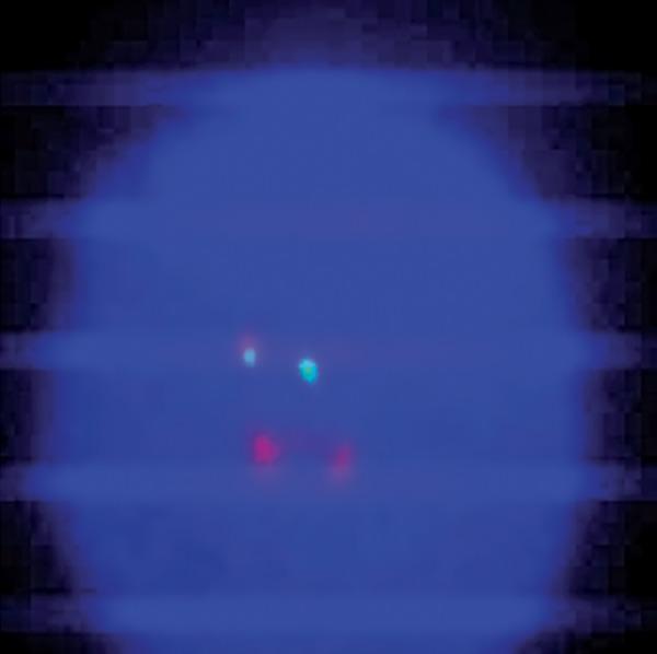
A diagnostic microphotograph of a peripheral blood interphase cell from a patient with Chronic Myeloid Leukemia (CML), analyzed via Fluorescence In Situ Hybridization (FISH). The image demonstrates an atypical BCR-ABL1 translocation pattern using a dual-color, dual-fusion probe. Against the dark blue DAPI-stained nuclear background, four distinct fluorescent signals are visible. The observed configuration is classified as 1F1G2R: one orange/yellow fusion signal (representing the BCR-ABL1 hybrid on the derivative chromosome 22), one green signal (representing the native BCR locus on chromosome 22), and two red signals (representing the native ABL1 locus on chromosome 9). This atypical pattern deviates from the standard 2F1G1R fusion signal usually seen in Philadelphia chromosome-positive cells, indicating clonal evolution or complex chromosomal rearrangements. This image serves as an educational example of cytogenetic variability in hematologic malignancies and the use of FISH for monitoring molecular response in patients undergoing tyrosine kinase inhibitor (TKI) therapy like imatinib.
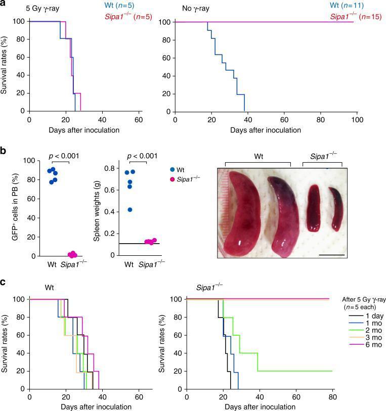
This composite figure presents research data on Chronic Myeloid Leukemia (CML) development in wild-type (Wt) and Sipa1 knockout (Sipa1-/-) mice following Bcr-Abl+ hematopoietic progenitor cell (HPC) injection. Panel (a) displays Kaplan-Meier survival curves showing that Sipa1-/- mice are completely resistant to CML in unirradiated conditions (100% survival), while gamma-irradiation abolishes this resistance. Panel (b) provides quantitative and visual evidence of disease progression: Wt mice exhibit massive splenomegaly (spleen weights 0.4-0.8g) and high peripheral blood GFP+ leukemic cell proportions (~80-90%), whereas Sipa1-/- mice maintain normal spleen weights (~0.1g) and negligible circulating leukemic cells. Clinical photographs show the striking morphological difference between the enlarged, dark-red spleens of Wt mice and the normal-sized spleens of Sipa1-/- mice. Panel (c) shows survival curves based on varying intervals after irradiation, indicating that the host resistance in Sipa1-/- mice is radiosensitive and requires several months to recover. This content illustrates the role of the host microenvironment and Sipa1 in leukemia progression and immune-mediated rejection.
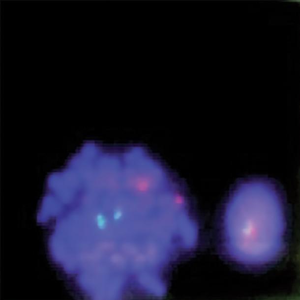
This diagnostic image displays a fluorescence in situ hybridization (FISH) analysis of human bone marrow cells, used primarily for detecting the BCR-ABL1 gene fusion associated with Chronic Myeloid Leukemia (CML). The image shows two blue-stained nuclei (DAPI) against a dark background. On the left, a larger metaphase cell exhibits a normal signal pattern with two distinct cyan/green signals and two distinct red signals (2G2R), representing the normal location of the ABL1 and BCR genes on their respective chromosomes. On the right, a smaller interphase cell demonstrates a fusion signal where a red and a cyan/green signal overlap or are closely apposed, indicative of a chromosomal translocation. This visual is a key educational tool for demonstrating molecular cytogenetics, specifically the dual-color, dual-fusion probe technique used in hematopathology to identify the Philadelphia chromosome.
Leukemias - Comprehensive Overview
Classification Framework
WHO Classification of Acute Leukemias
- AML with recurrent genetic abnormalities (t(8;21), inv(16), APL with PML-RARA, t(9;11), t(6;9), inv(3), t(1;22))
- AML with mutated NPM1, CEBPA (biallelic)
- AML with myelodysplasia-related changes
- Therapy-related myeloid neoplasms
- AML, not otherwise specified (NOS) - by differentiation (M0-M7)
- B-ALL with recurrent genetic abnormalities (t(9;22)/BCR-ABL1, t(v;11q23)/KMT2A, t(12;21)/ETV6-RUNX1, hyperdiploidy, hypodiploidy, t(5;14), t(1;19))
- B-ALL, NOS
- Goldman-Cecil Medicine, p. 2273
1. Acute Myeloid Leukemia (AML)
Epidemiology
Pathogenesis & Molecular Biology
| Translocation | Genes | Frequency | Prognosis |
|---|---|---|---|
| t(8;21) | RUNX1-RUNX1T1 | 10% | Favorable (CBF-AML) |
| inv(16) or t(16;16) | CBFB-MYH11 | 10% | Favorable (CBF-AML) |
| t(15;17) | PML-RARA | 7% | Favorable (APL) - responds to ATRA |
| t(9;11) | MLLT3-KMT2A | 5% | Poor (more common in pediatric AML) |
| t(6;9) | DEK-NUP214 | 1% | Very poor (basophilia, FLT3 ITD in 70%) |
| inv(3)/t(3;3) | GATA2, MECOM | 2% | Very poor |
| Normal karyotype | FLT3-ITD, NPM1, CEBPA | ~40% | Intermediate-variable |
-
Core binding factor (CBF) AMLs - t(8;21) and inv(16) - disrupt CBF-alpha and CBF-beta function, impairing differentiation. Both are diagnostic of AML regardless of blast count.
-
FLT3-ITD in normal-karyotype AML confers a poor prognosis and may benefit from HLA-matched allogeneic transplant.
-
Acute Promyelocytic Leukemia (APL/M3) with t(15;17) is a hematologic emergency - high risk of DIC; responds to ATRA + arsenic trioxide.
-
Goldman-Cecil Medicine, p. 2376-2378; Tietz Laboratory Medicine, p. 2810
Morphology
- Blasts: 12-20 μm, discrete nuclear chromatin, multiple nucleoli, azurophilic cytoplasmic granules
- Auer rods - slender fusiform cytoplasmic inclusions staining red with Wright-Giemsa - are virtually pathognomonic of AML
Immunophenotyping
-
10-20% of AML blasts aberrantly express B/T lineage markers (CD2, CD7, CD19, CD56) - does not change lineage assignment
-
Goldman-Cecil Medicine, p. 2270
Clinical Features
- Bone marrow failure symptoms: fatigue/pallor (anemia), infections (neutropenia), bleeding/petechiae (thrombocytopenia)
- Organomegaly (hepatosplenomegaly), leukostasis (WBC >100,000 - respiratory distress, confusion)
- Gingival hypertrophy in monocytic variants (KMT2A mutations)
- Skin infiltration (leukemia cutis)
- DIC - especially in APL
Treatment
- Good-risk (CBF AML): high-dose cytarabine (HiDAC) x 3-4 cycles
- Intermediate/poor-risk: allogeneic HSCT in CR1
- Allogeneic HSCT achieves cure in 40-70% in CR1 (vs. ~20-40% in CR2)
-
Midostaurin + chemotherapy for FLT3-mutated AML
-
Gilteritinib for relapsed/refractory FLT3-mutated AML
-
Venetoclax + azacitidine for older/unfit patients
-
Ivosidenib (IDH1-mutated), enasidenib (IDH2-mutated)
-
APL: ATRA + arsenic trioxide (non-high risk) or ATRA + chemotherapy
-
Goldman-Cecil Medicine, p. 2491-2525; Tietz Laboratory Medicine, p. 2810
2. Acute Lymphoblastic Leukemia (ALL)
Epidemiology
Classification: B-ALL vs T-ALL
| Subtype | Markers | Notes |
|---|---|---|
| Pro-B ALL | CD19, CD22 (no CD10) | ~10%, most immature |
| Common ALL (CALLA+) | CD19, CD22, CD10 | 50-60%, best prognosis in B-ALL |
| Pre-B ALL | CD19 + intracytoplasmic Ig | ~10% |
| Mature B-ALL | Surface Ig | <5%, Burkitt-like (t(8;14), MYC) |
- Early T-cell precursor ALL: CD7, no CD1a or surface CD3
- Thymic T-ALL: CD1a positive
- Mature T-ALL: surface CD3 positive
Key Cytogenetics in ALL
| Abnormality | Frequency | Significance |
|---|---|---|
| t(9;22) BCR-ABL1 (Ph+) | 5% pediatric, 25% adult | Poor - TKI + chemo |
| t(12;21) ETV6-RUNX1 | 25% pediatric | Favorable |
| Hyperdiploidy (>50 chr) | 25% pediatric | Favorable |
| Hypodiploidy (<44 chr) | 5% | Poor |
| t(v;11q23) KMT2A | More in infant ALL | Poor |
| t(8;14) MYC | Burkitt-like | Requires specific regimen |
- Goldman-Cecil Medicine, p. 2361-2362
Treatment
- Consolidation (6-8 intensive courses) + Maintenance (6-MP + methotrexate) for 2-3 years
- Ph+ ALL: TKI (imatinib/dasatinib/ponatinib) added to all phases; allogeneic HSCT in CR1
- Rituximab: improves outcome in CD20+ Ph-negative B-ALL
- Blinatumomab (bispecific CD3/CD19): superior to chemo in relapsed/refractory B-ALL
- Inotuzumab ozogamicin (CD22-ADC): effective in relapsed ALL
- CAR-T cell therapy (tisagenlecleucel): approved for pediatric/young adult relapsed/refractory B-ALL
- Goldman-Cecil Medicine, p. 2501-2527
3. Chronic Myeloid Leukemia (CML)
Epidemiology & Pathogenesis
- Philadelphia chromosome (t(9;22)) in ~95% - moves ABL from chr 9 to chr 22, creating BCR-ABL chimeric gene
- BCR-ABL encodes a constitutively active tyrosine kinase that drives uncontrolled proliferation of granulocytic precursors
- BCR-ABL fusion is present in granulocytic, erythroid, megakaryocytic, and B-cell precursors - indicating origin in a pluripotent HSC
- Remaining 5%: BCR-ABL created by complex cytogenetically cryptic rearrangements
Clinical Phases
| Phase | Blast Count | Features |
|---|---|---|
| Chronic | <10% blasts | Asymptomatic or mild sx; 3-5 year duration without therapy |
| Accelerated | 10-19% blasts | Progressive resistance, cytopenias, new cytogenetic changes |
| Blast Crisis | ≥20% blasts | AML-like (~70%) or ALL-like (~30%) transformation; median survival weeks-months |
Treatment
- Imatinib (Gleevec) - first-generation TKI; ~95% complete hematologic remission, major molecular response in majority
- Dasatinib, nilotinib - second-generation; faster/deeper responses; used first-line or after imatinib resistance
- Ponatinib, asciminib - third-generation; for T315I "gatekeeper" mutation
- Treatment-free remission (TFR) is achievable in ~40-50% of optimal responders after stopping TKI
- Robbins Basic Pathology, p. 406; Goldman-Cecil Medicine; Tietz Laboratory Medicine, p. 2810
4. Chronic Lymphocytic Leukemia (CLL)
Epidemiology
Pathogenesis
- Mature B-cell malignancy with shared immunoglobulin gene rearrangement
- No single defining mutation - most common mutations: TP53, NOTCH1, SF3B1, ATM (each 15-20%)
- Key: deletion 13q14 removes miR-15/-16 → BCR-2 overexpression → impaired apoptosis (most common recurrent genetic event)
- Unmutated IGHV (immunoglobulin heavy chain variable) = worse prognosis
- Mutated IGHV = better prognosis (post-germinal center origin)
Diagnosis and Staging
| Rai Stage | Features | Risk |
|---|---|---|
| 0 | Lymphocytosis only | Low |
| I | + Lymphadenopathy | Intermediate |
| II | + Splenomegaly/hepatomegaly | Intermediate |
| III | + Anemia (Hgb <11g/dL) | High |
| IV | + Thrombocytopenia (<100K) | High |
Clinical Features
-
75% asymptomatic at diagnosis - found incidentally on CBC
- Symptomatic: lymphadenopathy, splenomegaly, fatigue/dyspnea (anemia), bleeding (thrombocytopenia)
- B symptoms (fever, night sweats, weight loss) are unusual except in advanced disease
- Immunosuppression: Hypogammaglobulinemia + T-cell exhaustion → bacterial infections (sinus, pneumonia), herpes reactivation, increased COVID-19 mortality
- Autoimmune complications: AIHA (autoimmune hemolytic anemia), ITP (immune thrombocytopenia)
- Richter transformation: ~5-10% transform to diffuse large B-cell lymphoma → median survival <1 year
Treatment
- BTK inhibitors: Ibrutinib (1st gen), acalabrutinib, zanubrutinib (2nd gen) - target B-cell receptor signaling
- BCL-2 inhibitor: Venetoclax (± obinutuzumab/rituximab) - exploits BCL-2 overexpression
- Anti-CD20: Rituximab, obinutuzumab (combined with BTK/BCL2 inhibitors)
- Previous standard: FCR (fludarabine + cyclophosphamide + rituximab) - now primarily for young fit IGHV-mutated patients
- Goldman-Cecil Medicine, p. 2727-2960; Robbins Basic Pathology, p. 410-411
Comparative Summary Table
| Feature | AML | ALL | CML | CLL |
|---|---|---|---|---|
| Cell of origin | Myeloid progenitor | Lymphoid progenitor | Pluripotent HSC (BCR-ABL) | Mature B cell |
| Peak age | >60 years | Children (2-5y), adults | 25-60 years | >60 years (median 70) |
| Key genetic lesion | t(15;17), FLT3, NPM1 | t(9;22), t(12;21), hyperdiploidy | t(9;22) BCR-ABL | del(13q14), IGHV mutation status |
| Peripheral blood | Blasts + cytopenias | Blasts + cytopenias | Massive leukocytosis, full maturation | Mature lymphocytosis, smudge cells |
| Morphologic hallmark | Auer rods (AML) | Lymphoblasts (small, agranular) | Basophilia, full granulocytic spectrum | Small lymphocytes, smudge cells |
| Key surface markers | CD13, CD33, CD34, CD117 | CD10, CD19, CD22 (B); CD7, CD3 (T) | BCR-ABL (molecular) | CD5+, CD19+, CD23+ |
| Treatment cornerstone | 7+3 (cytarabine + anthracycline) | VCR + pred + L-asp ± anthracycline | TKI (imatinib/dasatinib) | BTKi (ibrutinib/acalabrutinib) or venetoclax |
| Curative option | Allogeneic HSCT | Allogeneic HSCT (high-risk/relapsed) | TKI (TFR possible); HSCT for blast crisis | HSCT (rarely); TKI-based control |
| Key emergency | DIC (APL), leukostasis | CNS leukemia, tumor lysis | Blast crisis | Richter transformation |
Special Topics
Hairy Cell Leukemia (HCL)
- Rare mature B-cell leukemia with characteristic "hairy" cytoplasmic projections on peripheral blood smear
- Pathognomonic: BRAF V600E mutation (present in virtually all cases)
- Positive for CD11c, CD25, CD103, CD123; tartrate-resistant acid phosphatase (TRAP) positive
- Pancytopenia with massive splenomegaly
- Treatment: cladribine (2-CDA) or pentostatin - single course → durable remission in >80%
Prolymphocytic Leukemia (PLL)
- More aggressive variant with large prolymphocytes (>55% of circulating lymphoid cells)
- B-PLL: often TP53 deletion/mutation; poor prognosis
- T-PLL: associated with ATM mutations; aggressive behavior
Clonal Hematopoiesis (Precursor State)
-
Acquired pathogenic mutations (DNMT3A, TET2 most common) in hematopoietic cells with normal blood counts
-
Incidence: ~10% at age 70, ~30% by age 90
-
~1% annual risk of progression to hematologic malignancy
-
Also associated with cardiovascular disease and inflammatory conditions through proinflammatory monocyte reprogramming
-
Robbins & Kumar Pathologic Basis of Disease, p. 578-580
Sources
- Goldman-Cecil Medicine International Edition, pp. 2251-2960 (Chapters 168, 169)
- Robbins & Kumar Basic Pathology, pp. 405-412 (Chapter 10)
- Robbins, Cotran & Kumar Pathologic Basis of Disease, pp. 578-580
- Tietz Textbook of Laboratory Medicine, 7th Edition, pp. 2810-2812 (Chapter 70)