
Anatomy of middle ear
Reading File
Now let me search the medical library for middle ear anatomy content in parallel.
Finding Sources
Finding Sources
Excellent. Let me read the most comprehensive sources in parallel - the Cummings Otolaryngology section and Gray's Anatomy, plus Scott-Brown's.
Reading File
Reading File
Reading File
Gray's Anatomy has excellent anatomy content. Let me read more of it and also read the KJ Lee's Essential Otolaryngology section in parallel.
Reading File
Reading File
Let me read the KJ Lee section on tympanic membrane and ossicles, plus continue the Gray's Anatomy section.
Reading File
Reading File
Now let me fetch the key anatomical diagrams and read the ossicles + muscles sections in Gray's and KJ Lee.
Reading File
Reading File
Loading Image
Now let me fetch a couple more key diagrams - the boundaries/walls diagram and the ossicles diagram.
Loading Image
Loading Image
I now have comprehensive information from multiple authoritative sources. Let me compile the full answer.
Anatomy of the Middle Ear
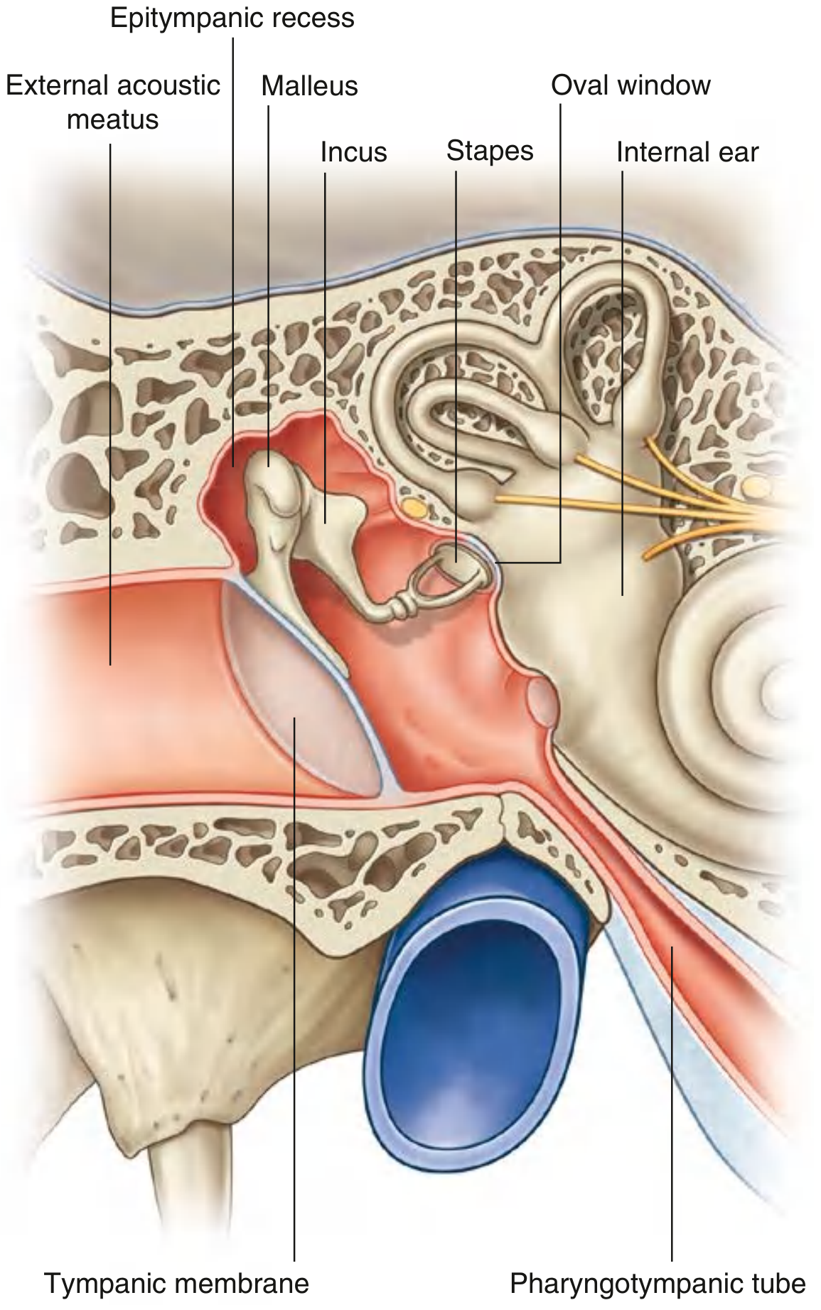
The middle ear is an air-filled, mucous membrane-lined space within the temporal bone, situated between the tympanic membrane laterally and the lateral wall of the internal ear medially. Its primary function is to transmit and amplify sound vibrations from the tympanic membrane to the oval window of the inner ear.
(Gray's Anatomy for Students, p. 1098)
Parts
The middle ear consists of two main parts:
- Tympanic cavity proper - the space immediately adjacent to the tympanic membrane
- Epitympanic recess (attic) - the superior extension above the level of the tympanic membrane, which houses the heads of the malleus and incus
Communications
- Posteriorly - via the aditus to the mastoid antrum, which communicates with the mastoid air cells
- Anteriorly - via the pharyngotympanic (Eustachian) tube to the nasopharynx
Walls / Boundaries
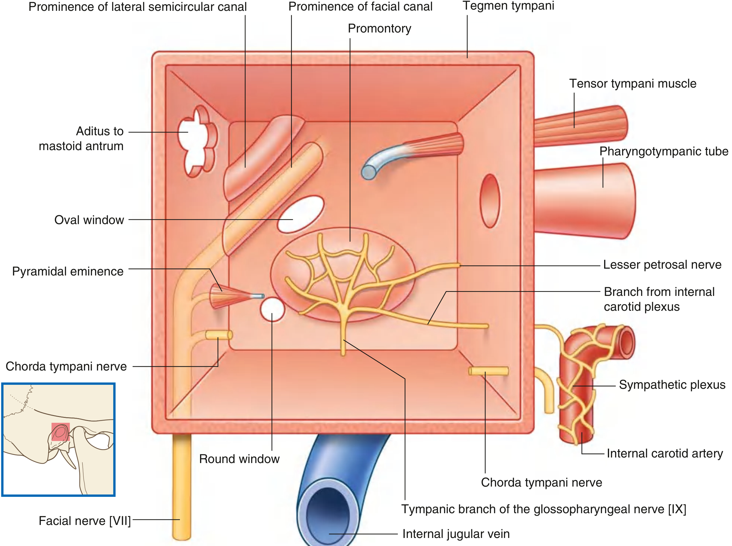
The middle ear has six walls:
| Wall | Also Called | Key Contents / Relations |
|---|---|---|
| Roof | Tegmental wall | Tegmen tympani (thin bone) separates the middle ear from the middle cranial fossa |
| Floor | Jugular wall | Thin bone over the internal jugular vein; tympanic branch of CN IX enters here |
| Lateral | Membranous wall | Largely formed by the tympanic membrane; upper part is bony (lateral wall of epitympanic recess) |
| Medial | Labyrinthine wall | Lateral wall of the inner ear; features the promontory, oval window, round window, prominence of facial canal, prominence of lateral semicircular canal |
| Anterior | Carotid wall | Separates tympanic cavity from internal carotid artery; has openings for the pharyngotympanic tube and tensor tympani canal |
| Posterior | Mastoid wall | Lower part separates tympanic cavity from mastoid air cells; superiorly opens into aditus ad antrum; contains pyramidal eminence (for stapedius tendon) and opening for chorda tympani |
(Gray's Anatomy for Students, p. 1098-1099)
Tympanic Membrane
The tympanic membrane separates the external from the middle ear and has four layers (from outside in):
- Squamous epithelium (outer)
- Radiating fibrous layer
- Circular fibrous layer
- Mucosal layer (inner)
- Average total area: 70-80 mm²
- Average vibrating surface: 55 mm²
- Divided into the pars tensa (the larger, taut portion) and the pars flaccida (Shrapnell's membrane) (the looser superior portion above the lateral process of the malleus)
(K.J. Lee's Essential Otolaryngology, p. 270)
Auditory Ossicles
Three tiny bones form a chain from the tympanic membrane to the oval window, transmitting sound vibrations:
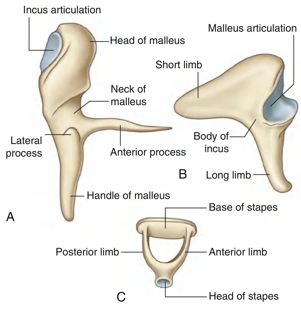
Malleus (largest ossicle)
- Head - in the epitympanic recess; articulates posteriorly with the incus
- Neck - constriction below the head
- Anterior process - attached by a ligament to the anterior wall
- Lateral process - attached to the malleolar folds of the tympanic membrane
- Handle (manubrium) - embedded in the tympanic membrane
- Receives tensor tympani tendon on the medial surface of its upper manubrium
Incus
- Body - in the epitympanic recess; articulates with the head of the malleus
- Long limb (process) - runs downward parallel to the manubrium of the malleus; ends in the lenticular process, which articulates with the stapes head
- Short limb (process) - runs posteriorly and is anchored by a ligament to the posterior incudal fossa
- The long process of the incus receives the least blood supply and is most frequently necrosed in chronic ear disease
Stapes (smallest bone in the body)
- Head - articulates with the long process of the incus
- Anterior and posterior crura - two bony limbs diverging from the head
- Footplate (base) - oval in shape (avg 1.41 mm × 2.99 mm); fits into the oval window, attached by the annular ligament
(Gray's Anatomy for Students, p. 1101-1102; K.J. Lee's Essential Otolaryngology, p. 271)
Ossicular Joints
- Malleoincudal joint - diarthrodial (synovial)
- Incudostapedial joint - diarthrodial (synovial)
- Stapediovestibular (stapediolabyrinthine) joint - syndesmotic (via annular ligament)
Muscles of the Middle Ear
| Muscle | Origin | Insertion | Innervation | Action |
|---|---|---|---|---|
| Tensor tympani | Cartilaginous pharyngotympanic tube, greater wing of sphenoid, its bony canal | Medial surface of upper manubrium of malleus (via cochleariform process) | Medial pterygoid nerve (branch of V3) | Pulls handle of malleus medially, tensing the tympanic membrane to dampen vibrations |
| Stapedius | Inside the pyramidal eminence | Posterior surface of neck of stapes | Branch of facial nerve (CN VII) | Pulls stapes posteriorly, dampening oscillation in response to loud sounds |
The contraction of both muscles in response to loud sounds is called the acoustic (stapedial) reflex, which protects the inner ear.
(Gray's Anatomy for Students, p. 1102)
Medial Wall - Key Landmarks
The labyrinthine (medial) wall contains several important structures:
- Promontory - rounded bulge formed by the basal coil of the cochlea; covered by the tympanic plexus of nerves
- Oval window (fenestra vestibuli) - posterosuperior to the promontory; accepts the stapes footplate; transmits vibrations to the perilymph of the scala vestibuli
- Round window (fenestra cochleae) - posteroinferior to the promontory; faces posteroinferiorly and is protected by an anterior lip; closed by the secondary tympanic membrane; allows pressure release
- Prominence of the facial canal - indicates the path of CN VII above the oval window
- Prominence of the lateral semicircular canal - above the facial canal prominence
(Gray's Anatomy for Students, p. 1099)
Nerves
Tympanic Plexus
Formed on the surface of the promontory. Contributions:
- Tympanic branch of CN IX (Jacobson's nerve) - major contributor; carries parasympathetics that continue as the lesser petrosal nerve to the parotid gland via the otic ganglion
- Auriculotemporal nerve (V3)
- Auricular branch of CN X
- Branches from the internal carotid sympathetic plexus
The plexus supplies the mucous membrane of the middle ear, mastoid cells, and pharyngotympanic tube.
Chorda Tympani (CN VII branch)
- Enters the middle ear through the posterior wall (via a canal near the pyramidal eminence)
- Crosses the middle ear between the handle of the malleus and the long process of the incus
- Exits through the anterior wall via the petrotympanic fissure (canal of Huguier)
- Carries taste from the anterior 2/3 of tongue and preganglionic parasympathetics to the submandibular and sublingual glands
(K.J. Lee's Essential Otolaryngology, p. 268; Gray's Anatomy for Students, p. 1100-1101)
Ligaments of the Ossicles
Malleus:
- Superior malleal ligament (head to tegmen/roof of epitympanum)
- Anterior malleal ligament (neck/anterior process to sphenoid via petrotympanic fissure)
- Lateral malleal ligament (neck to tympanic notch)
Incus:
- Superior incudal ligament (body to tegmen)
- Posterior incudal ligament (short process to incudal fossa)
Stapes:
- Annular ligament (footplate to margins of oval window)
Clinically Important Spaces
- Prussak's space - the space between Shrapnell's membrane (laterally) and the neck of the malleus (medially), bounded superiorly by the lateral malleal fold and inferiorly by the lateral process of the malleus. This is the most common site for acquired cholesteatoma formation.
- Aditus ad antrum - the channel connecting the epitympanic recess to the mastoid antrum
- Pouches of von Troeltsch - anterior and posterior recesses between the malleal folds and the tympanic membrane
Blood Supply
| Source | Supplies |
|---|---|
| Anterior tympanic artery (from maxillary a.) | Tympanic membrane |
| Posterior tympanic artery (from stylomastoid a.) | Middle ear posterior |
| Superior tympanic artery (from middle meningeal a.) | Tensor tympani, tegmen area |
| Inferior tympanic artery (from ascending pharyngeal a.) | Promontory |
| Internal carotid artery branches | Anterior middle ear |
Venous drainage - to the superior petrosal sinus and the pterygoid plexus.
Note: The long process of the incus has the most precarious blood supply and is most prone to ischemic necrosis.
(K.J. Lee's Essential Otolaryngology, p. 269-270)
Sources:
- Gray's Anatomy for Students, pp. 1098-1103
- K.J. Lee's Essential Otolaryngology, pp. 268-272
- Cummings Otolaryngology Head and Neck Surgery
This is a shared conversation. Sign in to Orris to start your own chat.