
Generate diagrams for three parts of the maxillary artery and its supplies in detail for an MBBS seminar presentation.
maxillary artery three parts branches supply anatomy MBBS
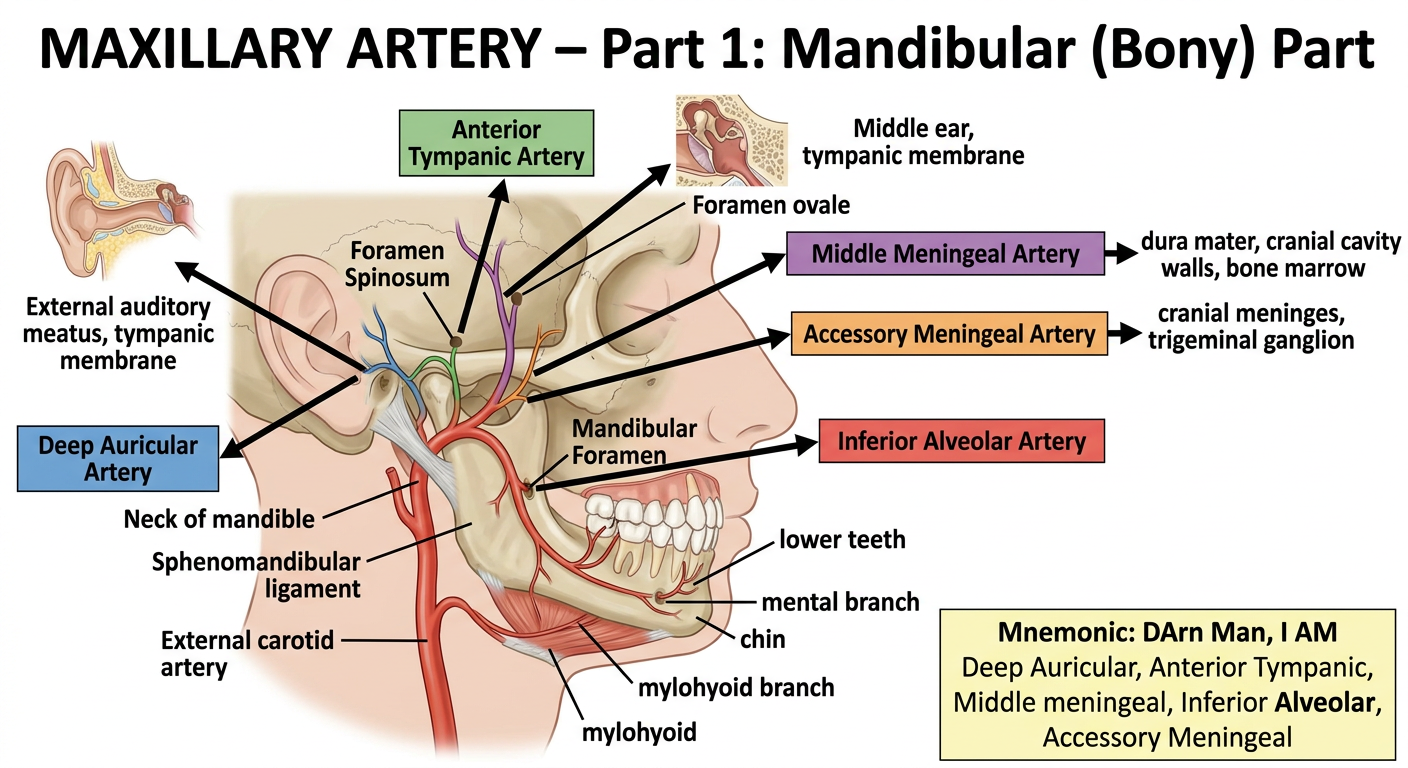
Detailed anatomical diagram of the FIRST PART (Mandibular Part) of the Maxillary Artery for an MBBS seminar presentation. Clean white background with bold title at top: "MAXILLARY ARTERY – Part 1: Mandibular (Bony) Part". Show the maxillary artery originating from the External Carotid Artery, passing between the neck of the mandible and sphenomandibular ligament. Label these 5 branches radiating outward with arrows and color-coded boxes: (1) Deep Auricular Artery → External auditory meatus, tympanic membrane; (2) Anterior Tympanic Artery → Middle ear, tympanic membrane; (3) Middle Meningeal Artery → through Foramen Spinosum → dura mater, cranial cavity walls, bone marrow; (4) Accessory Meningeal Artery → cranial meninges, trigeminal ganglion; (5) Inferior Alveolar Artery → through Mandibular Foramen → lower teeth, chin (mental branch), mylohyoid (mylohyoid branch). Include a mnemonic box: "DArn Man, I AM" → Deep Auricular, Anterior Tympanic, Middle meningeal, Inferior Alveolar, Accessory Meningeal. Use arterial red for vessels, anatomical bone beige for bony structures, clear black bold text labels, professional medical illustration style suitable for a lecture slide.
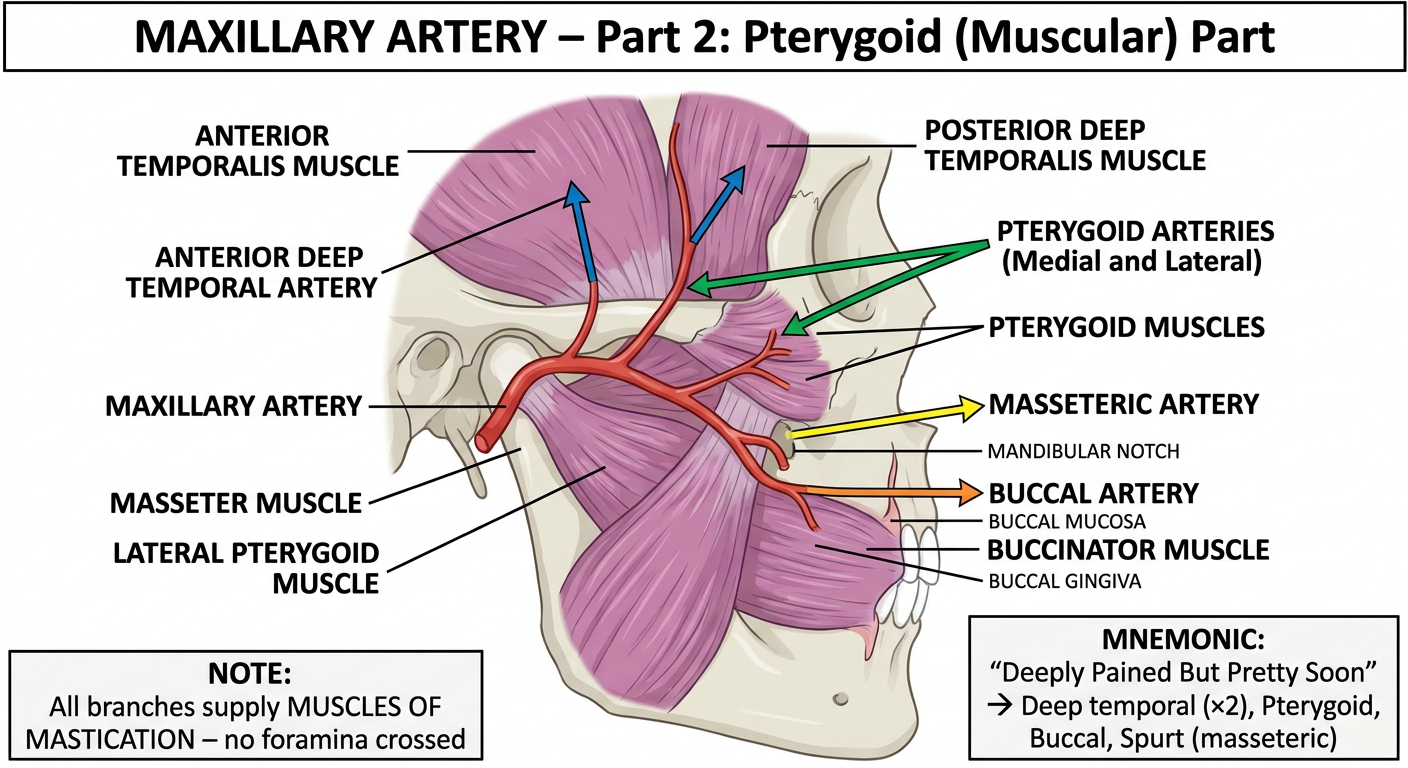
Detailed anatomical diagram of the SECOND PART (Pterygoid Part) of the Maxillary Artery for an MBBS seminar presentation. Clean white background with bold title: "MAXILLARY ARTERY – Part 2: Pterygoid (Muscular) Part". Show the maxillary artery coursing lateral (or medial) to the lateral pterygoid muscle in the infratemporal fossa. Label the lateral pterygoid muscle prominently. Show these 5 branches with color-coded arrows: (1) Anterior Deep Temporal Artery → anterior temporalis muscle; (2) Posterior Deep Temporal Artery → posterior temporalis muscle; (3) Pterygoid Arteries (Medial and Lateral) → pterygoid muscles; (4) Masseteric Artery → through mandibular notch → masseter muscle; (5) Buccal Artery → buccinator muscle, buccal mucosa, buccal gingiva. Include a note box: "All branches supply MUSCLES OF MASTICATION – no foramina crossed". Include mnemonic: "Deeply Pained But Pretty Soon" → Deep temporal (×2), Pterygoid, Buccal, Spurt (masseteric). Use arterial red for vessels, muscle purple/pink for muscular structures, bold black labels, clean professional medical education illustration style.
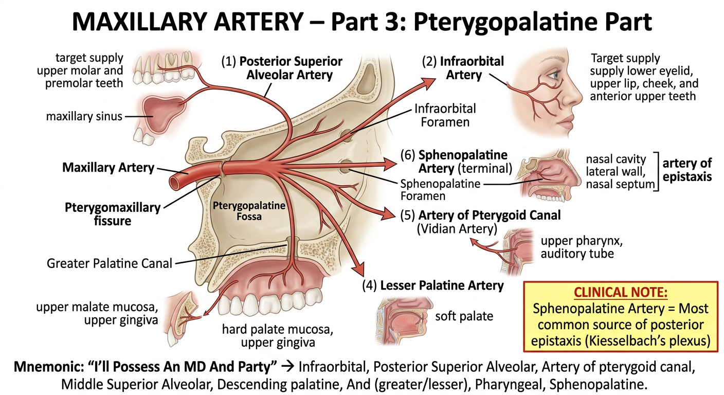
Detailed anatomical diagram of the THIRD PART (Pterygopalatine Part) of the Maxillary Artery for an MBBS seminar presentation. Clean white background with bold title: "MAXILLARY ARTERY – Part 3: Pterygopalatine Part". Show the maxillary artery entering the pterygopalatine fossa through the pterygomaxillary fissure. Label the pterygopalatine fossa. Show these 6 terminal branches with arrows fanning out to their targets: (1) Posterior Superior Alveolar Artery → upper molar and premolar teeth, maxillary sinus; (2) Infraorbital Artery → through Infraorbital Foramen → lower eyelid, upper lip, cheek, anterior upper teeth; (3) Greater Palatine Artery → through Greater Palatine Canal → hard palate mucosa, upper gingiva; (4) Lesser Palatine Artery → soft palate; (5) Artery of Pterygoid Canal (Vidian Artery) → upper pharynx, auditory tube; (6) Sphenopalatine Artery (terminal) → through Sphenopalatine Foramen → nasal cavity lateral wall, nasal septum – "artery of epistaxis". Include a highlighted clinical note box: "Sphenopalatine Artery = Most common source of posterior epistaxis (Kiesselbach's plexus)". Include mnemonic: "I'll Possess An MD And Party" → Infraorbital, Posterior Superior Alveolar, Artery of pterygoid canal, Middle Superior Alveolar, Descending palatine, And (greater/lesser), Pharyngeal, Sphenopalatine. Use arterial red for vessels, highlight clinical box in yellow with red border, professional medical lecture illustration style.
Maxillary Artery - Three Parts & Their Branches
Part 1 - Mandibular (Bony) Part
| Branch | Foramen | Supply |
|---|---|---|
| Deep Auricular | - | External auditory meatus, tympanic membrane |
| Anterior Tympanic | Petrotympanic fissure | Middle ear, inner surface of tympanic membrane |
| Middle Meningeal | Foramen spinosum | Dura mater, cranial vault bone, bone marrow |
| Accessory Meningeal | Foramen ovale | Trigeminal ganglion, meninges |
| Inferior Alveolar | Mandibular foramen | Lower teeth; mental branch (chin), mylohyoid branch |
Clinical pearl: The middle meningeal artery runs in a groove in the temporal bone. Rupture from a lateral head blow causes extradural haematoma - a neurosurgical emergency.
Part 2 - Pterygoid (Muscular) Part
| Branch | Supply |
|---|---|
| Anterior Deep Temporal | Anterior temporalis muscle |
| Posterior Deep Temporal | Posterior temporalis muscle |
| Pterygoid (medial + lateral) | Medial and lateral pterygoid muscles |
| Masseteric | Masseter muscle (via mandibular notch) |
| Buccal | Buccinator muscle, buccal mucosa, buccal gingiva |
Part 3 - Pterygopalatine Part
| Branch | Foramen | Supply |
|---|---|---|
| Posterior Superior Alveolar | Posterior alveolar canals | Upper molars/premolars, maxillary sinus |
| Infraorbital | Infraorbital foramen | Lower eyelid, cheek, upper lip, anterior upper teeth |
| Greater Palatine | Greater palatine canal/foramen | Hard palate mucosa, upper gingiva |
| Lesser Palatine | Lesser palatine foramen | Soft palate, tonsil |
| Artery of Pterygoid Canal (Vidian) | Pterygoid canal | Upper pharynx, auditory (Eustachian) tube |
| Sphenopalatine | Sphenopalatine foramen | Nasal cavity lateral wall, nasal septum |
| Pharyngeal | Palatovaginal canal | Nasopharyngeal roof, soft palate |
Clinical pearl: The sphenopalatine artery is the terminal branch and the most common source of severe posterior epistaxis. It anastomoses with branches of the facial and ophthalmic arteries at Kiesselbach's (Little's) plexus on the nasal septum - a classic surgical ligation target.
Full Mnemonic (all 17 branches)
- Deep auricular, Anterior tympanic, Middle meningeal, Inferior alveolar, Accessory meningeal (Part 1)
- Deep temporal (ant. + post.), Pterygoid, Buccal, Masseteric (Part 2)
- Posterior superior alveolar, Infraorbital, Greater/lesser palatine, Artery of pterygoid canal, Pharyngeal, Sphenopalatine (Part 3)