
What can you do
🔍 Research & Knowledge
- General questions across science, history, math, technology, law, finance, and more
- Medical & clinical questions - drugs, diagnoses, anatomy, physiology, procedures, pharmacology - backed by a library of authoritative medical textbooks
- Literature search - find peer-reviewed studies, clinical trials, and systematic reviews via PubMed
- Web search - real-time information, news, current events, and fact-checking
📄 Document & File Work
- Word documents (.docx) - create, read, edit, format professional reports, letters, memos
- PowerPoint (.pptx) - build slide decks, presentations, pitch decks; extract or edit existing slides
- Excel/spreadsheets (.xlsx, .csv) - create, clean, analyze, and format tabular data
- PDFs - read, extract text/tables, merge, split, rotate, watermark, OCR scanned pages
🖼️ Images & Visuals
- Generate images - create original diagrams, posters, flashcards, postcards, and visuals from a description
- Analyze images/videos - describe, extract text (OCR), or explain uploaded media
- Find medical images - search a library of clinical photos, histology slides, ECGs, anatomical diagrams
- Fetch and view images from URLs
💻 Code & Computation
- Write and run code in a Linux sandbox (Python and more)
- Data analysis - process files, compute statistics, generate charts
- Automate tasks - scripts, file manipulation, format conversion
🏥 Specialized Knowledge Domains
- Medical library - textbook-backed answers on clinical topics
- Indian taxation - direct taxes, income tax, transfer pricing (CA/CMA exam level)
- GST & indirect taxes - CA Final, ACCA prep via Navkar Institute materials
- Hip surgery - orthopaedic surgical videos and clinical guidelines (demo)
🗂️ Task Management
- Complex multi-step tasks - I break them into tracked steps and work through them systematically
- Parallel research - I can run multiple searches or sub-tasks simultaneously to save time
Detailed histology of Kidney
kidney histology nephron glomerulus tubules microscopy
Renal histopathology slide imaged under light microscopy after Hematoxylin and Eosin staining. The primary subject is human kidney cortex tissue displaying a glomerulus and adjacent renal tubules. The specimen type is a renal biopsy. The orientation appears as a transverse section with a glomerular tuft in the left-lower quadrant and tubular profiles dispersed through the interstitium; a large luminal space on the right is a ductule or artifact. The glomerulus contains densely packed nuclei and mesangial regions; capillary loops are variably visible. Interstitial stroma shows mild cellularity without confluent inflammatory infiltrate. Overall architecture is preserved with no overt necrosis, crescents, or significant sclerosis evident at this magnification. The stain highlights nuclei in deep purple and extracellular matrix in pink, with clear delineation between glomerular structures, tubules, and interstitium. Clinically, such a field could serve as baseline for diagnosing glomerular diseases, evaluating tubular integrity, and assessing nephron units in nephritis workups. Potential diagnostic considerations include primary glomerular disease, reactive changes, or incidental normal renal histology depending on clinical context. This image is educational for pathology residents and medical students to recognize basic renal microanatomy and H&E contrast characteristics. This representation supports familiarity with renal histology, including glomeruli, tubules, and vessels architecture
Renal cortical biopsy by light microscopy with Hematoxylin and Eosin staining. The image shows a globally sclerotic glomerulus surrounded by atrophic tubules within the renal cortex. The sclerotic tuft demonstrates obliteration of capillary lumina with increased mesangial matrix; tubulointerstitial compartment shows tubular atrophy and mild interstitial fibrosis. Vascular changes include hyaline arteriolosclerosis of small arteries and arterioles, compatible with chronic nephrosclerosis from long-standing hypertension. No acute inflammatory infiltrates or crescents are observed. This pattern is characteristic of benign hypertensive nephrosclerosis and contrasts with diabetic nephropathy or inflammatory glomerulonephritis. The accompanying tubules atrophy underscores chronic ischemic injury and reduced nephron mass. Clinically, such histology correlates with progressive decline in glomerular filtration rate (GFR), potential proteinuria, and risk of chronic kidney disease progression. Differential considerations include ischemic nephropathy due to vascular disease, age-related nephrosclerosis, and secondary hypertensive kidney injury. Understanding this morphology aids in etiologic assessment of CKD and guides management focusing on blood pressure control and renal protective strategies. This image is valuable for pathology education, nephrology training, and research on hypertensive vascular injury in the kidney. This pattern is commonly seen in chronic hypertensive patients.
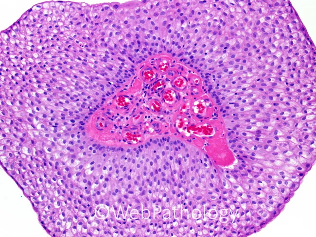
This is a brightfield light microscopy histology image of a renal cortex region, captured after standard hematoxylin and eosin staining. The primary subject is a renal glomerulus embedded in renal cortex, with Bowman's capsule and adjacent proximal/distal tubules visible in the peripheral parenchyma. The glomerular tuft consists of a dense network of capillary loops; erythrocytes appear as circular red cells within the capillary lumina, giving a pink-reddish core. The surrounding mesangial cells and basement membranes are discernible as pink-stained stroma, with purple-nuclei of endothelial and parietal epithelial cells. The tubules exhibit cuboidal epithelium with basophilic nuclei and clear luminal spaces. Overall architecture demonstrates typical renal cortical histology: a lobulated arrangement of tubules around a central tuft, with intact Bowman's capsule juxtaposed to the capillary network. There is no conspicuous sclerosis, crescents, or inflammatory infiltrates; no overt tubular atrophy or interstitial fibrosis evident. This image represents normal renal histology suitable for educational purposes and serves as a reference for pattern recognition. Clinically, it aids in teaching glomerular structure, filtration barrier components (endothelium, basement membrane, podocytes), and nephron organization. Potential use cases include medical education, comparative pathology, and radiology-pathology correlation studies for nephrology and urology training.
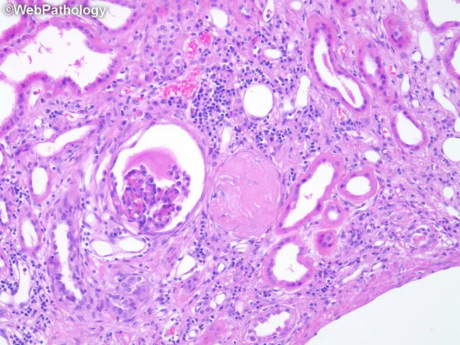
High-power brightfield histopathology of a renal cortical biopsy stained with Hematoxylin and Eosin (H&E) evaluated at approximately 400x magnification. The cortical nephron structures reveal changes typical of Autosomal Dominant Polycystic Kidney Disease (ADPKD) including a sclerotic, enlarged glomerulus with saccular expansion, indicating global or segmental sclerosis. Adjacent nephrons show distortion and dilatation of tubules with irregular luminal profiles and flattened, attenuated lining epithelium. The interstitial compartment displays mild inflammatory cells and periglomerular fibrotic changes, consistent with chronic injury and nephron loss. The overall architectural disruption reflects progressive cystic disease with reduced functional nephrons and compensatory remodeling, commonly seen in ADPKD. The image illustrates well the renal parenchymal consequences of polycystic disease—glomerulosclerosis, tubulomegaly, and tubulointerstitial fibrosis—contributing to decreased glomerular filtration rate. Clinically, these histologic features correlate with imaging-detected bilateral renal cysts and polycystic kidney disease progression, abnormal renal function tests, hypertension, and potential renal failure in affected adults. This slide is valuable for education, differential diagnosis against other glomerulopathies and cystic kidney diseases, and research into cystogenesis, nephron loss, and remodeling in polycystic kidney disease. Keywords for search include: ADPKD histology, glomerulosclerosis, tubulomegaly, polycystic kidney disease pathology, kidney cortex, renal biopsy, brightfield, H&E, renal cysts. educational, clinical, research, nephrology, nephron remodeling.
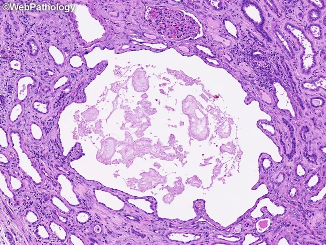
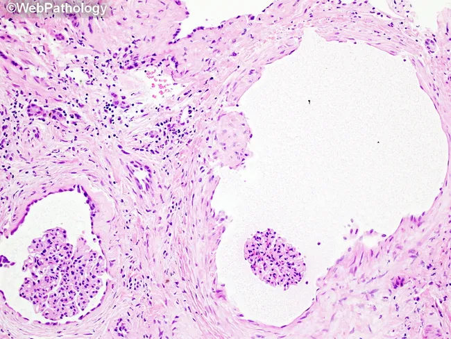
This renal histology image depicts cancellated cystic change characteristic of autosomal dominant polycystic kidney disease (ADPKD) examined with light microscopy. The tissue is stained with Hematoxylin and Eosin (H&E) and shows saccular dilation of tubular segments and Bowman’s capsule, producing multiple cysts of variable size within the cortical parenchyma. The surrounding nephron units display alternating regions of normal-appearing tubules and dilated tubules that contribute to a heterogeneous,lobulated architecture. The cyst linings are typically cuboidal to flattened epithelium, often contiguous with shared basement membranes, and the cyst contents appear pale or eosinophilic depending on the presence of proteinaceous material. This histologic pattern reflects cystogenesis beginning at various nephron levels with progressive expansion of tubules and glomerular capsule remnants, a hallmark of ADPKD. The pericystic stroma may demonstrate mild interstitial changes but is not prominent in this field. Clinically, these findings correlate with bilateral renal cyst formation and progressive parenchymal disruption, contributing to hypertension and chronic kidney disease over time. In isolation, these features should be interpreted alongside clinical history, imaging studies, and genetic testing to confirm ADPKD (PKD1/PKD2 mutations) and to differentiate from acquired or pediatric cystic diseases. These histologic cues guide prognosis and inform targeted genetic testing strategies for patients.
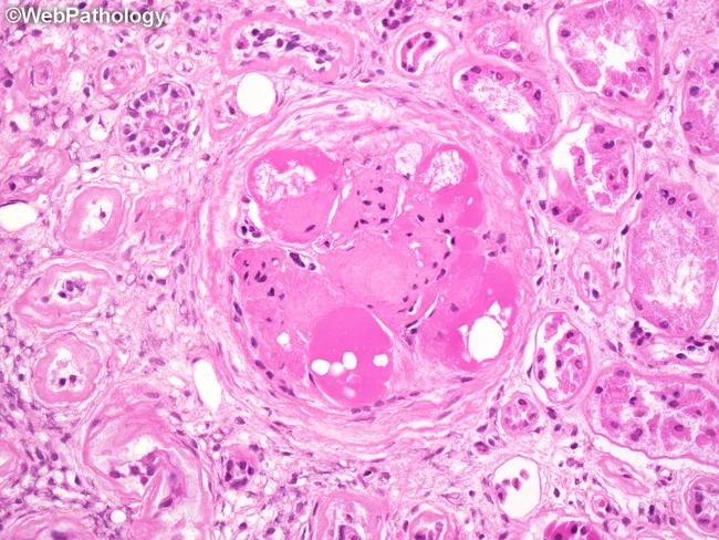
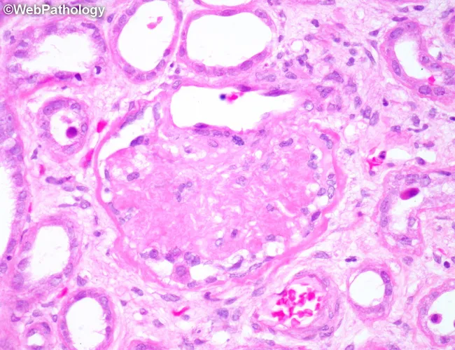
This is a renal biopsy histology image captured under bright-field light microscopy after Hematoxylin and Eosin staining. The focal structure is a glomerulus in the renal cortex showing near-complete global glomerulosclerosis with a markedly hyalinized tuft. Several hyaline caps outline the sclerotic core, appearing as pink, glassy deposits along capillary loops. Within these hyaline rims, lipid droplets and lipid-laden macrophages are evident, reflecting lipid accumulation associated with chronic diabetic microangiopathy. The surrounding tubulointerstitium displays mild interstitial fibrosis and preserved tubules in the periglomerular region. Collectively, the pattern is typical of advanced diabetic nephropathy with extensive glomerulosclerosis and hyaline arteriolosclerosis. The image highlights pathognomonic features such as noncellular sclerosis and lipid-laden inflammatory cells, which have prognostic implications for progressive renal insufficiency. Clinically, these findings correlate with long-standing diabetes mellitus, hypertension, proteinuria, and reduced GFR, indicating high risk for progression to end-stage kidney disease if glycemic control is poor. This specimen serves educational purposes in renal pathology, nephrology, and medical training to illustrate diabetic glomerulosclerosis, hyaline change, and lipid-related macrophage infiltration; it supports differential diagnosis with hypertensive nephrosclerosis and focal segmental glomerulosclerosis.
renal glomerulus Bowman capsule podocyte filtration barrier electron microscopy
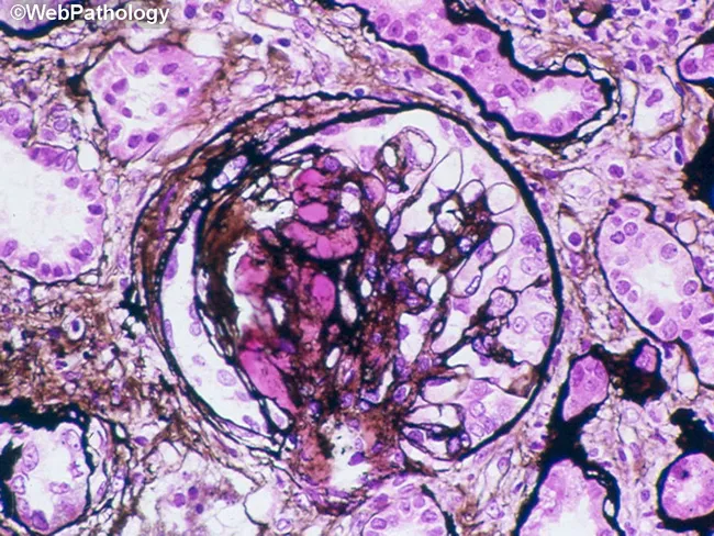
This renal histopathology image depicts a glomerulus in the renal cortex, examined under light microscopy after Hematoxylin and Eosin (H&E) staining. The glomerular tuft is round to ovoid, bounded by Bowman's capsule, and embedded in mild tubulointerstitial parenchyma. The capillary loops within the tuft show variable cellularity with modest mesangial proliferation and slight endothelial swelling. The basement membranes appear broadened in places, consistent with chronic or active injury. Some capillary walls harbor hyaline-like material that stains more eosinophilic, producing faint luminal narrowing. The surrounding tubular epithelium demonstrates typical cytoplasmic eosinophilia with preserved overall architecture but occasional luminal crowding. Overall, the image demonstrates a proliferative glomerular pattern with mild sclerosis potential, though global assessment is limited by single-field view. The observed morphology can be seen in glomerulonephritis and mesangial-dominant disorders, including early diabetic nephropathy, IgA nephropathy, or membranoproliferative patterns depending on additional staining and clinic. Clinically relevant, this pattern warrants correlation with proteinuria, serum creatinine, blood pressure, and serologies; immunofluorescence and electron microscopy would refine immune deposition, basement membrane thickening, and podocyte foot-process effacement—critical for diagnosis and prognosis planning.
This is a brightfield light microscopy histology image of a renal cortex region, captured after standard hematoxylin and eosin staining. The primary subject is a renal glomerulus embedded in renal cortex, with Bowman's capsule and adjacent proximal/distal tubules visible in the peripheral parenchyma. The glomerular tuft consists of a dense network of capillary loops; erythrocytes appear as circular red cells within the capillary lumina, giving a pink-reddish core. The surrounding mesangial cells and basement membranes are discernible as pink-stained stroma, with purple-nuclei of endothelial and parietal epithelial cells. The tubules exhibit cuboidal epithelium with basophilic nuclei and clear luminal spaces. Overall architecture demonstrates typical renal cortical histology: a lobulated arrangement of tubules around a central tuft, with intact Bowman's capsule juxtaposed to the capillary network. There is no conspicuous sclerosis, crescents, or inflammatory infiltrates; no overt tubular atrophy or interstitial fibrosis evident. This image represents normal renal histology suitable for educational purposes and serves as a reference for pattern recognition. Clinically, it aids in teaching glomerular structure, filtration barrier components (endothelium, basement membrane, podocytes), and nephron organization. Potential use cases include medical education, comparative pathology, and radiology-pathology correlation studies for nephrology and urology training.
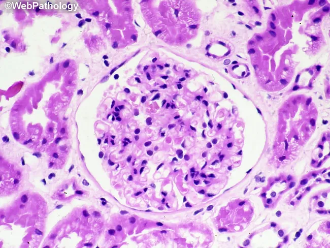
Renal biopsy histology image captured with light microscopy after Hematoxylin and Eosin staining. The section highlights a renal glomerulus embedded in renal cortex, with Bowman’s capsule clearly visible. Within the glomerulus there is marked cellular proliferation, with numerous densely packed nuclei suggestive of intraglomerular inflammation. A prominent cellular crescent occupies Bowman's space, formed by parietal epithelial cells and infiltrating leukocytes; this crescent partially compresses the capillary tuft. Surrounding tubulointerstitial tissue shows mild edema and scattered inflammatory cells. The basement membranes appear thickened in places, and there is focal endocapillary hypercellularity. Overall architecture suggests acute glomerular injury with crescent formation, compatible with rapidly progressive glomerulonephritis. The image serves as a morphologic correlate for nephritic syndrome and abrupt loss of renal function, guiding urgent diagnostic workup. Immunofluorescence and electron microscopy would assist etiologic classification (pauci-immune, anti-GBM, or immune complex), as would serologic testing for anti–neutrophil cytoplasmic antibodies, anti-GBM antibodies, and complements. Clinically, such histology is associated with rapidly progressive renal failure, hematuria, proteinuria, and hypertension. This representation supports differential diagnoses including anti-GBM disease, immune complex GN, and pauci-immune GN, and is valuable for education, research, and clinical correlation in nephrology. These features emphasize urgent management, biopsy interpretation, and multidisciplinary care planning strategies.
juxtaglomerular apparatus macula densa renin secreting cells kidney
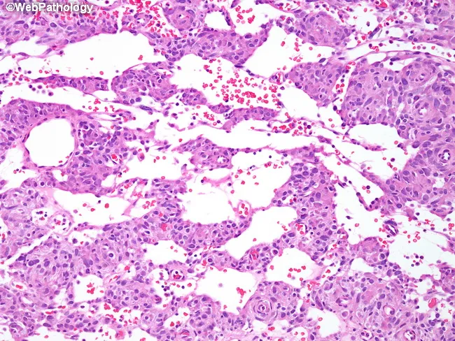
Juxtaglomerular cell tumor (reninoma) on bright-field light microscopy using Hematoxylin and Eosin staining. Modality: Histopathology; magnification high (40x objective). Localization: kidney, renal cortex, juxtaglomerular apparatus region; derived from juxtaglomerular cells; renin-producing. Morphology: highly vascular neoplasm composed of cords or nests of monomorphic tumor cells separated by prominent sinusoidal vascular spaces and interposed capillaries. The tumor demonstrates a pronounced angiovascular network with branching vessels that may resemble a stag-horn pattern typical of hemangiopericytoma; variably arranged small veins and muscular arterioles are present. The cells show uniform, round to ovoid nuclei with scant cytoplasm and minimal pleomorphism; mitotic activity is low. The vascular spaces contribute to a pseudovascular, labyrinthine architecture. Clinically, JGCTs are renin-secreting tumors causing secondary hypertension; radiology often reveals a small, well-circumscribed renal mass. Immunohistochemistry (if performed) may support juxtaglomerular lineage (e.g., renin positivity). Diagnostic significance: recognition of this histology supports diagnosis of juxtaglomerular cell tumor and informs surgical management; differential includes hemangiopericytoma-like renal tumors and other renal neoplasms with prominent vascularity. Potential clinical use cases: diagnostic education, pathology reference, case characterization, research on renin-angiotensin system tumors, and surgical planning. This image exemplifies the classic vascular architecture and cellular uniformity used to teach renal tumor histology and differential diagnosis skills.
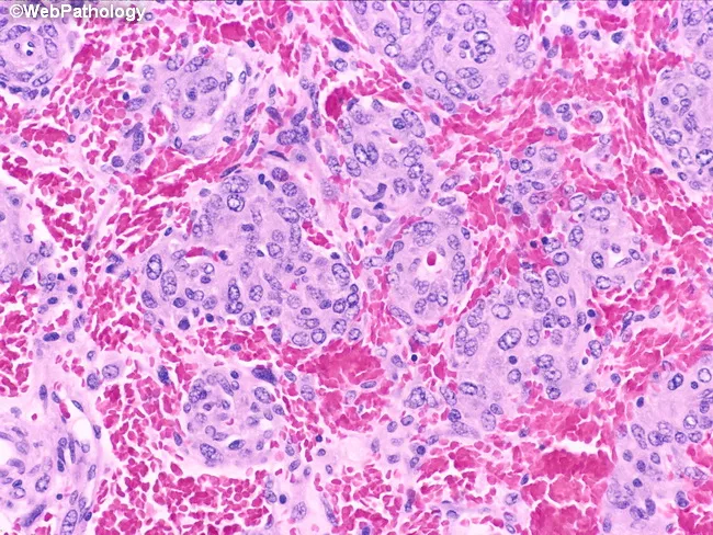
Imaging modality and technique: Light microscopy of hematoxylin and eosin stained renal tumor tissue (H&E). Specimen shows renal cortical parenchyma with neoplastic cells arranged in aggregates. Primary subject: Juxtaglomerular cell tumor (reninoma), a rare renin-secreting renal neoplasm. Anatomical localization: Kidney, renal cortex, juxtaglomerular apparatus region, perivascular renal cortex. Morphology: Epithelioid tumor cells are round to polygonal with moderate eosinophilic granular cytoplasm. Nuclei are uniform and vesicular with evenly dispersed chromatin and small nucleoli. Cells form nests and cords with occasional concentric rings around arterioles, reflecting juxtaglomerular origin. The stroma shows a delicate vascular network with interspersed red blood cells; mitotic activity is low; overall cellularity is variable. Notable features: concentric perivascular patterns around arterioles; cohesive arrangement; clear cytoplasmic granularity. Diagnostic significance: Histologic features are characteristic of juxtaglomerular cell tumor; correlation with clinical data (hypertension, elevated renin) supports diagnosis. Differential considerations: other renal neoplasms such as renal cell carcinoma (clear cell, papillary), oncocytoma, angiomyolipoma; however, perivascular concentric arrangement around arterioles favors juxtaglomerular lineage. Clinical correlation: typically presents in young adults with secondary hypertension; renin release may be elevated; treatment often includes nephron-sparing resection. These features aid pathologists in distinguishing juxtaglomerular tumors from mimics and guide surgical management in selected cases.
Detailed Histology of the Kidney
1. Overview: Cortex and Medulla
- Cortex - outer region, contains all renal corpuscles (glomeruli + Bowman's capsule), proximal convoluted tubules (PCT), distal convoluted tubules (DCT), and initial collecting ducts. Appears granular on gross section due to packed nephrons.
- Medulla - inner region, contains the loops of Henle, collecting ducts, and vasa recta. Organized into renal pyramids (8-12 per kidney). Each pyramid's apex (renal papilla) opens into a minor calyx.
- Medullary rays - pale streaks extending from medulla into cortex, containing straight tubules and collecting ducts.
- Outer medulla: outer stripe (contains thick descending and ascending limbs) + inner stripe (contains thin descending limbs and thick ascending limbs)
- Inner medulla: thin limbs of long loops and collecting ducts converging at papilla
2. The Renal Corpuscle (Malpighian Corpuscle)
- Vascular pole: where the afferent arteriole enters and efferent arteriole exits
- Urinary pole: where Bowman's space drains into the PCT

2a. The Glomerulus
2b. Glomerular Basement Membrane (GBM)
- Lamina rara interna - adjacent to endothelium
- Lamina densa - central, electron-dense, mainly type IV collagen and laminin
- Lamina rara externa - adjacent to podocyte foot processes, rich in heparan sulfate proteoglycans (negatively charged - restricts albumin filtration)
2c. The Filtration Barrier (Three-Layer)
| Layer | Structure | Pore Size | Key Feature |
|---|---|---|---|
| Fenestrated endothelium | Capillary wall with fenestrae | 50-100 nm | Prevents blood cells; covered by glycocalyx |
| GBM | Noncellular lamina densa | - | Charge barrier (heparan sulfate) |
| Podocyte slit diaphragms | Filtration slits between foot processes | 30-40 nm | Main filtration barrier |
2d. Glomerular Cell Types
- Highly fenestrated (unlike most endothelia)
- Covered by glycocalyx of negatively charged glycoproteins and glycosaminoglycans
- Produce VEGF receptors and are maintained by podocyte-derived VEGF
- Large cell bodies with primary processes extending around capillaries
- Secondary foot processes (pedicles) interdigitate with those from neighboring podocytes
- Foot processes contact the GBM; the gaps between them are the filtration slits (30-40 nm)
- Slit diaphragms span the filtration slits - key proteins: nephrin, podocin, CD2AP
- Podocytes cannot regenerate easily; injury leads to foot process effacement (proteinuria)
- Line the inner surface of Bowman's capsule
- Simple squamous epithelium at the parietal layer
- Reflect at the vascular pole to become visceral epithelium (podocytes)
- Highly proliferative; thought to provide a niche for podocyte replenishment after injury
- Form crescents in rapidly progressive glomerulonephritis (RPGN)
- Occupy the intercapillary mesangial space
- Functions:
- Structural support for glomerular capillaries
- Regulate GFR via contractility (altering filtration surface area)
- Produce and maintain mesangial matrix
- Phagocytic (remove macromolecules from mesangium)
- Secrete cytokines, prostaglandins, and repair factors
- Surrounded by mesangial matrix (not GBM)
3. Renal Tubules
Summary Table of Tubule Segments
| Segment | Epithelium | Location | Key Histologic Feature | Major Function |
|---|---|---|---|---|
| PCT | Simple cuboidal; well-stained | Cortex | Long brush border (microvilli); prominent basal labyrinth; many mitochondria; apical vacuoles/lysosomes | Reabsorption of all organic nutrients, proteins, ~67% water & Na+; H+ secretion |
| Proximal straight tubule (PST / S3) | Simple cuboidal | Outer stripe, medullary rays | Less brush border than PCT | Secretion of organic anions/cations, toxins, drugs |
| Thin descending limb (tDL) | Simple squamous | Inner stripe & inner medulla | Extremely flat epithelium; few organelles | Permeable to water; contains UT-A2 (urea recycling) |
| Thin ascending limb (tAL) | Simple squamous | Inner medulla | Heavily interdigitated; low organelles | Highly permeable to ions (Na+, Cl-); impermeable to water |
| Thick ascending limb / TAL (Distal straight tubule) | Simple cuboidal | Inner stripe & medullary rays | No brush border; many mitochondria; cells very large | Active NaCl reabsorption via NKCC2; water impermeable ("diluting segment") |
| DCT | Simple cuboidal; smaller cells | Cortex | Short stubby microvilli; prominent basolateral folds; many mitochondria; more open lumens | NaCl reabsorption; PTH-regulated Ca2+ reabsorption |
| Principal cells (CD) | Cuboidal to columnar; pale-staining | Medullary rays & medulla | Distinct cell membranes; few organelles | ADH-regulated water reabsorption; aldosterone-regulated Na+/K+ |
| Intercalated cells (CD) | Slightly darker; scattered | Medullary rays | Darker cytoplasm; carbonic anhydrase-rich | K+ reabsorption; acid-base balance (H+ / HCO3- secretion) |
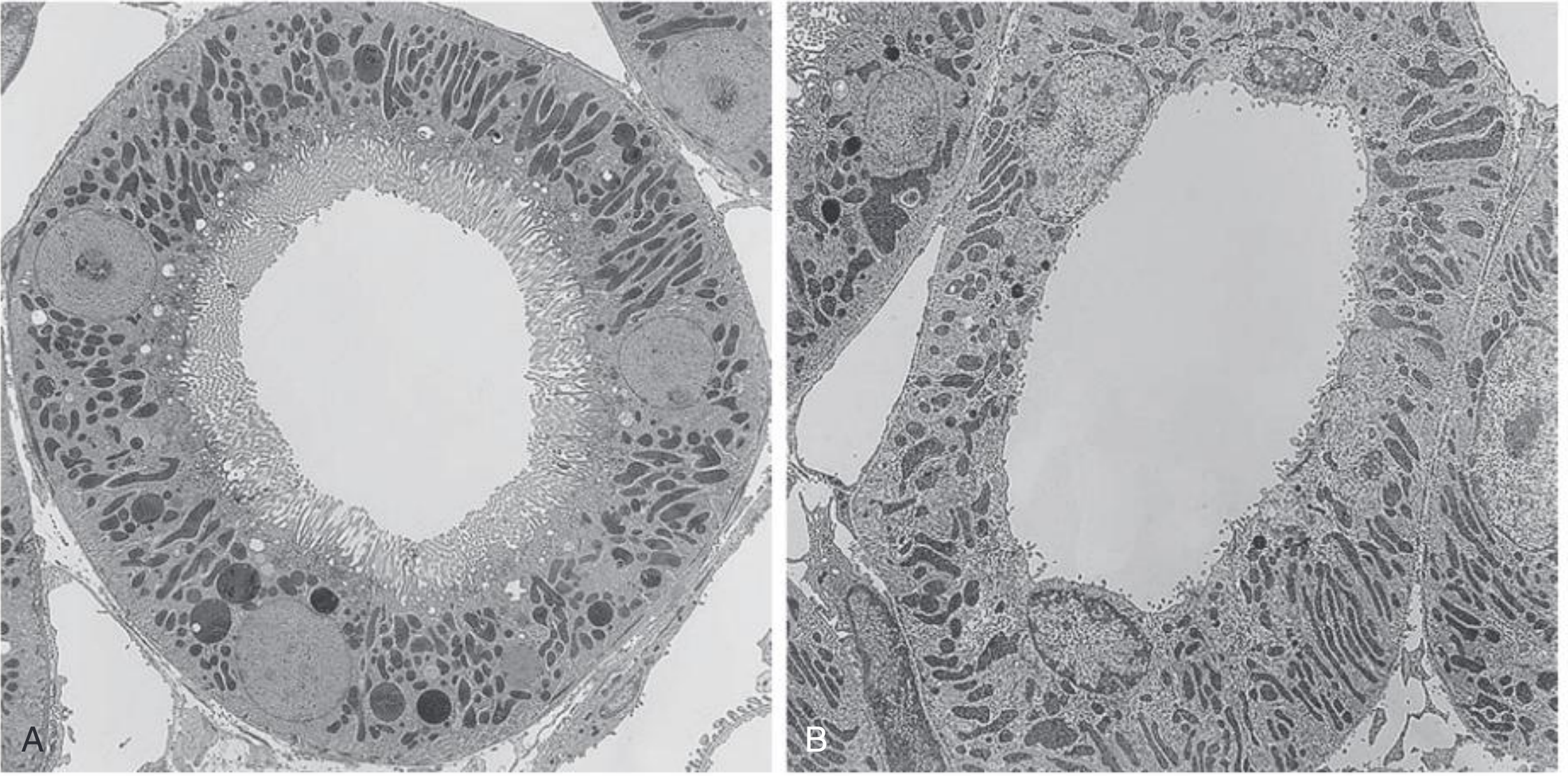
3a. Proximal Convoluted Tubule (PCT)
- Simple cuboidal epithelium, cells tall with eosinophilic cytoplasm (many mitochondria)
- Brush border - long, dense apical microvilli (greatly increases absorptive surface area)
- Basal labyrinth - extensive basolateral membrane infoldings housing rows of mitochondria (drive Na+/K+-ATPase activity)
- Apical endocytotic pits, vesicles, and lysosomes (handle protein reabsorption)
- Zonula occludens seal apical ends; tight junctions are relatively "leaky" (allow paracellular transport)
- In H&E sections: pink, granular cytoplasm; lumen often appears occluded or small due to the brush border
3b. Intermediate Tubule (Thin Limbs of Henle)
- Simple squamous epithelium - extremely flat, nuclei bulge into lumen
- Short-loop tDL: Water-permeable initial segment + UT-A2 urea transporter in distal part
- Long-loop tDL (inner medulla): Loses salt permeability, becomes water permeable
- Thin ascending limb (tAL): Heavily interdigitated epithelium; highly permeable to Na+, Cl-; completely impermeable to water - critical for countercurrent multiplication
3c. Thick Ascending Limb (TAL) / Distal Straight Tubule
- Simple cuboidal epithelium, no brush border, very abundant mitochondria
- Completely water-impermeable - called the "diluting segment"
- Key transporter: NKCC2 (Na+-K+-2Cl- cotransporter) - site of action of loop diuretics (furosemide)
- Produces Tamm-Horsfall protein (uromodulin) - secreted into tubular lumen
3d. Distal Convoluted Tubule (DCT)
- Simple cuboidal epithelium; cells smaller than PCT cells
- No true brush border - only short, stubby microvilli
- Lumens appear more open than PCT on H&E
- Large mitochondria interdigitated with prominent basolateral folds
- Na+ reabsorbed via NCC (Na+-Cl- cotransporter) - site of action of thiazide diuretics
- PTH-regulated Ca2+ reabsorption (TRPV5 channels)
3e. Collecting Duct System
- Cuboidal to columnar, pale-staining cytoplasm
- Distinct cell membranes visible
- Few organelles; relatively sparse microvilli
- Express: ENaC (epithelial Na+ channel, regulated by aldosterone), Aquaporin-2 (regulated by ADH/AVP), Aquaporin-3/4 basolaterally
- Slightly darker cytoplasm than principal cells (more mitochondria)
- Two subtypes:
- Type A IC cells: Secrete H+ into lumen via H+-ATPase (apical); regulate acid excretion
- Type B IC cells: Secrete HCO3- into lumen; regulate alkalosis
- Rich in carbonic anhydrase II
4. The Juxtaglomerular Apparatus (JGA)
| Component | Cell Type | Location | Function |
|---|---|---|---|
| Macula densa | Modified DCT epithelium | Wall of DCT at vascular pole | Senses NaCl concentration in tubular fluid; signals JG cells |
| Juxtaglomerular (JG) cells | Modified smooth muscle cells | Wall of afferent arteriole | Contain secretory granules; produce and secrete renin |
| Extraglomerular mesangium (Lacis cells) | Interstitial cells | Between macula densa and glomerulus | Signal transduction between macula densa and JG cells |
- Macula densa cells are taller and more densely packed with closely spaced nuclei ("dense spot") - detectable on H&E at the vascular pole
- JG cells appear as large, pale, granular smooth muscle cells in the afferent arteriole wall - granules contain renin (PAS-positive)
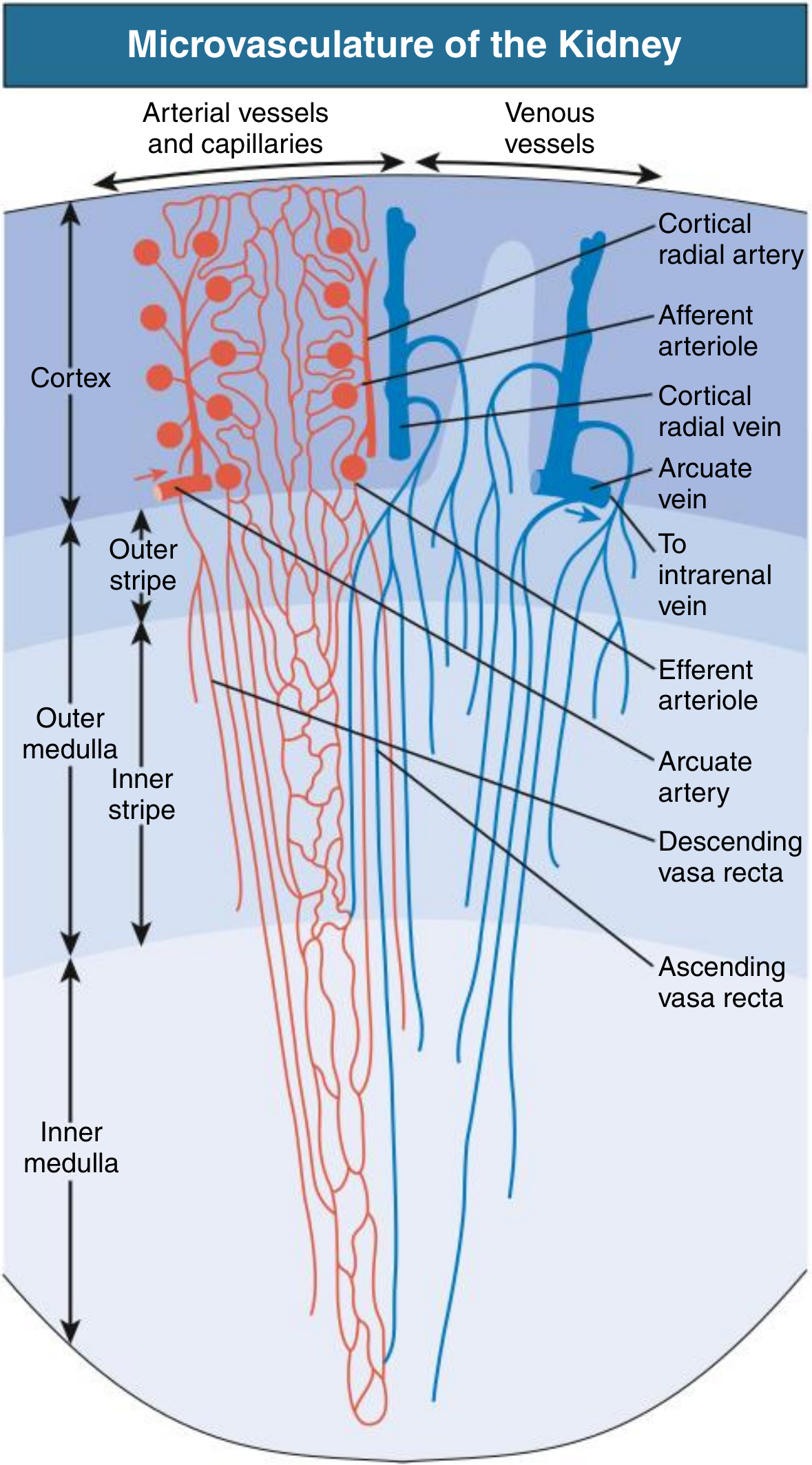
- The JGA regulates renin release and hence the renin-angiotensin-aldosterone system (RAAS); also mediates tubuloglomerular feedback
5. Renal Interstitium
- Cortical interstitium: sparse, ~5-7% of cortical volume. Contains fibroblasts (produce EPO), dendritic cells, and a small amount of extracellular matrix.
- Medullary interstitium: increases in volume toward the papilla (up to 30-40% in inner medulla). Contains:
- Type I interstitial cells (lipid-laden "drooping mustache" cells) - produce prostaglandins (PGE2), important in regulating vasa recta blood flow
- Type II interstitial cells - similar to pericytes
- Rich in glycosaminoglycans that help maintain the osmotic gradient
6. Renal Vasculature (Histologic Overview)
- Efferent arterioles of cortical nephrons → peritubular capillary network (dense mesh around cortical tubules, facilitates reabsorption)
- Efferent arterioles of juxtamedullary nephrons → vasa recta (hairpin loops descending into and ascending from medulla, maintain medullary osmotic gradient via countercurrent exchange)
- Afferent arterioles: muscular walls; JG cells present near glomerulus
- Efferent arterioles: thinner walls; no elastic lamina
- Vasa recta: thin-walled, simple squamous endothelium; bundles in medulla
7. Quick Stain Reference
| Stain | What it highlights in kidney |
|---|---|
| H&E | General architecture; PCT (pink, granular); DCT (paler, open lumen); glomerulus (purple nuclei, pink matrix) |
| PAS (Periodic Acid-Schiff) | GBM, mesangial matrix, tubular basement membranes (magenta); thickening in DM |
| Silver (Jones methenamine) | GBM and mesangial matrix (black); best for detecting GBM spikes or holes |
| Masson's Trichrome | Collagen/fibrosis (blue); useful in interstitial fibrosis assessment |
| Congo Red | Amyloid deposits (apple-green birefringence under polarized light) |
| Immunofluorescence (IF) | Immune complex deposits (IgA, IgG, C3, C1q) in glomerular disease |
Key Clinical-Histologic Correlations
- Minimal change disease: Normal LM and IF; foot process effacement on EM → nephrotic syndrome
- Diabetic nephropathy: GBM thickening + mesangial expansion on PAS; Kimmelstiel-Wilson nodules
- IgA nephropathy: Mesangial IgA deposits on IF; mesangial proliferation on LM
- FSGS: Focal (some glomeruli) and segmental (part of tuft) sclerosis; podocyte injury
- Acute tubular necrosis (ATN): Flattened PCT epithelium, loss of brush border, cell sloughing into lumen
- Acute interstitial nephritis: Interstitial edema + lymphocyte/eosinophil infiltrate; tubulitis
Midgut rotation embryology for 1st year MBBS
midgut rotation embryology stages 270 degrees intestinal herniation umbilical cord
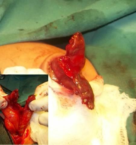
This clinical photograph and surgical inset document a rare case of a high umbilical cord (HUC) hernia associated with congenital loss of the midgut. The main image shows a segment of bowel protruding through an umbilical defect, characterized by its dark reddish-brown and vivid red coloration, suggesting a collapsed and potentially ischemic or highly vascularized state. The tissue exhibits a glistening serosal surface with irregular folds. The inset image provides a closer view of the surgical manipulation, where the remnant gut is being held open to reveal its internal morphology. It demonstrates the extremely short length of the available bowel, specifically showing a few inches of proximal jejunum and distal sigmoid colon, with an absence of the intervening midgut structures. The exposed tissues in the inset are bright red and show disorganized, lobed projections. This visual is significant for pediatric surgery and embryology, illustrating a severe intestinal atresia or congenital absence of the midgut within an umbilical cord hernia.
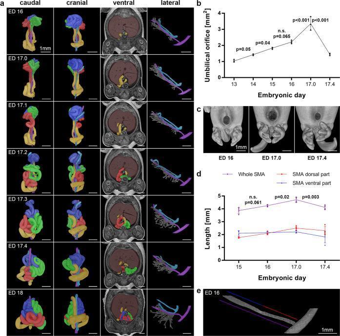
Educational medical composite illustrating the embryological 'midgut shift' in a rat model, representing mammalian intestinal development from Embryonic Day (ED) 16 to ED 18. Panel (a) presents chronological 3D reconstructions from caudal, cranial, ventral, and lateral views. Intestinal segments are color-coded by cluster (Red, Green, Blue, Yellow), and the Superior Mesenteric Artery (SMA) is highlighted in purple. The sequence captures the transition from physiological herniation (ED 16) through progressive retraction into the abdominal cavity (ED 17.0–17.3) to the final internal positioning (ED 17.4–18). Ventral views show the relationship with the liver (brown) and abdominal wall. Panels (b) and (d) provide morphometric quantitative data, graphing the umbilical orifice area and SMA length. The data indicate that the umbilical orifice reaches peak expansion at ED 17.0 before closing, while SMA length increases during the shift's tension phase and decreases upon completion (ED 17.4), signifying a 'relaxed' configuration. Panel (c) provides surface reconstructions of the umbilical region, while (e) shows isolated vessel analysis.
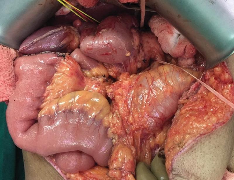
This clinical photograph is an intraoperative view of the abdominal cavity during a surgical procedure, demonstrating classic findings of intestinal nonrotation. The image shows the small bowel loops clumped primarily within the right upper quadrant of the abdomen, rather than following the standard peripheral distribution. Centrally and near the midline, the cecum and hepatic flexure are visible, indicating an arrest in the normal 270-degree counterclockwise rotation during embryological development. The duodenum appears vertically oriented, lacking its typical C-loop configuration, with the duodenojejunal flexure positioned to the right of the midline. Surgical retractors and laparotomy pads are visible at the periphery of the operative field, exposing the mesenteric attachments and the atypical positioning of the midgut structures. This visual provides high educational value for surgical anatomy, embryology, and the diagnosis of congenital malrotation in an adult clinical context.
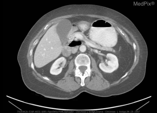
**Imaging Modality:** Contrast-enhanced Computed Tomography (CT) of the abdomen. **Anatomical Region:** Axial section at the level of the upper abdomen, displaying the liver, gallbladder, pancreas, kidneys, and mesenteric vasculature. **Observed Pathology:** The image demonstrates findings consistent with intestinal malrotation. A primary diagnostic cue is the inversion of the normal anatomical relationship between the superior mesenteric artery (SMA) and the superior mesenteric vein (SMV). **Characteristic Visual Features:** * **Vascular Inversion:** The SMV is positioned to the left of the SMA, rather than its normal position to the right. * **SMV Sign:** The mesenteric vessels show an altered orientation, which is a classic radiologic indicator of malrotation. * **Organ Positioning:** While the liver and kidneys appear gross-anatomically normal in this slice, the midgut structures demonstrate abnormal mesenteric anchoring. * **Absence of Volvulus:** No "whirlpool sign" is immediately visible in this specific axial plane, though the vascular transposition highly suggests underlying malrotation. **Clinical Significance:** This congenital anomaly results from the failure of the midgut to undergo the normal 270-degree counterclockwise rotation during embryogenesis, predisposing the patient to midgut volvulus and internal herniation.
Midgut Rotation - Embryology (1st Year MBBS)
What is the Midgut?
| Derivative | Final Structure |
|---|---|
| Distal duodenum (beyond ampulla) | Duodenum (2nd-4th parts) |
| Cranial limb of loop | Jejunum + ileum |
| Caudal limb of loop | Cecum, appendix, ascending colon, right 2/3 of transverse colon |
Why Does the Midgut Herniate?
The Midgut Loop
- Cranial limb (pre-arterial) - gives rise to the jejunum and ileum (elongates and forms multiple coils)
- Caudal limb (post-arterial) - gives rise to the cecum, appendix, ascending colon, and right 2/3 of transverse colon (shows minimal coiling)
The Three Stages of Midgut Rotation
Stage 1 - Herniation + Initial Rotation (Week 6, 90° rotation)
- The midgut loop herniates into the umbilical cord
- While outside the abdomen, the loop rotates 90° counterclockwise around the SMA axis
- This brings the cranial limb to the right and the caudal limb to the left
- The cranial limb elongates massively, forming the coiled loops of jejunum and ileum
- Net result: Cranial limb = right side; caudal limb = left side; loop is horizontal
Stage 2 - Retraction into the Abdomen (Week 10, 180° rotation)
- During week 10, the intestines return to the abdomen (reduction of hernia)
- Favoured by: enlargement of the abdominal cavity + relative decrease in liver/kidney size
- The small intestine returns first, passing posterior to the SMA, occupying the central and left abdomen
- As the large intestine returns, it undergoes a further 180° counterclockwise rotation
- The cecum temporarily lies in the right upper quadrant below the right lobe of the liver (subhepatic position)
- Total rotation at this point = 90° + 180° = 270° counterclockwise
Stage 3 - Descent of the Cecum and Fixation (Weeks 10-11)
- The cecum descends from the right upper quadrant to the right iliac fossa
- This places the ascending colon and hepatic flexure on the right side
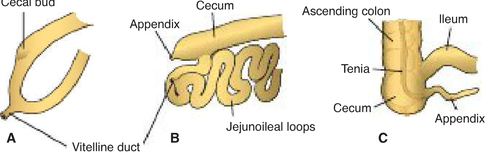
- During descent, the distal end of the cecal bud grows slowly, forming the vermiform appendix
- Mesenteries fuse with posterior abdominal wall peritoneum → retroperitoneal fixation of duodenum, ascending colon, descending colon
Summary Table: The 270° Rotation at a Glance
| Stage | Timing | Rotation | Location | Key Event |
|---|---|---|---|---|
| 1 | Week 6 | 90° CCW | In umbilical cord | Cranial limb moves right; caudal limb moves left |
| 2 | Week 10 | 180° CCW | Returning to abdomen | Small bowel enters first (left); cecum ends subhepatic |
| 3 | Weeks 10-11 | Fixation | Abdominal cavity | Cecum descends to RIF; mesenteries fuse |
| Total | 270° CCW | Normal adult intestinal position achieved |
Mesenteric Fixation (What Becomes Retroperitoneal)
- Duodenum (2nd-4th parts) - becomes retroperitoneal (mesentery fuses with posterior wall); rotation of stomach + duodenum pushes pancreas/duodenum to the right
- Ascending colon - its mesentery fuses with parietal peritoneum → retroperitoneal
- Descending colon - same mechanism → retroperitoneal
- Jejunum, ileum, transverse colon, sigmoid colon - retain free mesentery (remain intraperitoneal)
Development of Cecum and Appendix
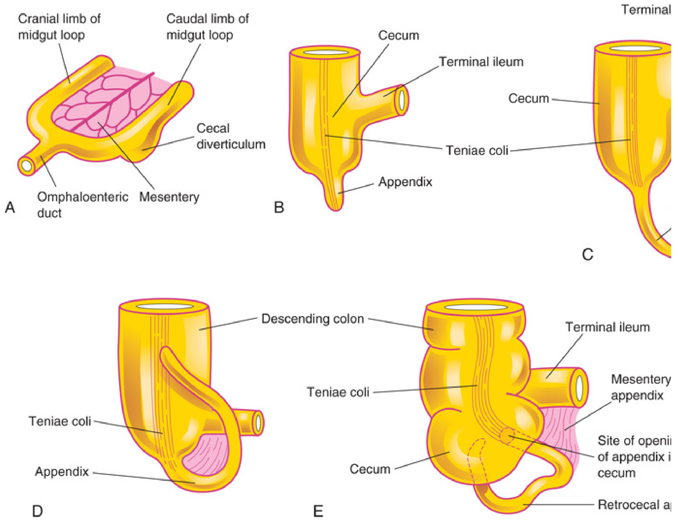
- 6th week: Cecal swelling (cecal bud) appears as a conical dilation on the antimesenteric border of the caudal limb
- The apex of the cecal bud grows slower than the rest - this underdeveloped apex becomes the appendix
- At birth: appendix is a relatively long tube arising from the distal/inferior end of cecum
- After birth: unequal growth of cecal wall shifts the appendix to enter the medial side of the cecum
- In ~64% of people: appendix is retrocecal (its commonest position)
Clinical Correlates (Anomalies of Midgut Rotation)
1. Non-rotation (Failure of Stage 2)
- Intestine does not rotate as it re-enters the abdomen
- Caudal limb returns first (opposite of normal)
- Small intestine lies on the right side; entire large intestine on the left
- Cecum and appendix lie just below the pylorus - called subhepatic cecum
- Prone to midgut volvulus (twisting around narrow SMA pedicle)
2. Malrotation (Incomplete Rotation)
- Most common rotation anomaly
- Failure to complete the final 90° - cecum arrested below pylorus/liver
- Ladd's bands: peritoneal bands from cecum crossing over the duodenum → cause duodenal obstruction
- Narrow mesenteric pedicle predisposes to midgut volvulus
- Classic presentation: bilious vomiting in neonate (bilious emesis = surgical emergency)
- CT sign: SMA and SMV relationship reversed - SMV lies to the LEFT of SMA (normally SMV is to the right)
3. Midgut Volvulus
- Twisting of the entire midgut around the SMA pedicle
- Can obstruct the SMA → intestinal infarction and gangrene
- Treatment: Ladd's procedure (emergency surgery: detorsion + division of Ladd's bands + appendectomy)
4. Omphalocele vs. Gastroschisis
| Feature | Omphalocele | Gastroschisis |
|---|---|---|
| Defect | Enlarged umbilical ring | Lateral abdominal wall defect (right side) |
| Covering | Peritoneal sac covers herniated organs | No sac; bowel directly exposed |
| Contents | Liver, intestines, stomach, spleen possible | Intestines only |
| Associated anomalies | Common (cardiac, chromosomal) | Less common |
| Cause | Failure of intestinal retraction into abdomen | Vascular accident / rupture of umbilical ring |
5. Meckel's Diverticulum (Vitelline Duct Remnant)
- Persistence of the vitelline (omphalomesenteric) duct - normally obliterated by week 7-8
- Rule of 2s: 2% incidence, 2 inches long, 2 feet from ileocecal valve, 2 types of ectopic mucosa (gastric most common, pancreatic), usually presents in first 2 years of life
- On the antimesenteric border of the ileum
- May contain ectopic gastric mucosa → acid secretion → ulceration → bleeding (painless rectal bleeding in a child)
Memory Tricks for Exams
- "270° CCW" = the magic number for normal midgut rotation
- "Cranial limb = Small bowel; Caudal limb = Cecum/Colon"
- "Small bowel returns first, goes posterior to SMA"
- "Cecum: right upper → right iliac" (descends post-rotation)
- Malrotation = Bilious vomiting in neonate → Upper GI contrast series → Ladd's procedure
- Meckel's: Rule of 2s
- Retroperitoneal structures post-fixation: Duodenum (2nd-4th), ascending colon, descending colon, pancreas head