
Medullary controll of the cardiovascular system in detail with each and every step
medulla cardiovascular control
medullary cardiovascular center NTS CVLM RVLM baroreflex physiology
Topic map
-
Organization of the medullary cardiovascular control network
- Nucleus tractus solitarius (NTS)
- Caudal ventrolateral medulla (CVLM)
- Rostral ventrolateral medulla (RVLM)
- Nucleus ambiguus and dorsal motor nucleus of vagus
- Spinal intermediolateral cell column
-
Afferent input to the medulla
- Carotid sinus and aortic arch baroreceptors
- Carotid and aortic body chemoreceptors
- Cardiopulmonary low-pressure receptors
- Higher brain centers, respiratory system, pain, emotion, and exercise input
-
Efferent autonomic pathways
- Sympathetic pathway to heart, arterioles, veins, and kidneys
- Parasympathetic vagal pathway to the heart
-
Baroreceptor reflex, step by step
- Response to increased arterial pressure
- Response to decreased arterial pressure, such as standing suddenly or hemorrhage
-
Chemoreceptor and cardiopulmonary reflexes
-
Integration with respiration and higher centers
-
Physiological and clinical relevance
- Orthostatic hypotension, neurogenic shock, vasovagal syncope, and medullary lesions
NTS is a part of which system like sympathetic nervous system or parasympathetic nervous system?
nucleus tractus solitarius autonomic
Nucleus tractus solitarius autonomic integration sympathetic parasympathetic NCBI
What does the NTS do?
- Glossopharyngeal nerve (CN IX): from carotid sinus and carotid body
- Vagus nerve (CN X): from aortic arch, aortic bodies, heart, lungs, and abdominal viscera
- Arterial blood pressure
- Blood gases: O₂, CO₂, H⁺
- Blood volume and atrial stretch
- Heart and lung reflexes
Relation to sympathetic and parasympathetic systems
| NTS connection | Effect |
|---|---|
| NTS → nucleus ambiguus and dorsal motor nucleus of vagus | Increases parasympathetic (vagal) output to the heart |
| NTS → CVLM → inhibits RVLM | Decreases sympathetic output to heart and blood vessels |
| Reduced NTS firing | Less vagal output and less inhibition of RVLM, so sympathetic activity rises |
Simple example: rise in blood pressure
- Blood pressure rises.
- Carotid sinus and aortic arch baroreceptors fire more.
- Their signals reach the NTS via CN IX and CN X.
- NTS stimulates vagal nuclei, especially the nucleus ambiguus.
- Vagal activity to the SA node increases, causing heart rate to fall.
- NTS activates the CVLM.
- CVLM inhibits the RVLM.
- RVLM-driven sympathetic outflow falls.
- Heart contractility, arteriolar constriction, and venous constriction decrease.
- Blood pressure returns toward normal.
NTS is an autonomic sensory integration nucleus, not purely sympathetic or parasympathetic. It receives afferent input and regulates both parasympathetic vagal and sympathetic outputs.
How NTs is helpful in, अ, increment of sympathetic system, अ...
nucleus tractus solitarius sympathetic RVLM
NTS decreased baroreceptor input sympathetic activation RVLM physiology
When blood pressure falls: how sympathetic activity increases
-
Blood pressure falls
Example: standing suddenly, hemorrhage, dehydration. -
Less stretch of carotid sinus and aortic arch baroreceptors
Less stretch means these receptors fire fewer action potentials. -
Less sensory input reaches the NTS
- Carotid sinus signals travel through CN IX
- Aortic arch signals travel through CN X
-
NTS activity decreases
-
The NTS now sends less excitatory stimulation to the CVLM
Normally:
NTS excites CVLM -
CVLM activity decreases
CVLM neurons are inhibitory and release GABA onto the RVLM. -
Therefore, there is less GABA inhibition of the RVLM
This is called disinhibition of the RVLM. -
RVLM becomes more active
RVLM is the major medullary source of tonic excitatory drive to sympathetic preganglionic neurons. -
RVLM sends more excitatory signals to the intermediolateral cell column (IML) in thoracic spinal cord, mainly T1-L2.
-
Sympathetic nerves become more active:
- Heart: heart rate increases and contractility increases
- Arterioles: vasoconstriction increases total peripheral resistance
- Veins: venoconstriction increases venous return
- Kidney: renin release increases
- Cardiac output and total peripheral resistance rise, so arterial blood pressure rises toward normal.
Pathway in one line
Important distinction
- NTS activated in high BP: decreases sympathetic activity and increases vagal activity.
- NTS less activated in low BP: sympathetic activity increases because inhibition of RVLM is removed.
Explain the NTS–CVLM–RVLM pathway with a labeled diagram
labeled baroreceptor reflex diagram nucleus tractus solitarius caudal ventrolateral medulla rostral ventrolateral medulla
A clean, medically accurate labeled teaching diagram of the medullary baroreceptor reflex pathway. Show carotid sinus and aortic arch baroreceptors with afferent cranial nerves IX and X to the NTS in dorsomedial medulla. Show NTS excitatory glutamate arrow to CVLM, then CVLM inhibitory GABA arrow to RVLM. Show RVLM excitatory glutamate descending arrow to thoracic spinal cord IML sympathetic preganglionic neurons and onward to heart, arterioles, and veins. Include a side branch from NTS to nucleus ambiguus with vagus to the SA node. Clearly label excitation with green plus signs and inhibition with red minus signs. Include two small panels: high blood pressure activates NTS and lowers sympathetic output; low blood pressure reduces NTS activity and disinhibits RVLM, raising sympathetic output.
baroreflex medulla NTS RVLM
NTS CVLM RVLM baroreflex pathway physiology
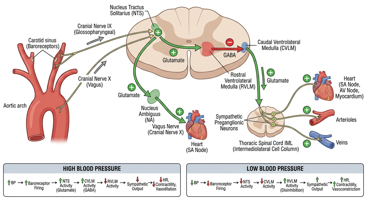
NTS-CVLM-RVLM pathway
CAROTID SINUS and AORTIC ARCH BARORECEPTORS
                │
                │ Increased stretch when BP rises
                │ Increased afferent firing
                ▼
CN IX (carotid sinus nerve) and CN X (aortic depressor/vagal fibers)
                │
                │ Glutamate (+)
                ▼
NTS: Nucleus Tractus Solitarius
Dorsomedial medulla
                │
      ┌─────────┴─────────────────────┐
      │                               │
      │ Glutamate (+)                 │ Glutamate (+)
      ▼                               ▼
CVLM                              Nucleus ambiguus
Caudal ventrolateral medulla       and dorsal motor nucleus
      │                               │
      │ GABA (-)                       │ Vagus nerve
      ▼                               ▼
RVLM                            Heart, especially SA node
Rostral ventrolateral medulla       ↓ Heart rate
      │                               ↓ AV-node conduction
      │ Glutamate (+)
      ▼
IML, thoracic spinal cord
Intermediolateral cell column
(sympathetic preganglionic neurons)
      │
      ▼
Sympathetic nerves to:
heart, arterioles, veins, kidneys
      │
      ▼
↓ HR, ↓ contractility, ↓ vasoconstriction,
↓ venous tone, ↓ renin release
      │
      ▼
↓ Arterial blood pressure
Important signs
- (+) = excitatory
- (-) = inhibitory
- NTS → CVLM is excitatory, mainly glutamatergic.
- CVLM → RVLM is inhibitory, mainly GABAergic.
- RVLM → IML is excitatory, mainly glutamatergic.
During high blood pressure
Step-by-step
- Blood pressure increases.
- Carotid sinus and aortic arch are stretched more.
- Baroreceptors fire more frequently.
- Afferent impulses reach the NTS:
- Carotid sinus via glossopharyngeal nerve, CN IX
- Aortic arch via vagus nerve, CN X
- NTS activates the CVLM.
- CVLM releases GABA onto the RVLM and inhibits it.
- RVLM sends less excitatory drive to the IML in the spinal cord.
- Sympathetic outflow falls.
- Simultaneously, NTS activates the nucleus ambiguus, increasing vagal activity to the heart.
- Heart rate and contractility decrease; arterioles and veins dilate.
- Blood pressure falls back toward normal.
Short formula
↑ BP → ↑ baroreceptor firing → ↑ NTS → ↑ CVLM
→ inhibition of RVLM → ↓ sympathetic outflow → ↓ BP
During low blood pressure
↓ BP → ↓ baroreceptor firing → ↓ NTS → ↓ CVLM
→ less GABA inhibition of RVLM → ↑ RVLM activity
→ ↑ IML sympathetic outflow → ↑ HR, contractility, vasoconstriction
→ ↑ BP
Step-by-step
- Blood pressure falls, for example after standing quickly or blood loss.
- Baroreceptors are less stretched.
- Their firing to the NTS decreases.
- NTS activates CVLM less.
- CVLM gives less GABA-mediated inhibition to RVLM.
- RVLM is therefore disinhibited and fires more.
- RVLM increases activation of thoracic IML sympathetic preganglionic neurons.
- Sympathetic effects occur:
- Heart: increased heart rate and contractility
- Arterioles: vasoconstriction, increasing total peripheral resistance
- Veins: venoconstriction, increasing venous return
- Kidneys: increased renin release
- Blood pressure rises toward normal.
One high-yield concept
Reduced NTS activity removes the CVLM-mediated inhibitory brake on RVLM.