
Baffle closure of ASD
Reading File
Baffle closure (also called the baffle repair or intra-atrial baffle technique) is a method to close certain types of atrial septal defects (ASDs), particularly sinus venosus ASDs, by redirecting pulmonary venous blood flow rather than simply patching a hole.
Searching sources...
Good results found. Now let me read the detailed content from the most relevant sections.
Reading File
Reading File
Reading File
Finding Sources
Finding Sources
Finding Sources
Reading File
Reading File
Reading File
Loading Image
I now have comprehensive information from multiple authoritative surgical textbooks. Here is a detailed, structured answer:
Baffle Closure of ASD
Overview
"Baffle closure" is a surgical technique used specifically for sinus venosus ASDs (not secundum or primum ASDs). Unlike a simple patch, the baffle does not just close a hole - it redirects (baffles) anomalously draining pulmonary venous blood across the ASD into the left atrium, correcting both the septal defect and the partial anomalous pulmonary venous return (PAPVR) simultaneously.
Why a Baffle is Needed (Not a Simple Patch)
Sinus venosus ASDs are not true defects of the atrial septum. They represent a malalignment between either:
- The superior vena cava (SVC) and right atrial junction + right upper pulmonary vein(s) [most common - superior type], or
- The inferior vena cava (IVC) and right atrial junction + right lower pulmonary veins [inferior type]
Because anomalous pulmonary veins drain into the SVC or IVC rather than the left atrium, simple patch closure would leave pulmonary venous return still entering the right side. A baffle is therefore needed to channel this flow to the left atrium.
- These defects are not amenable to catheter-based device closure, and surgical repair is always required.
- Sabiston Textbook of Surgery, p. 2564; Mulholland & Greenfield's Surgery, p. 4403
Types of Sinus Venosus ASD and Repair Strategy
1. Superior Sinus Venosus ASD (most common)
The anomalous pulmonary veins (typically right upper ± middle lobe veins) drain into the SVC below the right pulmonary artery.
Intracardiac Patch Baffle Technique:
-
Cardiopulmonary bypass (CPB) with bicaval cannulation
-
Right atriotomy is performed
-
A patch (pericardial or synthetic) is positioned inside the right atrium/SVC to create a tunnel (baffle)
-
The baffle directs pulmonary venous blood behind the patch, through the sinus venosus defect, and into the left atrium
-
The superior vena cava is kept widely patent anterior to the baffle to drain systemic venous blood into the right atrium
-
Care is taken to avoid obstruction of the pulmonary veins or SVC (the SVC is usually dilated and provides ample room)
-
Schwartz's Principles of Surgery, p. 781; Mulholland & Greenfield's Surgery, p. 4403
2. When the Anomalous Vein Connects Cranially to the Right Pulmonary Artery - The Warden Procedure
If the anomalous pulmonary vein(s) connect to the SVC cranial to (above) the right pulmonary artery, a simple intracardiac baffle risks SVC obstruction. The Warden procedure is then used:
Steps:
- The SVC is transected cranial to the anomalous vein connection
- The caudal stump of the SVC is oversewn (closed)
- The cranial end of the SVC is anastomosed to the right atrial appendage - this redirects upper body systemic venous return into the right atrium
- Inside the atrium, a patch baffle is sewn to cover the SVC-right atrial junction and redirect anomalous pulmonary venous blood through the sinus venosus defect into the left atrium
This effectively creates two separate channels: pulmonary venous blood enters the left atrium via the baffle, while systemic venous blood (from the head/upper limbs) drains into the right atrium via the SVC-to-appendage anastomosis.
- Schwartz's Principles of Surgery, p. 781-782; Sabiston Textbook of Surgery, p. 2564
3. Inferior Sinus Venosus ASD (scimitar syndrome)
- Associated with the scimitar vein (anomalous pulmonary vein draining to the IVC)
- Repair may involve a patch baffle within the intrahepatic IVC, which is technically more complex
- May require hypothermic circulatory arrest
- Sabiston Textbook of Surgery, p. 2564
Key Surgical Principles
| Point | Detail |
|---|---|
| CPB configuration | Bicaval cannulation, normothermia, antegrade cardioplegia |
| Approach | Median sternotomy or limited sternotomy |
| Patch material | Autologous pericardium or polytetrafluoroethylene (PTFE) |
| Critical step | Verify positions of coronary sinus, all systemic + pulmonary veins before placing baffle |
| SVC patency | Must be confirmed after baffle; obstruction is a serious complication |
| Pulmonary vein patency | Baffle must not kink or obstruct anomalous veins |
Outcomes and Complications
- Operative mortality approaches zero in the absence of pulmonary hypertension
- Postoperative SVC obstruction - a major concern unique to baffle repair (not seen with simple patch)
- Pulmonary vein obstruction - can cause pulmonary hypertension and is avoided by careful patch sizing
- Atrial arrhythmias - occur in 10-40%, particularly in older patients (>40 years) or those with pre-existing arrhythmias; not completely mitigated by repair
- Baffle dehiscence - late complication; a dehisced patch after Warden procedure may be amenable to percutaneous closure using an ASD occluder (e.g., 44-mm Gore Cardioform device), guided by 3D CMR modeling
- Schwartz's Principles of Surgery, p. 782; Sabiston Textbook of Surgery, p. 2565
Key Comparison: Types of ASD and Closure Method
| ASD Type | Repair Method |
|---|---|
| Secundum ASD | Device closure (preferred if anatomy favorable) or surgical patch/primary suture |
| Primum ASD | Surgical patch (surgical always) + AV valve repair |
| Sinus venosus ASD (superior, low) | Intracardiac patch baffle to redirect pulmonary veins to LA |
| Sinus venosus ASD (superior, high/cranial) | Warden procedure (SVC transection + baffle) |
| Sinus venosus ASD (inferior/scimitar) | Complex baffle, possibly with circulatory arrest |
| Coronary sinus ASD | Surgical closure required |
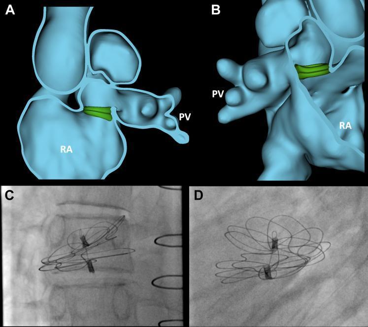
Above: 3D cardiac segmentation (A, B) showing virtual ASD occluder placement to repair a dehisced pulmonary venous baffle after a Warden procedure, alongside intraprocedural fluoroscopy (C, D) showing the deployed 44-mm Gore Cardioform device.
Sources: Sabiston Textbook of Surgery, pp. 2563-2565 | Schwartz's Principles of Surgery, pp. 781-782 | Mulholland & Greenfield's Surgery, pp. 4402-4403 | Harrison's Principles of Internal Medicine 22E, p. 2114
What is ASD
ASD stands for Atrial Septal Defect - a hole in the wall (septum) between the two upper chambers (atria) of the heart. It is one of the most common congenital heart defects, causing a left-to-right shunt and right heart volume overload.
Searching sources...
Finding Sources
Searching Images
atrial septal defect types diagram anatomy
Reading File
Reading File
Loading Image
Reading File
Atrial Septal Defect (ASD)
Definition
An ASD is an abnormal, fixed opening in the wall (septum) between the left and right atria of the heart, allowing blood to communicate between the two upper chambers. It is one of the most common congenital heart defects and should not be confused with a patent foramen ovale (PFO), which is a failure of the normal fetal channel to close after birth (not a true structural defect).
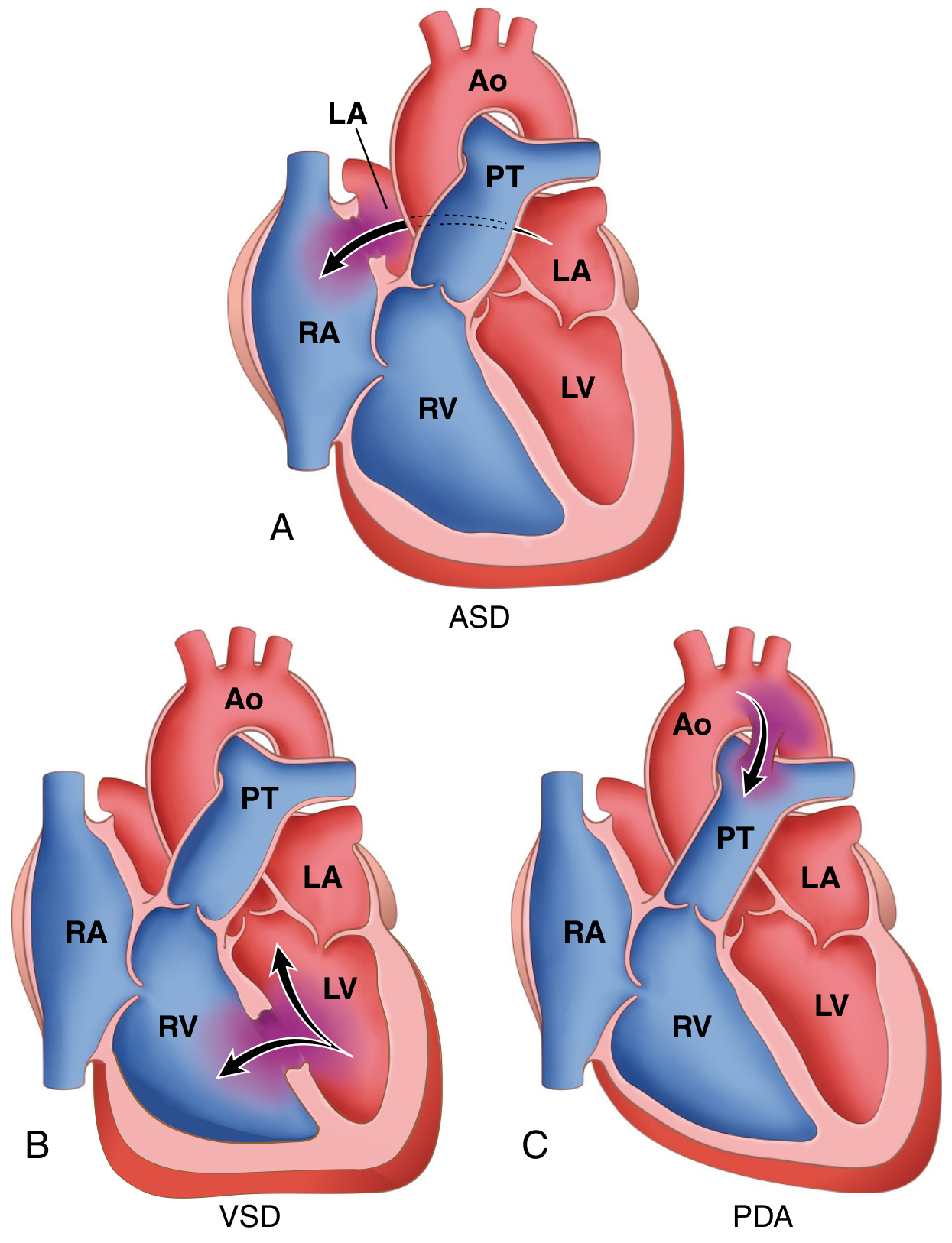
Embryological Basis
Normal atrial septum development involves two overlapping membranes:
- Septum primum - a crescent-shaped ingrowth that partially divides the atria; the gap in front (ostium primum) closes as the septum grows, but a new opening (ostium secundum) forms posteriorly
- Septum secundum - grows to the right of septum primum and covers the ostium secundum, leaving a small channel called the foramen ovale, which normally closes at birth when lung expansion drops right atrial pressure below left atrial pressure
An ASD results when this developmental process is incomplete or abnormal.
- Robbins, Cotran & Kumar Pathologic Basis of Disease, p. 504
Types of ASD
| Type | Frequency | Location | Notes |
|---|---|---|---|
| Secundum ASD | ~90% | Centre of atrial septum (fossa ovalis region) | Deficient septum secundum; may be single, multiple, or fenestrated; most amenable to device closure |
| Primum ASD | ~5% | Adjacent to AV valves (atrioventricular canal) | Associated with cleft mitral valve and AV valve abnormalities; always needs surgery |
| Sinus venosus ASD | ~5% | Near SVC-atrial or IVC-atrial junction | Associated with partial anomalous pulmonary venous return (PAPVR); always needs surgery |
| Coronary sinus ASD | Rare | Coronary sinus-left atrial junction | Rare; surgical repair required |
- Robbins, Cotran & Kumar, p. 504; Harrison's Principles of Internal Medicine 22E, p. 2114
Pathophysiology
- Normal left atrial (LA) pressure > right atrial (RA) pressure, driving a left-to-right shunt through the ASD
- Pulmonary vascular resistance is much lower than systemic, and the right ventricle is more distensible - both factors amplify the shunt
- Pulmonary blood flow may be 2 to 8 times normal (Qp:Qs up to 8:1)
- This causes right heart volume overload - right atrium and right ventricle dilate
- Over time: pulmonary hypertension, right heart failure, atrial arrhythmias
- Risk of paradoxical embolism (clot crossing from right to left via the ASD, causing stroke)
- Robbins & Kumar, p. 505
Clinical Features
Symptoms
- Usually asymptomatic until adulthood (most patients reach their 30s before symptoms)
- Exercise intolerance, dyspnea on exertion, fatigue
- Palpitations, syncope (from atrial arrhythmias)
- Stroke (from paradoxical embolism)
-
70% of patients become impaired by the fifth decade if untreated
Physical Examination Signs
-
Wide and fixed splitting of S2 - the hallmark sign; the split does not vary with respiration (because increased venous return during inspiration partially equalises the shunt and offsets phasic changes)
-
Soft midsystolic ejection murmur at left upper sternal border - due to increased flow across the pulmonary valve (not across the ASD itself, which is too large to generate turbulence)
-
Mid-diastolic murmur at lower left sternal border - increased tricuspid flow in large shunts
-
Right ventricular heave (parasternal lift)
-
Dilated pulmonary artery may be palpable at the second left intercostal space
-
Goldman-Cecil Medicine, p. 984
Investigations
| Investigation | Finding |
|---|---|
| ECG | Incomplete right bundle branch block (rSr' in V1); right axis deviation; prolonged PR interval; atrial fibrillation/flutter in older patients |
| Chest X-ray | Cardiomegaly; dilated RA, RV, and pulmonary artery; pulmonary plethora (increased vascular markings bilaterally) |
| Echocardiography (TTE/TEE) | Diagnostic - directly visualises defect, estimates shunt ratio (Qp:Qs), measures pulmonary pressures. Sinus venosus ASD can be missed on TTE - must specifically be sought. Bubble study (agitated saline) helps detect right-to-left shunting |
| Cardiac catheterisation | Used when pulmonary hypertension is suspected; quantifies Qp:Qs and pulmonary vascular resistance (PVR); needed if PVR is elevated in older patients |
Indications for Closure
Closure is recommended when:
- Significant left-to-right shunt (Qp:Qs > 1.5:1) with or without symptoms
- Right heart enlargement (RA/RV dilation) on echocardiography
- Symptomatic patients at any age
- Usually performed at 3-5 years of age in children
Closure is contraindicated if PVR is fixed and severely elevated (>12 Wood units) - Eisenmenger syndrome.
Treatment Options
1. Percutaneous Device Closure (Transcatheter)
- Suitable for secundum ASDs with adequate rims and no associated anomalous veins
- Defects up to 3.5 cm can be closed
- Most commonly used device: Amplatzer Septal Occluder
- Currently accounts for >60% of all ASD interventions
- Avoids sternotomy and cardiopulmonary bypass
2. Surgical Closure
-
Required for primum, sinus venosus, and coronary sinus ASDs (all need surgery)
-
Also for large secundum ASDs not suitable for device closure
-
Methods:
- Primary suture closure - for small defects
- Patch closure - pericardium or PTFE, sutured with polypropylene
- Baffle repair - for sinus venosus ASDs with PAPVR (redirects anomalous pulmonary veins to LA)
-
Mortality approaches zero in the absence of pulmonary hypertension
-
Long-term outcomes excellent; postoperative survival comparable to normal population
-
Goldman-Cecil Medicine, p. 1000; Mulholland & Greenfield's Surgery, p. 4403
Key Points Summary
- ASD = abnormal opening in the interatrial septum (not the same as PFO)
- Causes left-to-right shunt → right heart volume overload
- Most common form: secundum ASD (90%)
- Classic sign: wide, fixed splitting of S2
- ECG: incomplete RBBB pattern
- Secundum ASD - device closure preferred; all others require surgery
- Untreated, leads to right heart failure, pulmonary hypertension, arrhythmias, and paradoxical embolism
Sources: Robbins, Cotran & Kumar Pathologic Basis of Disease, pp. 504-505 | Goldman-Cecil Medicine, pp. 982-986 | Harrison's Principles of Internal Medicine 22E, p. 2114 | Mulholland & Greenfield's Surgery, p. 4402
BDG procedure
The BDG (Bidirectional Glenn) procedure is a palliative cardiac surgery where the superior vena cava (SVC) is connected directly to the pulmonary artery, allowing deoxygenated blood from the upper body to flow passively into the lungs - bypassing the right ventricle. It is used in single-ventricle heart defects (e.g., tricuspid atresia, hypoplastic left heart syndrome) as the second stage of a three-stage Fontan palliation.
Searching sources...
Finding Sources
Finding Sources
Searching Images
bidirectional Glenn procedure SVC pulmonary artery anastomosis Fontan palliation
Reading File
Reading File
Reading File
Reading File
Reading File
Finding Sources
Loading Image
Reading File
Reading File
I now have comprehensive content from multiple textbooks. Here is a complete answer:
Bidirectional Glenn (BDG) Procedure
What It Is
The Bidirectional Glenn (BDG) is a palliative cardiac surgery in which the superior vena cava (SVC) is anastomosed end-to-side to the right pulmonary artery (RPA), allowing deoxygenated blood from the upper body to flow passively and directly into both pulmonary arteries (hence "bidirectional") - bypassing the heart entirely. It is also called the Bidirectional Cavopulmonary Connection (BCPC) or hemi-Fontan (a related but slightly different variant).
Historical Context: Glenn Shunts
| Type | Era | Anastomosis |
|---|---|---|
| Classic (unidirectional) Glenn | 1962 - 1980s | SVC to the divided right PA (end-to-end) - blood flowed only to the right lung |
| Bidirectional Glenn | Current standard | SVC to undivided RPA (end-to-side) - blood flows to both lungs |
The classic Glenn was largely abandoned because it only perfused one lung and caused differential pulmonary arteriovenous malformations from exclusion of hepatic blood from the lungs.
- Textbook of Clinical Echocardiography, p. classification table
Context: The Three-Stage Fontan Palliation
The BDG is the Stage 2 of a three-stage surgical plan for single-ventricle congenital heart defects. The logic is to gradually offload the single ventricle and prepare the circulation for a final passive pulmonary circuit.
| Stage | Procedure | Age | Goal | SpO₂ |
|---|---|---|---|---|
| Stage 1 | Norwood / mBTT shunt / Sano / Hybrid palliation | Neonatal period | Establish controlled pulmonary blood flow; secure systemic output | 75-85% |
| Stage 2 | Bidirectional Glenn / Hemi-Fontan | 3-6 months | Redirect SVC blood to pulmonary arteries; reduce single-ventricle volume overload | 80-85% |
| Stage 3 | Fontan (lateral tunnel or extracardiac conduit) | 18-24 months | Redirect IVC blood to pulmonary arteries; complete cavopulmonary separation | >92% |
- Harriet Lane Handbook 23rd Ed, p. 257; Mulholland & Greenfield's Surgery, p. 4436
Indications (Diseases Requiring BDG)
Any functionally single-ventricle lesion where a biventricular repair is not possible:
-
Tricuspid atresia (prototype single-ventricle lesion)
-
Hypoplastic Left Heart Syndrome (HLHS) - most common indication today
-
Double-inlet left ventricle
-
Pulmonary atresia with intact ventricular septum (with severely hypoplastic RV)
-
Heterotaxy syndromes
-
Also used as "one-and-a-half ventricle repair" when the RV is judged too small or dysfunctional to support full pulmonary circulation (paired with a biventricular repair)
-
Braunwald's Heart Disease; Sabiston Textbook of Surgery, p. 2584
Surgical Technique
- Cardiopulmonary bypass may or may not be used (some centres perform it off-pump)
- The SVC is transected at its junction with the right atrium; the SVC-RA junction is oversewn
- The free end of the SVC is anastomosed end-to-side to the right pulmonary artery, creating a direct passive flow pathway from the upper body to both lungs
- Any prior systemic-to-pulmonary shunts (mBTT, Sano) placed at Stage 1 are taken down at this time
- The azygos vein is typically ligated to prevent a competing venous pathway that would cause desaturation
The result: SVC blood flows passively through both pulmonary arteries to the lungs, while IVC blood still mixes with pulmonary venous blood in the single ventricle (accounting for the persistent mild cyanosis at SpO₂ 80-85%).
- Grainger & Allison's Diagnostic Radiology, p. 309; Sabiston Textbook of Surgery, p. 2588
Hemi-Fontan vs. Bidirectional Glenn
These two procedures achieve the same physiological goal but differ technically:
| Feature | BDG | Hemi-Fontan |
|---|---|---|
| SVC-RPA connection | End-to-side SVC to RPA | Patch enlargement of SVC-RPA junction (leaves a large anastomosis) |
| SVC-RA junction | Oversewn | Temporarily occluded with a patch (easier to take down at Fontan completion) |
| Advantage | Simpler, can be off-bypass | Better fluid dynamics; easier Fontan conversion; some evidence of improved haemodynamics |
- Mulholland & Greenfield's Surgery, p. 4436
Why Stage 2 is Done at 3-6 Months
- By 3-6 months, pulmonary vascular resistance (PVR) has naturally fallen enough to allow passive (non-pulsatile) SVC flow to the lungs without a driving ventricle
- At birth, PVR is too high for passive pulmonary flow - a shunt with a ventricular pump source is still needed
- Delaying beyond 6 months prolongs the volume overload on the single ventricle, risking ventricular dysfunction
Imaging Surveillance After BDG (Pre-Fontan Assessment)
Before proceeding to Stage 3 (Fontan), imaging must confirm:
| Parameter | Goal |
|---|---|
| Branch PA anatomy | Adequate size, no stenosis at SVC-PA anastomosis |
| Systemic venous return | SVC and IVC connections defined |
| Ventricular function | Normal or near-normal systolic function |
| AV valve regurgitation | Absent or trivial |
| Atrial communication | Adequate (non-restrictive) |
| Collateral vessels | Veno-venous collaterals (SVC-to-IVC territory) are common and can cause desaturation |
| Pulmonary arterial pressure | Must be <20 mmHg (measured by transducing internal jugular vein pressure at time of anaesthesia) |
- Grainger & Allison's Diagnostic Radiology, p. 309-310
Prerequisites for Proceeding to Fontan (Stage 3)
After a successful BDG, the following must be met to safely perform Stage 3:
-
Pulmonary artery pressure < 20 mmHg
-
PVR < 2 Wood units/m²
-
Normal ventricular systolic function
-
Normal or acceptable AV valve function (no significant regurgitation)
-
Adequate pulmonary artery size
-
Mulholland & Greenfield's Surgery, p. 4436
Complications of BDG / Living with Glenn Circulation
- Veno-venous collaterals - common; SVC-to-IVC collaterals form and shunt deoxygenated blood away from the lungs, worsening cyanosis
- Systemic-to-pulmonary collaterals - develop because the upper body is at high venous pressure; can cause pulmonary overcirculation and volume overload
- Pulmonary arteriovenous malformations (PAVMs) - can develop over time, especially if hepatic venous blood (which contains a hepatic factor that prevents PAVMs) is excluded from the pulmonary circulation. This was the major drawback of the classic unidirectional Glenn
- Superior vena cava syndrome - if the anastomosis becomes obstructed
- Pleural effusions - from elevated SVC pressure
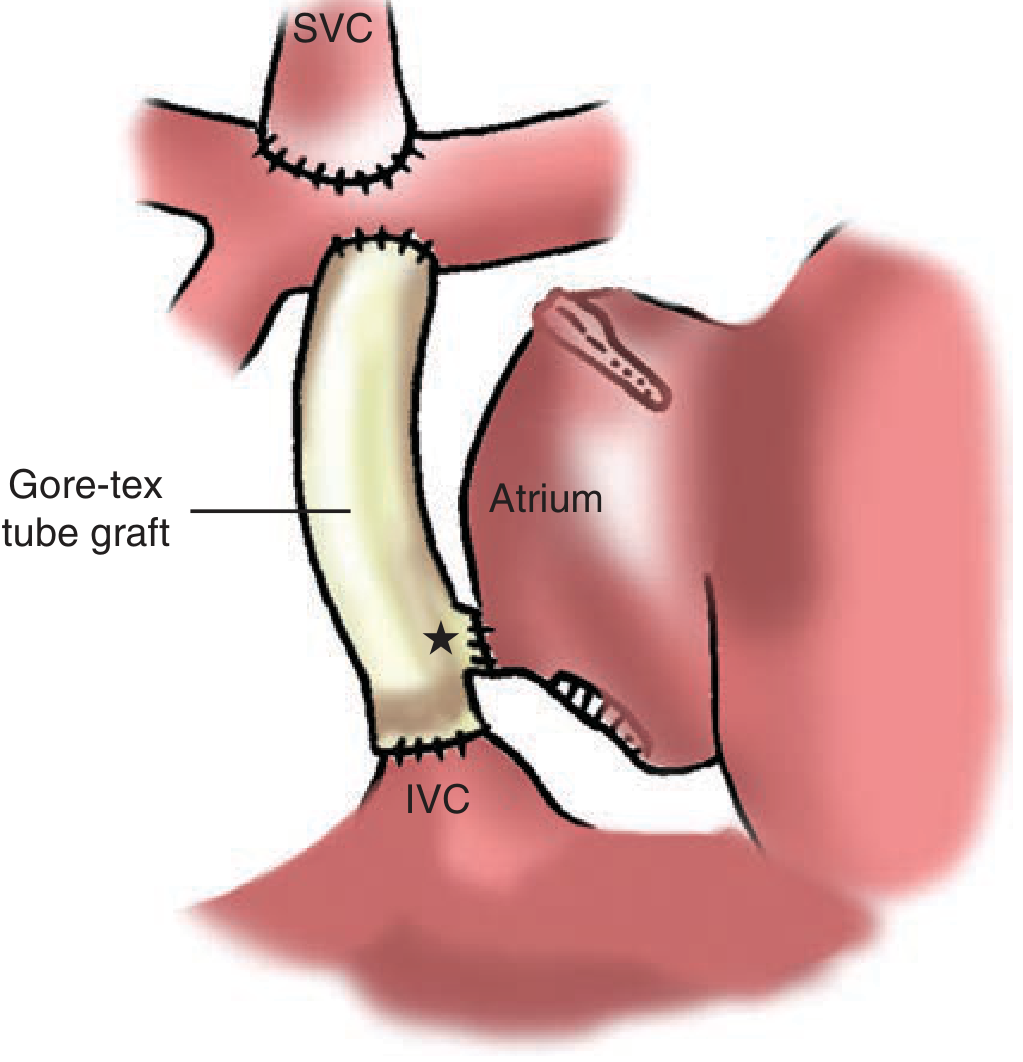
Key Diagram
Summary in One Sentence
The BDG connects the SVC end-to-side to the RPA, directing upper-body venous return passively to both lungs, thereby reducing single-ventricle volume overload and preparing the patient for complete cavopulmonary separation (Fontan) at 18-24 months.
Sources: Sabiston Textbook of Surgery, pp. 2584-2589 | Mulholland & Greenfield's Surgery, pp. 4435-4437 | Grainger & Allison's Diagnostic Radiology, pp. 309-310 | Harriet Lane Handbook 23rd Ed, p. 257 | Braunwald's Heart Disease
This is a shared conversation. Sign in to Orris to start your own chat.