

IMG_8894.jpg
Explain seminoma in very easy language and easy to understand and learn
Reading File
analyse_media
Finding Sources
Searching Images
seminoma testis gross and microscopic histology lymphocytic infiltration
Reading File
Loading Image
Reading File
Here is a complete, easy-to-understand explanation of Classic Seminoma based on your notes and authoritative textbooks:
🎯 Seminoma - Made Simple
🔷 What is Seminoma?
Think of it like this: your testicle normally produces sperm from special "germ cells." When these germ cells go rogue and become cancerous, the resulting tumor is called a Germ Cell Tumor (GCT). Seminoma is the most common type of such cancer.
| Simple fact | Details |
|---|---|
| What is it? | Most common malignant (cancerous) tumor of the testis |
| How common? | ~45-50% of ALL germ cell tumors |
| Female equivalent? | Dysgerminoma (same type of cancer in the ovary) |
| Age group? | Usually 30-40 year old men (rarely in children or elderly) |
👁️ What Does It Look Like? (Gross Appearance)
Imagine cutting open the tumor - here is what you see:
- The entire testis is replaced by the tumor (normal tissue is gone)
- The cut surface looks homogeneous (uniform, no weird holes or patches)
- Color = grey-white (some books describe it as creamy-pink)
- It has a lobulated (divided into lobes/sections) appearance
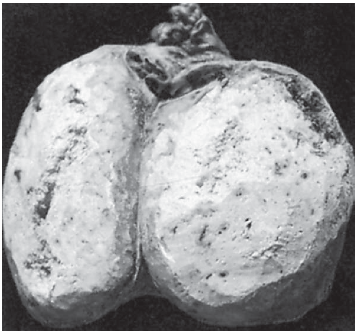
Gross specimen of a seminoma - notice the uniform, lobulated white cut surface (Bailey & Love's Surgery)
🔬 What Does It Look Like Under the Microscope?
This is the most important part for exams. Remember it in 3 layers:
1. How cells are ARRANGED
Cells sit in lobules (clusters/nests), separated by fibrous walls (septa)
2. What the CELLS look like (Cytology)
Think of "UCWGN" to memorize:
| Feature | What it means (simple) |
|---|---|
| Uniform in size | All cancer cells look the same size - neat and uniform |
| Clear cytoplasm | The fluid inside the cell looks clear/empty |
| Well-defined borders | Each cell has sharp, visible edges |
| Glycogen positive (PAS+) | Cytoplasm contains glycogen (sugar) → stains pink with PAS stain |
| Nuclei - big & central | Large nucleus, sitting in the center, with 1-2 prominent nucleoli (the dark dots inside) |
3. The STROMA (background tissue)
The tissue between the lobules shows lymphocytic infiltration - immune cells (lymphocytes) come flooding in. This is a GOOD sign - it means the body is fighting back!
🧪 Tumor Markers (Blood Tests)
This is a high-yield exam topic. Seminoma has a very specific marker pattern:
| Marker | Seminoma Result | Why it matters |
|---|---|---|
| KIT (CD117) | ✅ POSITIVE | Key diagnostic marker |
| OCT4 | ✅ POSITIVE | Confirms germ cell origin |
| PLAP (Placental Alkaline Phosphatase) | ✅ POSITIVE | Classic seminoma marker |
| AFP (Alpha-fetoprotein) | ❌ NEGATIVE | If AFP is positive → it's NOT pure seminoma |
| HCG (Human Chorionic Gonadotropin) | ❌ NEGATIVE | A few cases may have mild elevation, but typically negative |
Exam Tip: AFP negative + HCG negative = think SEMINOMA. If AFP is raised, you are dealing with a non-seminomatous tumor (e.g., yolk sac tumor).
📡 How Does It Spread?
Seminoma spreads mainly through lymphatics (lymph channels), not blood vessels.
- First stop: Para-aortic lymph nodes (near the spine, at the level of the kidneys)
- Inguinal (groin) lymph nodes are only involved if the scrotum skin is also involved
- Blood-borne spread (haematogenous) is uncommon - Bailey & Love's Surgery, p. 1437
🌟 Prognosis (Outcome)
This is where seminoma earns a "good reputation" among cancers:
- Highly radiosensitive - Radiotherapy works extremely well on it (it is one of the most radiation-sensitive tumors)
- Better prognosis than all other germ cell tumors
- Even when it spreads to lymph nodes, it can often be cured
🧓 Spermatocytic Seminoma (Bonus - A Different Entity!)
This is a SEPARATE tumor that was once grouped with seminomas but is now considered distinct:
| Feature | Spermatocytic Seminoma |
|---|---|
| Age | Elderly men (>50 years) |
| Metastasis | ❌ Does NOT metastasize |
| Prognosis | Excellent - just remove the testis (orchidectomy) |
| Risk if untreated | Can rarely develop into sarcoma |
🗂️ Quick Memory Table (Exam Revision)
| Topic | Key Point |
|---|---|
| Type | Most common malignant testicular tumor (~45-50% GCTs) |
| Female equivalent | Dysgerminoma |
| Gross | Homogeneous, grey-white, lobulated |
| Cells | Uniform, clear cytoplasm, PAS+, large nuclei, 1-2 nucleoli |
| Stroma | Lymphocytic infiltration (good sign) |
| Positive markers | KIT, OCT4, PLAP |
| Negative markers | AFP, HCG |
| Spread | Lymphatics → para-aortic nodes |
| Treatment sensitivity | Highly radiosensitive |
| Prognosis | Best among GCTs |
| Spermatocytic variant | Old age, no mets, excellent prognosis |
Sources: Your uploaded notes + Bailey and Love's Short Practice of Surgery 28th Ed., p. 1407-1490

IMG_8895.jpg
Explain BPH in very easy language and easy to understand and learn
analyse_media
Finding Sources
Your credits are exhausted. You can purchase a top-up or subscribe to a plan to continue using Orris. If you're already subscribed, your daily credits will reset tomorrow.
This is a shared conversation. Sign in to Orris to start your own chat.