
Differences between central line and ijv
A "central line" is a broad term for any catheter placed in a large central vein, while the internal jugular vein (IJV) is one specific site where a central line can be placed. Let me pull up the detailed clinical information from the medical library.
Searching sources...
Reading File
Finding Sources
Finding Sources
Reading File
Reading File
Finding Sources
Reading File
Finding Sources
Reading File
Reading File
Reading File
Reading File
Reading File
Now I have enough detailed information to compose a thorough answer. Let me also fetch the neck anatomy image found in Tintinalli's for visual clarity.
Loading Image
Here is a clear, structured breakdown of the differences between a central line (CVC) and the internal jugular vein (IJV) as an access site.
Central Line vs. Internal Jugular Vein (IJV) - Key Differences
The Core Conceptual Distinction
| Central Line (CVC) | Internal Jugular Vein (IJV) | |
|---|---|---|
| What it is | A catheter (device) | A blood vessel (anatomy) |
| Type | Equipment / procedure | One of several possible access sites |
| Relationship | Can be placed INTO the IJV | Is one option for inserting a CVC |
In other words, a central line is what you insert; the IJV is one place you can insert it. The IJV line is a subset of central lines.
Central Venous Catheter (CVC) - Overview
A central venous catheter is a catheter placed so its tip rests in a large central vein (typically the superior vena cava or right atrium). It can be inserted at several sites:
Common CVC sites:
- Internal jugular vein (IJV) - neck
- Subclavian vein - under the clavicle
- Femoral vein - groin
- External jugular vein (technically peripheral access to central circulation)
- Antecubital veins (PICC line approach)
Indications for CVC (any site)
- No peripheral access available
- CVP monitoring (sepsis, heart failure, pericardial effusion)
- Vasopressors, concentrated electrolytes, chemotherapy, hyperalimentation
- Pulmonary artery catheter insertion, transvenous pacing, hemodialysis
- Aspiration of air emboli; repeated blood sampling (Miller's Anesthesia, 10e - Box 32.5; Tintinalli's EM - Table 31-4)
IJV as a CVC Site - Specific Characteristics
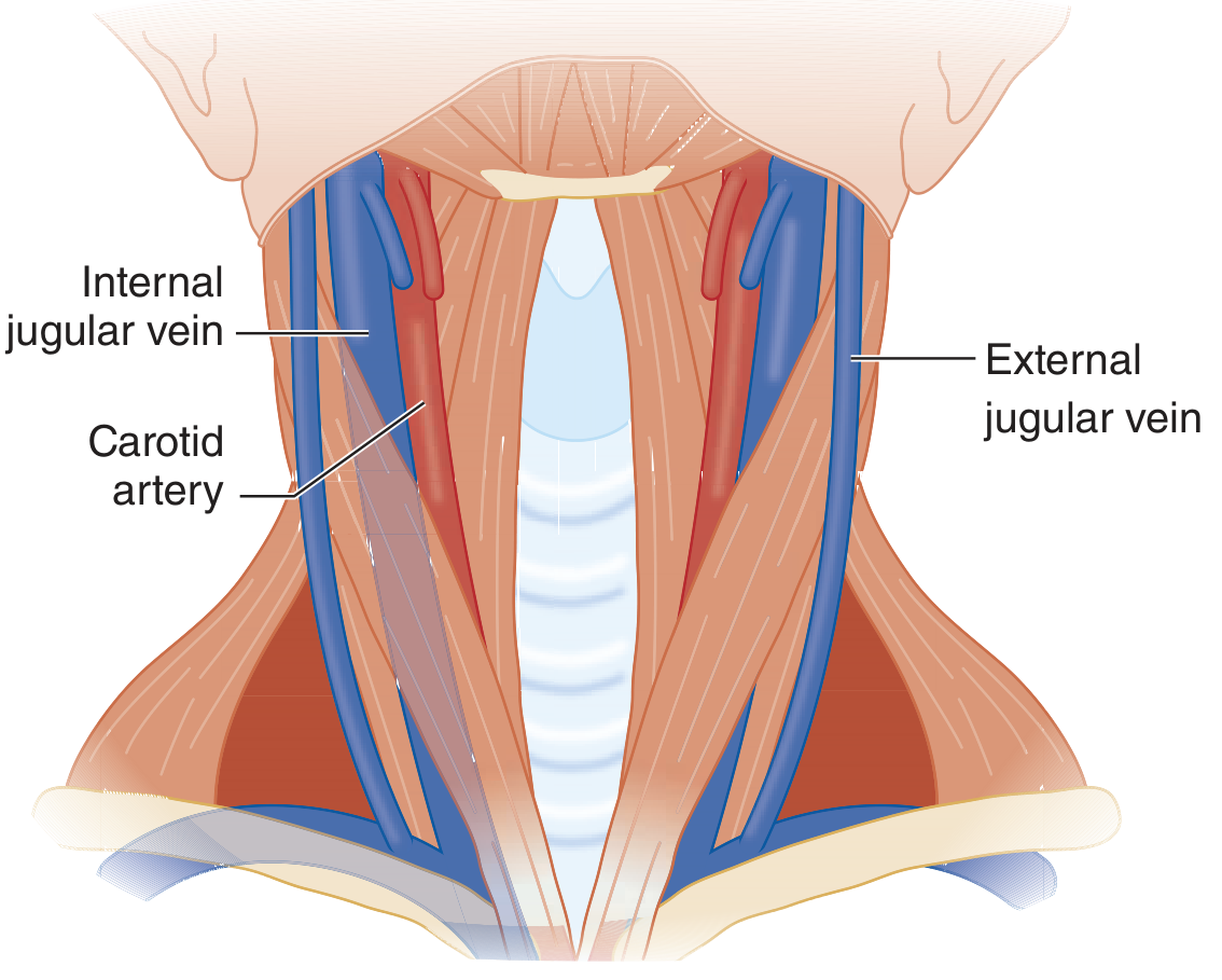
Anatomy
- The IJV lies lateral and superficial to the carotid artery inside the carotid sheath
- Located at the apex of the triangle formed by the two heads of the sternocleidomastoid (SCM)
- More oval/thin-walled than the carotid artery; compresses with gentle pressure
- Size increases with Valsalva maneuver and decreases in hypovolemia
- The IJV joins the subclavian vein to form the brachiocephalic vein
- The right IJV provides the most direct route to the right ventricle (preferred for transvenous pacing) (Roberts & Hedges' Clinical Procedures in Emergency; Tintinalli's EM)
Why the Right IJV is Preferred
- Accessible from the head of the operating table (favored by anesthesiologists)
- Predictable anatomy with high success rates in adults and children
- Most direct path to SVC - requires a shorter catheter than the left IJV (3-5 cm less than left-sided approach)
- Left IJV risks damaging the thoracic duct and is harder to navigate at the jugular-subclavian junction (Barash Clinical Anesthesia, 9e)
Comparison: IJV vs. Other CVC Sites
| Feature | IJV | Subclavian | Femoral |
|---|---|---|---|
| Infection risk | Moderate | Lowest (CDC preference) | Highest |
| Thrombosis risk | Moderate | Low | Highest (~20% in some studies) |
| Pneumothorax risk | Lower than subclavian | Higher | None |
| Bleeding control | Compressible | NOT compressible | Compressible |
| Use in coagulopathy | Preferred | Avoid | Acceptable |
| Use in emphysema | Preferred | Avoid | Acceptable |
| Emergency CPR access | Difficult | Difficult | Easiest |
| Neck trauma/immobilization | Avoid (use femoral/subclavian) | Preferred | Preferred |
| Accidental arterial puncture | Carotid artery (manageable) | Subclavian artery (NOT compressible - dangerous) | Femoral artery |
| Ultrasound guidance | Strongly recommended | Recommended | Recommended |
(Miller's Anesthesia, 10e; Barash Clinical Anesthesia, 9e; Tintinalli's EM)
Complications Unique to or Prominent with IJV CVCs
- Carotid artery puncture - most feared immediate complication
- Hematoma in the neck
- Air embolism (less so than subclavian due to position)
- Thoracic duct injury on the left side
- Catheter-associated bloodstream infection (CLABSI)
- Pneumothorax (less common than subclavian)
- Hemothorax (~1.3-1.5%, similar to subclavian) (Murray & Nadel's Respiratory Medicine; Roberts & Hedges')
Ultrasound Guidance for IJV
Real-time ultrasound is now strongly recommended by all major professional societies for IJV cannulation. On US:
- IJV appears oval/triangular, thin-walled, compressible
- Carotid artery appears round, thick-walled, pulsatile, non-compressible
- IJV lies lateral and slightly superficial to carotid artery (though anatomic variation exists) (Roberts & Hedges'; Miller's Anesthesia, 10e)
Summary in One Line
A central line is the device/catheter; the IJV is one anatomical site (in the neck) where it is commonly placed. The IJV CVC is preferred in coagulopathic patients, for transvenous pacing (right side), and when pneumothorax risk must be minimized, but carries risk of carotid artery puncture and has moderate infection risk compared to subclavian.
This is a shared conversation. Sign in to Orris to start your own chat.