
Dengue
What aspect of dengue are you interested in?
dengue fever rash clinical presentation
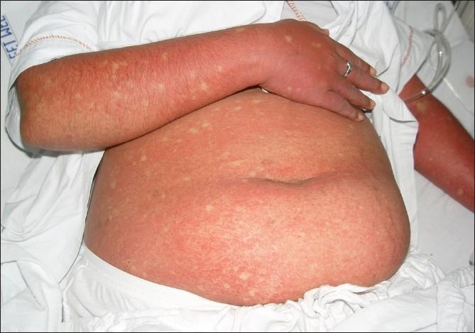
This clinical photograph displays a patient's abdomen and arm presenting a generalized, confluent erythematous rash, a characteristic cutaneous manifestation of Dengue Fever. The exanthem is marked by widespread, intense redness resulting from capillary dilatation and coalesced maculopapular lesions. A diagnostic hallmark visible here is the presence of 'islands of sparing'—small, rounded, or oval-shaped patches of normal-colored skin scattered throughout the erythematous background, often described as 'white islands in a sea of red'. These spared areas are randomly distributed across the affected trunk and limbs. The image serves as a key educational reference for recognizing the secondary rash phase of Dengue viral infection, typically occurring 3 to 6 days after the onset of fever. The visual presentation is essential for clinical diagnosis and differentiation from other viral exanthems, highlighting the specific immunologic response patterns seen in Dengue Fever (DF) and Dengue Hemorrhagic Fever (DHF).
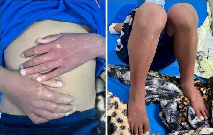
This clinical photograph displays a pediatric patient with a classic dermatological presentation of the recovery phase of Dengue Fever. Two side-by-side images show the dorsal surfaces of the hands, forearms, lower legs, and feet. The primary finding is a diffuse, confluent, erythematous rash covering the extremities. This background erythema is punctuated by multiple small, irregular macules and patches of pale, normal-appearing skin. This characteristic morphological pattern is classically described in clinical medicine as 'white islands in a sea of red.' The distribution is symmetric across the upper and lower limbs. Clinically, this convalescent rash typically appears after the defervescence of fever in dengue virus infection and is a key diagnostic physical sign indicating the recovery phase of the disease. The image is a valuable educational resource for infectious disease and pediatric dermatology, illustrating the morphological distinction between confluent erythematous lesions and spared areas of skin.
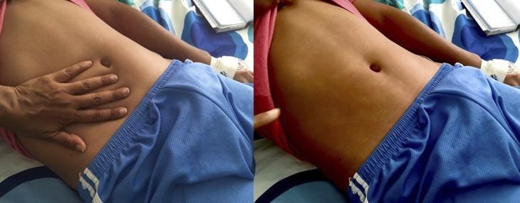
This clinical photograph displays a diffuse, erythematous maculopapular rash on the abdomen of a patient, characteristic of Dengue fever. The image is presented in a two-part sequence to demonstrate clinical blanching. On the left, a clinician's hand is shown applying direct pressure to the right side of the patient's abdomen. On the right, after the hand is removed, a clear, pale handprint-shaped outline remains where the pressure was applied, indicating that the redness fades (blanches) as blood is temporarily displaced from the superficial vessels. This finding confirms the rash is inflammatory rather than hemorrhagic (petechial/purpuric). The rash is widespread across the torso but typically spares the palms and soles in this clinical presentation. This image serves as a teaching tool for medical students and clinicians to distinguish between vascular inflammatory rashes and non-blanching hemorrhagic lesions in the context of viral hemorrhagic fevers and infectious diseases.
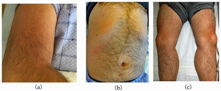
A series of three clinical photographs demonstrating cutaneous manifestations in a patient with dengue fever. (a) Right upper arm showing confluent, blanchable erythema with a diffuse distribution. (b) Anterior trunk (abdomen and chest) displaying a patchy, mottled erythematous rash interspersed with areas of normal skin tone, characteristic of the 'islands of white in a sea of red' pattern often seen in viral exanthems. (c) Lower limbs showing localized, more intense erythema focused around the knees and pretibial regions. The visual findings illustrate the secondary maculopapular rash typical of the recovery or late febrile phase of dengue virus infection. These images serve as an educational resource for identifying the clinical presentation of viral hemorrhagic fever rashes and their variable morphology across different anatomical regions, including the trunk, extremities, and periarticular areas.
"dengue"[MeSH Terms] AND "management"[MeSH Terms]
dengue hemorrhagic fever thrombocytopenia petechiae
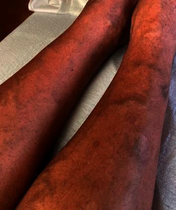
This clinical photograph shows the bilateral lower extremities of a 47-year-old male presenting with a diffuse dermatologic manifestation of thrombocytopenia. The skin demonstrates widespread, non-blanching petechiae, appearing as numerous pinpoint-sized (1-2 mm), reddish-purple macules. These lesions are densely scattered across the anterior and lateral aspects of the shins and calves. In addition to the petechial rash, several larger, coalescent purpuric lesions and subtle ecchymoses are visible, indicating small-vessel bleeding into the dermis. The background skin exhibits generalized erythema. This visual finding is characteristic of severe thrombocytopenia, in this clinical context associated with a viral hemorrhagic fever such as dengue. The image serves as an educational example of the cutaneous signs of low platelet counts and the systemic progression of infectious diseases like dengue fever in a hospital setting.
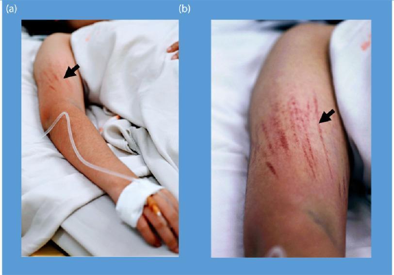
Clinical photograph of a patient's arm demonstrating bleeding manifestations characteristic of Dengue Shock Syndrome (DSS). The image is divided into two panels: (a) shows the full arm in a clinical setting with an intravenous (IV) line secured to the hand, and (b) provides a close-up view of the upper arm. Both panels highlight linear petechiae (indicated by black arrows), which are small, red to purplish hemorrhagic spots arranged in distinct linear patterns. These lesions represent microvascular fragility and are shown following the inflation of a blood pressure cuff (Hess test or capillary fragility test), a common diagnostic maneuver for assessing vascular permeability in dengue fever. The presence of an IV line and hospital bedding contextualizes the severity of the illness, illustrating the systemic plasma leakage and hemostatic dysfunction associated with severe viral hemorrhagic fevers. Key educational concepts include endothelial dysfunction, capillary fragility, and clinical signs of thrombocytopenia or vasculopathy.
1. Etiology & Epidemiology
- Primary vector: Aedes aegypti (day-biting mosquito); less commonly Aedes albopictus and Aedes polynesiensis
- Transmission cycle: human → mosquito → human (no direct human-to-human transmission)
- Incubation period: 4-7 days
- Rare routes: vertical (mother to neonate, ~20% rate), blood/organ transfusion, needlestick, breastfeeding
-
~400 million infections annually; ~100 million symptomatic; ~21,000-40,000 deaths/year
-
Endemic in >100 countries across tropical/subtropical zones: Southeast Asia, Western Pacific, Central/South America, Africa, the Caribbean
-
Considered the most common flavivirus causing human infection worldwide
-
Sherris & Ryan's Medical Microbiology, 8th Ed., p. 646
-
Robbins, Cotran & Kumar Pathologic Basis of Disease, p. 336
2. Pathogenesis
- Antibody-Dependent Enhancement (ADE): Cross-reactive non-neutralizing antibodies from the first infection actually enhance uptake of the second-infecting serotype into macrophages via Fc receptors, dramatically increasing viral replication and leading to severe disease
- DENV-2 is the serotype most associated with severe disease in secondary infections
- Severe dengue also occurs in infants with transplacental maternal antibodies - consistent with the ADE model
- Robbins, Cotran & Kumar Pathologic Basis of Disease, p. 336
- Harrison's Principles of Internal Medicine 22E (2025), p. 3901
3. Clinical Classification (WHO 2009)
Phase 1 - Febrile Phase (Days 1-3)
- Abrupt high fever (2-7 days)
- Severe headache, retro-orbital pain
- Severe myalgia and arthralgia - hence the term "breakbone fever"
- Facial erythema, injected oropharynx
- Macular/maculopapular rash
- Leukopenia, mild petechiae
Phase 2 - Critical Phase (Days 3-7, around defervescence)
- Plasma leakage due to increased vascular permeability
- Hemoconcentration (rising hematocrit)
- Platelet count drops sharply
- Duration: 24-48 hours
- Warning signs of progression appear here
Phase 3 - Convalescent Phase
- Gradual improvement, fluid reabsorption
- Risk of fluid overload during reabsorption
- Bradycardia may occur
| Category | Criteria |
|---|---|
| Dengue without warning signs | Fever + 2 of: nausea/vomiting, rash, aches, leukopenia, positive tourniquet test |
| Dengue with warning signs | Above + any of: abdominal pain/tenderness, persistent vomiting, clinical fluid accumulation, mucosal bleeding, lethargy/restlessness, liver >2 cm |
| Severe dengue | Severe plasma leakage → shock/respiratory distress; OR severe bleeding; OR severe organ damage (AST/ALT ≥1000 IU/L, impaired consciousness, organ failure) |
- Red Book 2021, Committee on Infectious Diseases, p. 520
4. Dengue Hemorrhagic Fever (DHF) & Dengue Shock Syndrome (DSS)
- Fever lasting 2-7 days
- Hemorrhagic tendency or spontaneous bleeding
- Thrombocytopenia (platelets ≤100,000/mm³)
- Evidence of increased vascular permeability: pleural effusion, ascites, or hemoconcentration (hematocrit ≥20% above baseline)
- Rosen's Emergency Medicine, p. 2630
- Red Book 2021, p. 520
5. Clinical Rash
6. Diagnosis & Laboratory Findings
Laboratory Findings
| Finding | Significance |
|---|---|
| Leukopenia | Characteristic of febrile phase |
| Thrombocytopenia | Key marker; <100,000 suggests DHF |
| Elevated hematocrit | Hemoconcentration = plasma leakage |
| Elevated AST/ALT | Dengue hepatitis; ≥1000 IU/L = severe dengue |
| Coagulopathy (PT/aPTT prolonged) | In DHF |
Confirmatory Diagnostic Tests
- RT-PCR for dengue viral RNA - most sensitive early; positive from Day 1 until Day 7-10
- NS1 antigen ELISA - detects non-structural protein 1; also positive from Day 1 to Day 7-10; highly specific
- Anti-dengue IgM antibody (EIA) - detectable from Day 3-5; 99% seroconvert by Day 10; IgM declines over 2-3 months
- Anti-dengue IgG - rises during convalescence and remains elevated for life; a ≥4-fold rise between acute and convalescent samples confirms recent infection
Testing NS1 antigen + IgM on a single specimen collected during days 1-10 identifies ≥90% of both primary and secondary dengue cases.
- Red Book 2021, p. 522-523
- Henry's Clinical Diagnosis and Management by Laboratory Methods
Tourniquet Test (Capillary Fragility Test)
7. Differential Diagnosis
-
Malaria (most important, especially in returned travelers)
-
Chikungunya
-
Zika virus
-
Leptospirosis
-
Rickettsial infections
-
Other viral hemorrhagic fevers (Ebola, Marburg, yellow fever, bunyaviruses)
-
Measles - especially important in returned travelers from dengue-endemic areas that are also measles-endemic
-
Rosen's Emergency Medicine, p. 2630
8. Management
No specific antiviral therapy exists. Treatment is supportive.
- Rest and adequate oral hydration
- Paracetamol (acetaminophen) for fever and pain
- Avoid: Aspirin, ibuprofen, and all NSAIDs (increase bleeding risk and platelet dysfunction)
- Avoid: Salicylate-containing compounds
- Monitor for warning signs, especially at defervescence (Days 3-7)
- IV fluid replacement therapy - crystalloids first line
- Close monitoring of hematocrit, platelet count, hemodynamic status
- Monitor for occult bleeding and plasma leakage
- Intensive IV fluid resuscitation - cornerstone of therapy
- Refractory shock: IV colloids and/or blood products after crystalloid trial
- Hemorrhagic complications: blood product transfusions as indicated
- Key monitoring: Watch for fluid overload during convalescent reabsorption phase (hematocrit falls due to dilution)
- Steroids: evaluated in several studies but evidence is inconclusive - NOT currently recommended
- Early recognition of shock + intensive supportive care reduces case fatality from 5-10% to <1%
-
Dengue fever without warning signs: outpatient management possible
-
Dengue with warning signs / DHF: hospital admission
-
DSS: ICU admission
-
Rosen's Emergency Medicine, p. 2630-2631
-
Red Book 2021, p. 513-515
9. Prevention & Vaccines
- Eliminate standing water (mosquito breeding sites)
- Screens, air conditioning, insecticide-treated bed nets
- Personal protective measures (DEET, long sleeves)
- Novel approach: deliberate release of Wolbachia-infected Aedes mosquitoes reduces dengue cases by 95-97% in studied Colombian populations
-
Dengvaxia (CYD-TDV): Recombinant live-attenuated tetravalent dengue vaccine; 3-dose schedule (0, 6, 12 months); approved in the US for ages 9-45 with laboratory-confirmed prior dengue infection
- Warning: can cause severe dengue in seronegative recipients (those without prior infection) - pre-vaccination serology testing required
-
Additional vaccines are in development; concern remains that vaccines might potentiate more severe disease via ADE in naive recipients
-
Red Book 2021, p. 521-524
10. Summary Table
| Feature | Detail |
|---|---|
| Virus | DENV 1-4 (Flavivirus, ssRNA) |
| Vector | Aedes aegypti (primary) |
| Incubation | 4-7 days |
| Classic triad | High fever, severe myalgia/arthralgia, rash |
| Rash | "White islands in a sea of red"; blanches; appears Day 3-6 |
| Key labs | Leukopenia, thrombocytopenia, hemoconcentration, raised transaminases |
| Diagnosis | RT-PCR (early); NS1 antigen (early); IgM (after Day 5) |
| Severe disease mechanism | ADE during secondary heterotypic infection |
| DHF criteria | Fever + bleeding + thrombocytopenia + plasma leakage |
| DSS | DHF + circulatory shock |
| Treatment | Supportive; IV fluids; NO aspirin/NSAIDs |
| Vaccine | Dengvaxia - only for previously infected (seropositive) individuals |