
Explain anatomy of right atrium
Reading File
Now let me search the medical library for right atrium anatomy content in parallel.
Finding Sources
Finding Sources
Excellent results. Now let me read the most detailed sources in parallel - Gray's Anatomy and Fuster's Heart textbook sections.
Reading File
Reading File
Reading File
Now let me fetch the key anatomical image from Gray's and read any remaining content from the Fuster's text, plus look for the sinoatrial node and fossa ovalis content:
Loading Image
Reading File
Reading File
I now have comprehensive content from multiple authoritative sources. Let me compile the full answer.
Anatomy of the Right Atrium
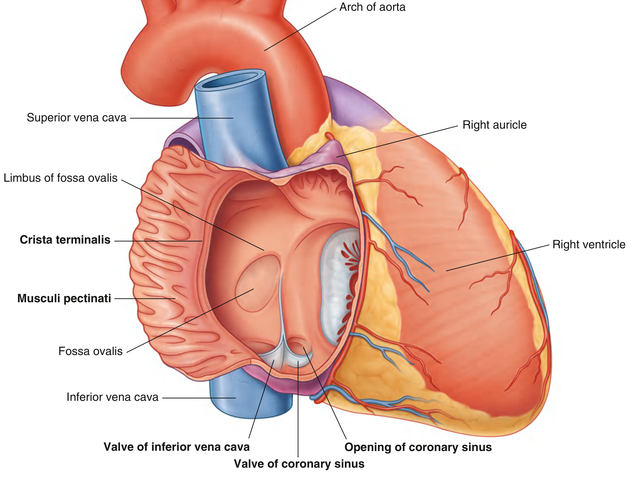
Position and External Appearance
The right atrium (RA) forms the right border of the heart in the anatomical position and contributes to the right portion of the heart's anterior surface. From the epicardial aspect, it is dominated by its large, triangular-shaped appendage (right auricle), which extends anteriorly and laterally, overlapping the ascending aorta.
Externally, a shallow vertical groove called the sulcus terminalis cordis runs from the right side of the superior vena cava opening to the right side of the inferior vena cava opening. This groove is typically filled with epicardial fat and marks the junction of the smooth-walled sinus venarum with the trabeculated atrial appendage. It also serves as a macroscopic landmark for the sinoatrial (SA) node.
Inlets - Venous Drainage
Three vessels deliver blood to the right atrium:
| Vessel | Entry Point |
|---|---|
| Superior vena cava (SVC) | Upper posterior portion |
| Inferior vena cava (IVC) | Lower posterior portion |
| Coronary sinus | Just medial to the IVC opening |
- The superior and inferior venae cavae together return deoxygenated blood from the systemic circulation.
- The coronary sinus returns blood from the walls of the heart (most cardiac veins), guarded by the small crescentic thebesian valve (Valve of the coronary sinus), which is often fenestrated.
Outlet
Blood exits the right atrium into the right ventricle through the right atrioventricular (tricuspid) orifice, which faces forward and medially and is closed during ventricular contraction by the tricuspid valve.
Internal Divisions - Two Compartments
The interior is divided into two continuous spaces by the crista terminalis, which is the internal counterpart to the sulcus terminalis:
1. Sinus Venarum (Sinus of Venae Cavae) - Posterior, Smooth-Walled
- Located posterior to the crista terminalis
- Walls are smooth and thin
- Receives both venae cavae
- Embryologically derived from the right horn of the sinus venosus
2. Atrium Proper + Right Auricle - Anterior, Trabeculated
- Located anterior to the crista terminalis
- Walls are covered by musculi pectinati (pectinate muscles) - muscular ridges that fan out from the crista like the teeth of a comb
- The right auricle is an ear-like, conical muscular pouch extending from this space
- Embryologically derived from the primitive atrium
Crista Terminalis (Terminal Crest)
This is a smooth muscular ridge that:
- Begins on the roof of the atrium just in front of the SVC opening
- Curves anteriorly around the entrance of the SVC
- Descends along the posterolateral wall toward the anterior lip of the IVC
- Gives off an array of pectinate muscles terminating at the smooth-walled vestibule leading to the tricuspid valve
The crista terminalis is clinically significant as a frequent source of focal atrial tachycardia and as the landmark for the SA node.
Interatrial Septum
The interatrial septum faces forward and to the right because the left atrium lies posteriorly and to the left. The septum contains:
- Fossa ovalis - a shallow oval depression marking the site of the embryonic foramen ovale. In fetal life, the foramen ovale allowed oxygenated blood entering from the IVC to pass directly into the left atrium, bypassing the non-functional lungs.
- Limbus fossa ovalis - the raised muscular rim surrounding the fossa ovalis.
Valves Associated with the Right Atrium
| Structure | Location | Function |
|---|---|---|
| Valve of the IVC (Eustachian valve) | Guards IVC opening | In fetal life, directed oxygenated blood toward the foramen ovale; variably developed in adults |
| Thebesian valve (Valve of coronary sinus) | Guards coronary sinus orifice | May be fenestrated or absent; a complete imperforate valve can prevent coronary sinus cannulation |
The Eustachian valve in adults is usually a triangular fibrous flap. In some individuals it is large and muscular, potentially obstructing catheter passage. When net-like or perforated, it is called a Chiari network.
Triangle of Koch (Key Clinical Landmark)
This is a triangular region on the septal aspect of the right atrium that contains the atrioventricular (AV) node. Its three borders are:
- Tendon of Todaro (continuation of the free border of the Eustachian valve) - superiorly, inserting into the central fibrous body
- Septal leaflet of the tricuspid valve - anteriorly
- Coronary sinus ostium - inferiorly
The paraseptal isthmus (between the coronary sinus ostium and tricuspid valve) is targeted for ablation of the slow pathway in AV nodal reentrant tachycardia (AVNRT).
Cavotricuspid Isthmus
The anteroinferior wall between the IVC/Eustachian valve and the tricuspid valve annulus forms the cavotricuspid (cavo-tricuspid) isthmus. This is the critical target for catheter ablation of typical atrial flutter (counterclockwise reentrant circuit).
Sinoatrial Node
The SA node is located at the junction of the SVC and the right atrium, in the region of the sulcus terminalis. The right atrial musculature often extends a short distance into the SVC, which can be a source of focal atrial arrhythmias.
Additional Features
- Foramina of venae cordis minimae (Thebesian foramina) - numerous small openings scattered along the walls through which small cardiac veins drain directly into the right atrium.
- The vestibule - the smooth-walled zone immediately surrounding the tricuspid orifice, continuous with the inferior part of the atrial wall.
Embryological Summary
| Adult Structure | Embryological Precursor |
|---|---|
| Sinus venarum (smooth part) | Right horn of sinus venosus |
| Atrium proper + auricle (trabeculated part) | Primitive atrium |
| Fossa ovalis | Closed foramen ovale (with septum primum forming the floor) |
| Eustachian and thebesian valves | Valve of embryonic sinus venosus |
Sources:
- Gray's Anatomy for Students, pp. 232-233
- Fuster and Hurst's The Heart, 15th Edition, pp. 1063-1064
- Textbook of Clinical Echocardiography, p. (RA section)
This is a shared conversation. Sign in to Orris to start your own chat.