
Ectopic pregnancy
ectopic pregnancy fallopian tube diagram sites of implantation
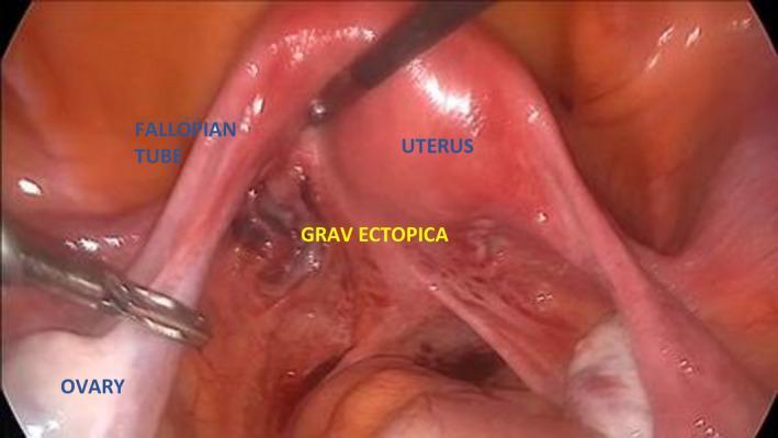
This laparoscopic clinical photograph depicts an intra-abdominal view of the female pelvic anatomy during a diagnostic procedure. The central feature is the uterus, which appears as a pinkish, smooth, and glistening organ. Extending laterally is the fallopian tube, which maintains a thin, tubular morphology. An ectopic pregnancy, labeled 'GRAV ECTOPICA', is visible as a dark, reddish-brown, irregular heteroechoic mass located at the base of the posterior lamina of the broad ligament, adjacent to the uterus. The ovary is situated distally and is being manipulated by a laparoscopic grasper. The image illustrates a rare site for ectopic implantation while highlighting the contrast between the healthy, vascularized uterine tissue and the darker, congested appearance of the ectopic gestation. This visual is significant for teaching the laparoscopic identification of adnexal masses and the surgical anatomy of the broad ligament in the context of early pregnancy complications.
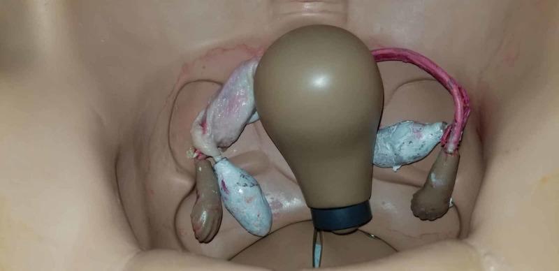
This photograph depicts an anatomical diagram within a gynecologic surgical simulation mannequin, viewed from a superior perspective. The setup illustrates the pelvic cavity with a central, pear-shaped uterine model and bilateral adnexal structures. To simulate a tubal ectopic pregnancy for obstetric surgical training, the mannequin's fallopian tubes and ovaries have been modified with organic and synthetic materials. On the left side of the image, the simulated tube appears pale and translucent with fleshy textures, while the right side features a reddish tubular structure and irregular, bluish-grey masses representing adnexal pathology. These modifications are designed to provide postgraduate resident physicians with a realistic visual and tactile representation of tissue for practicing laparoscopic salpingostomy or salpingectomy. The visual contrast between the tan plastic mannequin and the moist, textured simulated biological tissues emphasizes the target surgical site. This educational resource is used in high-fidelity simulation to bridge the gap between theoretical knowledge of ectopic pregnancy and operative skill acquisition.
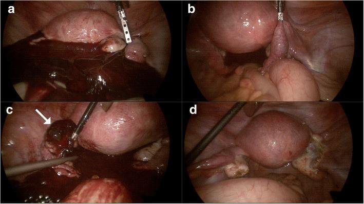
This composite of four laparoscopic images (a-d) illustrates the surgical findings and management of a rare bilateral ectopic pregnancy involving both a tubal and an ovarian implantation. Image (a) shows the initial pelvic cavity inspection, revealing significant hemoperitoneum with dark clotted and fresh blood, indicative of a rupture. Image (b) focuses on the right adnexa, demonstrating an unruptured tubal ectopic pregnancy characterized by a localized distention of the fallopian tube. Image (c) identifies the source of the bleeding: a ruptured ovarian ectopic pregnancy on the left ovary, indicated by a hemorrhagic, dark mass (white arrow) and adjacent hematoma being manipulated with surgical instruments. Image (d) displays the postoperative pelvic cavity following a right salpingectomy and resection of the left ovarian mass, showing a cleared surgical field with visible residual anatomical changes. This case highlights the clinical presentation of hemoperitoneum in gynecological emergencies and the importance of thorough laparoscopic exploration of both adnexa.
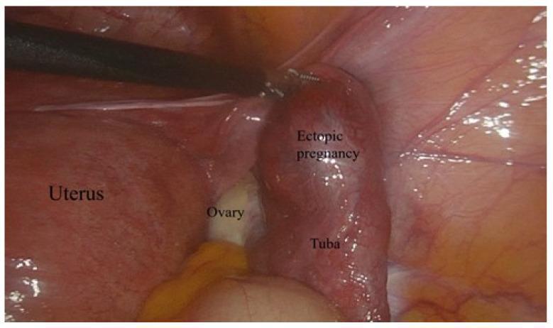
This clinical photograph displays a laparoscopic view of an unruptured ectopic pregnancy within the fallopian tube (tuba). The image highlights the pelvic anatomy, showing the uterus on the left, a partially visible ovary in the center, and the distended fallopian tube on the right. The fallopian tube exhibits significant bulbous swelling and intense erythema, indicating vascular engorgement and inflammation characteristic of an ampullary tubal pregnancy. The tubal surface appears stretched but intact, without visible rupture or active hemorrhage. A laparoscopic surgical instrument is positioned near the affected tube, demonstrating the intraoperative diagnostic and management setting. The educational focus is on the laparoscopic identification of ectopic gestations and the anatomical relationship between the adnexal structures and the uterus in gynecological surgery.
"ectopic pregnancy"[MeSH Terms] AND management
Ectopic Pregnancy
Definition
- Robbins & Kumar Basic Pathology, p. 907
- Berek & Novak's Gynecology, p. 1749
Sites of Implantation
| Site | Frequency |
|---|---|
| Fallopian tube (ampulla most common) | ~82% |
| Ovary | ~5% |
| Cesarean scar | ~1% |
| Cervix | ~1% |
| Intra-abdominal / peritoneal | ~1.4% |
| Interstitial (cornual) | Rare, but high rupture risk |
| Bilateral EPs | Very rare (1:750-1580) |
In IVF pregnancies, the EP rate rises to 2.1-8.6%, and up to 11% if there is an underlying fallopian tube abnormality.
- Grainger & Allison's Diagnostic Radiology, p. 914
Pathophysiology
- Robbins & Kumar, p. 1450
Risk Factors
-
Prior salpingitis / pelvic inflammatory disease (PID) - most important; damages tubal epithelium and cilia
-
Prior ectopic pregnancy (~15% recurrence rate, range 4-28%)
-
Intrauterine device (IUD) in situ
-
Prior tubal ligation or tubal surgery
-
Prior pelvic/abdominal surgery (adhesions)
-
Endometriosis
-
Assisted reproductive technology (IVF)
-
Cigarette smoking
-
Multiple sexual partners / STIs
-
Roberts & Hedges' Clinical Procedures, p. 1382; Berek & Novak, p. 1756
Clinical Features
Classic Triad (present together in only ~50% of cases)
- Amenorrhea (missed period)
- Pelvic/lower abdominal pain - often unilateral, colicky or sharp
- Vaginal bleeding - typically scanty, dark ("spotting")
Additional Features
- Adnexal mass or tenderness
- Cervical motion tenderness
- Shoulder-tip pain (diaphragmatic irritation from hemoperitoneum)
- Syncope / dizziness
- Rectal or back pain
Signs of Rupture (Emergency!)
- Sudden, severe abdominal pain
- Signs of hypovolemic shock: hypotension, tachycardia
- Peritoneal signs (guarding, rebound tenderness)
- Note: Bradycardia in the presence of significant intraperitoneal bleeding is not unusual (vagal response)
- Normal vital signs do NOT exclude significant hemoperitoneum
In one series of 300 consecutive ectopic pregnancies, 50% of patients received medical evaluation at least twice before the correct diagnosis was made.
- Roberts & Hedges, p. 1382
Diagnosis
1. Urine / Serum Beta-hCG
- Urine beta-hCG: sensitive at ≥20 mIU/mL; positive 98% of the time in early pregnancy
- Serum beta-hCG: sensitive to 5 mIU/mL - negative virtually excludes pregnancy
- Serial testing is key: serum beta-hCG should approximately double every 48 hours in a normal IUP; a plateau or subnormal rise is suspicious
- The discriminatory zone is beta-hCG ≥1,500-2,000 mIU/mL - at this level, a normal IUP should be visible on transvaginal ultrasound (TVUS)
2. Transvaginal Ultrasound (TVUS) - Investigation of Choice
- No intrauterine gestational sac when beta-hCG ≥2,000 mIU/mL
- Adnexal mass separate from the ovary - most specific (98%) and sensitive (84%) feature
- "Tubal ring" or "ring of fire" sign - hyperechoic ring with surrounding increased vascularity on Doppler (specificity 99.5-100%, but sensitivity only 20-65%)
- Visible yolk sac or embryo in the adnexa (pathognomonic if seen)
- Echogenic free fluid in the abdomen - suggests hemoperitoneum (96% specific for EP)
An extrauterine mass may NOT be visible in up to 35% of cases.
Special Ultrasound Scenarios:
- Interstitial EP: Eccentrically positioned gestational sac with ≤5 mm surrounding myometrium; presents later, rupture causes catastrophic hemorrhage
- Cervical EP: "Figure-of-8" appearance of the uterus
- Cesarean scar EP: Sac implanted within the myometrial scar
3. MRI
4. Laparoscopy
5. Culdocentesis (Historical)
6. Uterine Decidual Cast
- Roberts & Hedges, p. 1383
Management
A. Emergency Stabilization (Ruptured EP)
- Two large-bore IV lines, aggressive IV fluid resuscitation
- Type and crossmatch
- Urgent surgical intervention - do NOT delay for stabilization
- Rh status and Rho(D) immune globulin if Rh-negative
B. Medical Management - Methotrexate (MTX)
- Hemodynamically stable
- Unruptured EP confirmed (or strongly suspected)
- No contraindications to MTX
- Reliable for follow-up
- Beta-hCG typically <5,000 mIU/mL (higher levels - lower success)
- No fetal cardiac activity on US
- Immunodeficiency
- Hepatic/renal disease
- Active peptic ulcer
- Breastfeeding
- Fetal cardiac activity present
- Hemoperitoneum
- Single-dose: MTX 50 mg/m² IM on Day 1; beta-hCG checked on Day 4 and Day 7. Success if Day 7 level drops ≥15% from Day 4 level
- Two-dose: MTX on Day 1 and Day 4
- Multi-dose: MTX alternating with leucovorin (folinic acid rescue)
- Subsequent ectopic pregnancy risk: ~10%
- Tubal patency is variably affected
- Berek & Novak, p. 349-455
C. Surgical Management
Laparoscopic approach (preferred when surgery indicated)
- Salpingostomy (linear salpingostomy): Incision over the ectopic, removal of pregnancy tissue, tube left open to heal - preferred when future fertility is desired and tube is not severely damaged; residual trophoblast (persistent ectopic) risk requires serial beta-hCG monitoring
- Salpingectomy: Complete removal of the affected tube - preferred when tube is severely damaged, bleeding is uncontrolled, or patient has completed childbearing
Indications for surgery over MTX:
- Hemodynamic instability or rupture
- Contraindication to MTX
- Fetal cardiac activity
- Beta-hCG >5,000 mIU/mL
- Large adnexal mass (>3.5-4 cm)
- Patient unreliable for follow-up
D. Expectant Management
- Reserved for highly selected patients: very low and declining beta-hCG, no symptoms, minimal free fluid
- Requires intensive follow-up with serial beta-hCG
Special Types
Interstitial (Cornual) EP
- Located at the intramural portion of the tube within the uterine wall
- Presents later (8-16 weeks) due to distensible myometrium
- Higher risk of rupture with catastrophic hemorrhage
- Treatment: MTX (often first-line) or cornual resection/hysteroscopic management
Cesarean Scar EP
- Implantation within the fibrous scar of a prior cesarean section
- Incidence rising with increasing cesarean rates
- Can lead to uterine rupture and life-threatening hemorrhage
- Treatment: MTX, suction evacuation under US guidance, or hysteroscopic removal
- Berek & Novak, p. 558-567
Ovarian EP
- Meets Spiegelberg criteria: tube must be intact, gestational sac in ovarian position, sac connected to uterus by ovarian ligament, ovarian tissue histologically present in sac wall
- Treatment: ovarian cystectomy or partial oophorectomy (preserve ovary if possible)
Abdominal/Peritoneal EP
- Most progress from a secondary implantation after tubal abortion
- Rare primary abdominal pregnancies exist
- High maternal and fetal morbidity; management is surgical
Cervical EP
- Very rare; presents with painless profuse bleeding
- "Figure-of-8" uterus on US
- Treatment: MTX or hysteroscopic removal; hysterectomy in refractory cases
Differential Diagnosis
| Condition | Distinguishing Features |
|---|---|
| Threatened/incomplete abortion | IUP on US, products in os |
| Appendicitis | Right iliac fossa, fever, no amenorrhea typically |
| Ovarian torsion | Unilateral pain, enlarged ovary on US, beta-hCG negative |
| Ruptured ovarian cyst | Beta-hCG negative, simple cyst on US |
| PID / salpingitis | Bilateral tenderness, fever, cervical discharge, negative beta-hCG |
Prognosis and Future Fertility
- After a single ectopic, risk of recurrence is approximately 10-15%
- After two ectopics, risk rises to 25-30%
- Future intrauterine pregnancy rates: ~60-70% after salpingostomy; similar or slightly lower after salpingectomy depending on contralateral tube status
- Salpingectomy does NOT impair ovarian reserve from the ipsilateral ovary, as ovarian blood supply is maintained via the ovarian artery
- Berek & Novak, p. 451-455
Key Points Summary
- Ectopic = implantation outside the uterus - fallopian tube (ampulla) is the most common site (~82%)
- Classic triad: amenorrhea + pelvic pain + vaginal bleeding (only ~50% have all three)
- Diagnosis: serial beta-hCG + TVUS; most specific sign is an adnexal mass separate from the ovary
- Discriminatory zone: beta-hCG ≥2,000 mIU/mL without an IUP on TVUS = ectopic until proven otherwise
- Ruptured EP = surgical emergency - do not delay
- Methotrexate is first-line medical therapy for stable, unruptured EPs
- Laparoscopic salpingostomy or salpingectomy are the surgical options
- Interstitial and cesarean scar EPs present late, rupture with massive hemorrhage
- Chronic salpingitis (from PID/STIs) is the most important preventable risk factor