
Adenomyosis examination
I'll start by reading the medical library skill, then search for adenomyosis examination content in parallel.
Reading File
Now let me search the library and images in parallel.
Finding Sources
Finding Sources
Searching Images
adenomyosis uterus histology gross pathology
Good results. Now let me read the key sections from multiple textbooks in parallel.
Reading File
Reading File
Reading File
Reading File
Excellent content. Let me read the Berek & Novak's Gynecology section on adenomyosis examination and the Bailey & Love table, plus fetch the MRI image, in parallel.
Reading File
Reading File
Loading Image
Searching PubMed
adenomyosis diagnosis imaging criteria
I now have comprehensive content from multiple authoritative sources. Let me also fetch the second MRI image and get the Berek & Novak content on adenomyosis physical findings.
Loading Image
Reading File
I now have comprehensive content from multiple authoritative textbooks. Here is a detailed, well-structured answer on the examination of adenomyosis.
Adenomyosis: Examination & Diagnosis
Definition
Adenomyosis is a benign uterine disorder characterised by the presence of ectopic endometrial glands and stroma within the myometrium, accompanied by smooth muscle hypertrophy or hyperplasia. The ectopic tissue may be distributed diffusely or focally. When focal, the lesion is termed an adenomyoma.
- Robbins & Kumar Basic Pathology, p. 608; Bailey and Love's Surgery 28th Ed., p. 1604
Clinical Presentation (History)
Symptoms are often non-specific and overlap with fibroids and endometriosis. The classic triad is:
| Symptom | Notes |
|---|---|
| Dysmenorrhea | Secondary dysmenorrhea; pain typically begins 1-2 weeks before menses and persists until a few days after cessation |
| Abnormal uterine bleeding (menorrhagia) | Heavy or prolonged menstrual bleeding |
| Chronic pelvic pain | Can be constant or cyclical |
Other symptoms include:
-
Subfertility / infertility
-
Dyspareunia
-
Uterine enlargement and tenderness
-
33% of cases are asymptomatic (found incidentally on imaging or at hysterectomy)
-
Predominantly affects women in the 4th and 5th decades of life
-
Grainger & Allison's Diagnostic Radiology, p. 898; Bailey and Love's Surgery, p. 1604
Physical Examination Findings
Bimanual Pelvic Examination
The key finding is a uniformly enlarged, globular, tender uterus:
- Uterine enlargement: Symmetrically enlarged, often described as "boggy" - the myometrium is thickened due to reactive smooth-muscle hypertrophy from the ectopic tissue
- Uterine tenderness: Particularly noticeable just prior to and during menstruation
- Globular configuration: Unlike fibroid-related enlargement which is typically irregular/nodular, adenomyosis produces a more uniformly round, smooth enlargement
- No adnexal masses unless coexisting endometriosis (endometrioma) is present
The examination is often normal or non-specific, which explains why imaging is required for diagnosis. Adenomyosis cannot be reliably diagnosed on physical examination alone.
- Berek & Novak's Gynecology, p. 576; Bailey and Love's Surgery, p. 1604
Risk Factors (Relevant to Clinical Assessment)
-
Increased/prolonged oestrogen exposure (early menarche ≤10 years, short cycles ≤24 days, elevated BMI, tamoxifen use)
-
Multiparity; spontaneous miscarriage
-
Prior uterine instrumentation or surgery: caesarean section, surgical termination of pregnancy, endometrial curettage
-
Coexisting leiomyomas, endometriosis, or endometrial hyperplasia
-
Smoking
-
Bailey and Love's Surgery, p. 1604
Imaging Examination
The diagnosis of adenomyosis is now primarily made by imaging. Histology at hysterectomy remains the definitive gold standard, but imaging has largely replaced surgical diagnosis for initial evaluation.
1. Transvaginal Ultrasound (TVUS) - First-Line
Accuracy: 68-86% for diffuse adenomyosis.
The MUSA (Morphological Uterus Sonographic Assessment) group recommends commenting on 8 features: presence, location, differentiation, cystic vs. non-cystic, myometrial layer involvement, extent, lesion size, and vascularity.
Key ultrasound features:
| Feature | Description |
|---|---|
| Enlarged, globular uterus | Often with asymmetry of anterior/posterior walls |
| Myometrial cysts | 2-6 mm subendometrial cysts, present in ~50%; represent haemorrhagic foci |
| Echogenic subendometrial lesions | Diffuse echogenic nodules and linear striations |
| Hyperechogenic islands | Scattered echogenic foci in the myometrium |
| Irregular junctional zone | Poor definition of the endomyometrial interface |
| Fan-shaped shadowing | Also called "rain shower" appearance - multiple fine areas of attenuation throughout |
| Colour Doppler | Speckled pattern of increased vascularity within heterogeneous areas |
At least 3 sonographic criteria are usually required to make a diagnosis.
- Grainger & Allison's Diagnostic Radiology, p. 898; Bailey and Love's Surgery, p. 1604
2. MRI - Problem-Solving and Definitive Imaging
MRI is the most accurate non-invasive method and is particularly valuable in distinguishing adenomyosis from fibroids, which has direct management implications.
MRI is not technique-dependent and has low interobserver variability compared with TVUS, SIS, and hysteroscopy. - Berek & Novak's Gynecology, p. 508
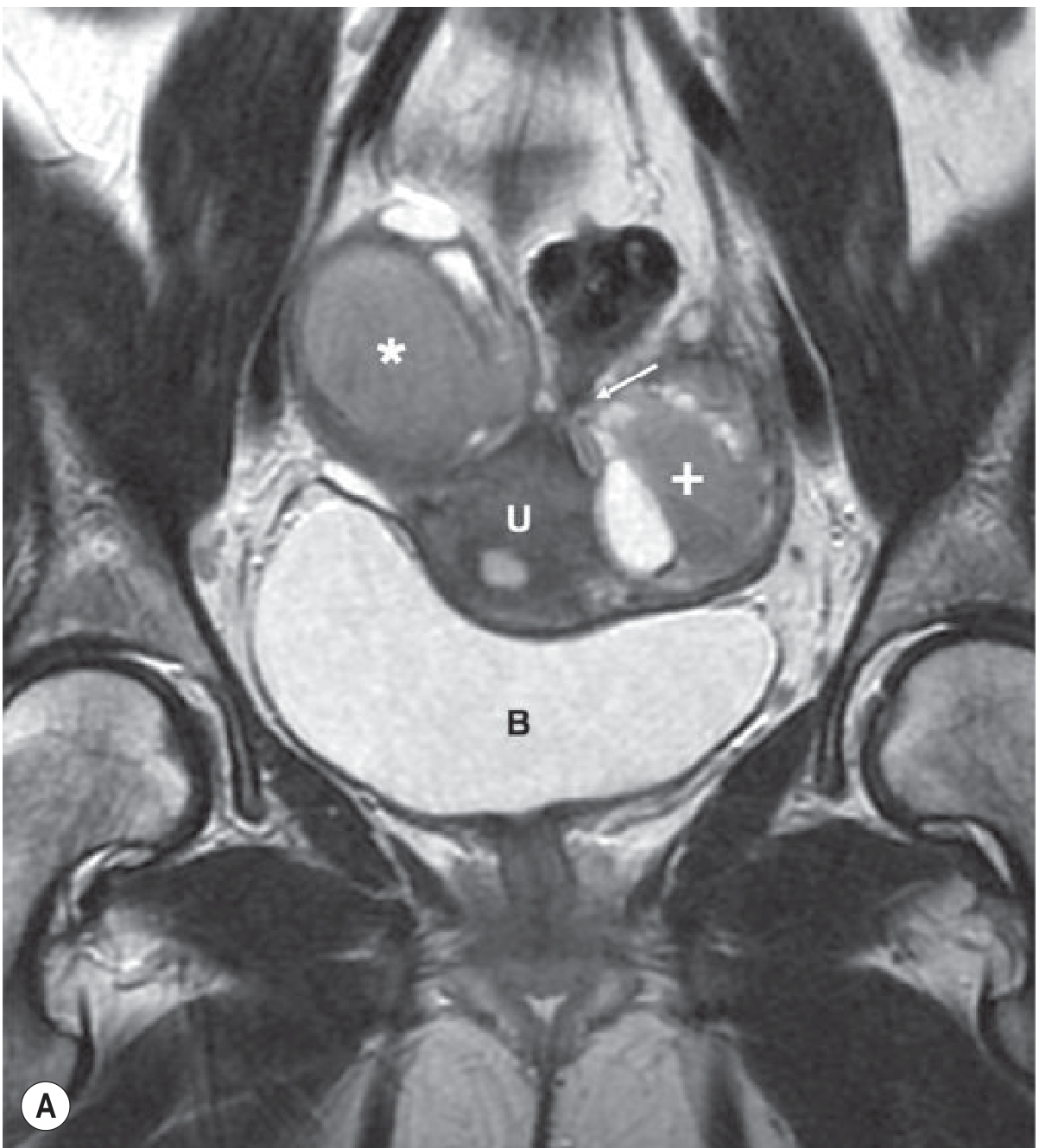
Key MRI features on T2-weighted imaging:
| Finding | Significance |
|---|---|
| Junctional zone (JZ) thickening ≥ 12 mm | Predicts adenomyosis with high accuracy (diffuse) |
| JZ ≤ 8 mm | Excludes adenomyosis with high accuracy |
| JZ 8-12 mm | Indeterminate - use ancillary criteria below |
| High T2 signal foci within low-signal myometrium | Islands of ectopic endometrial tissue |
| Cystic dilatation of glands in myometrium | Ancillary criterion |
| High T2 linear striations (finger-like projections) extending from endometrium into myometrium | Represents direct myometrial invasion |
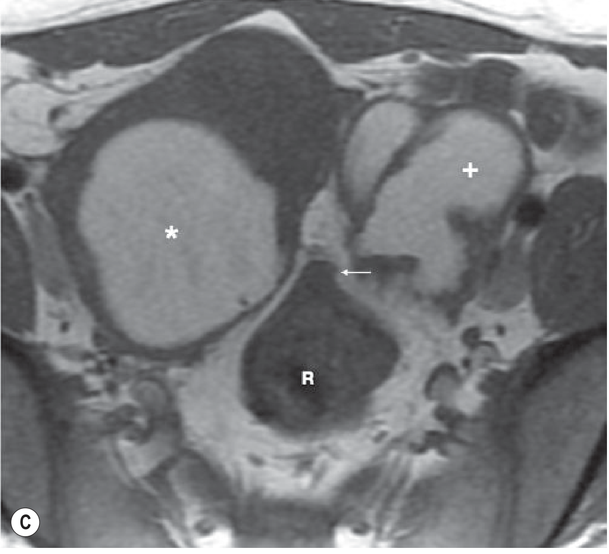
| High T1 signal foci (punctate haemorrhage) | 95% positive predictive value for adenomyosis |
| High T1 + intermediate-low T2 foci | Cystic adenomyosis (extensive bleeding) |
- Grainger & Allison's Diagnostic Radiology, p. 898
MRI of adenomyosis - coronal view (U = uterus, B = bladder):
MRI - axial view showing adenomyosis with bilateral ovarian endometriomas (asterisk = endometrioma, + = adenomyotic uterus, R = rectum):
3. Hysteroscopy
Abnormal findings may include:
- Irregular endometrium with endometrial defects
- Cystic haemorrhagic lesions
- Altered vascularisation
- "Strawberry-like" appearance
4. Histology (Gold Standard)
Classically obtained at hysterectomy. Diagnostic criteria:
-
Endometrial glands and/or stroma found deep in the myometrium, interposed between muscle bundles
-
Reactive hypertrophy of surrounding smooth muscle
-
Limited in women wishing to preserve fertility
-
Robbins & Kumar Basic Pathology, p. 608
Differential Diagnosis
| Condition | Distinguishing Features |
|---|---|
| Uterine fibroids (leiomyomas) | Irregular, nodular uterine contour; well-defined lesions on MRI with T2 low-signal, distinct margins |
| Endometriosis | Adnexal masses, uterosacral nodularity, fixed retroverted uterus on exam; normal-sized uterus |
| Endometrial polyp | Focal lesion on sonohysterography |
| Endometrial carcinoma | Postmenopausal bleeding; abnormal endometrial thickness |
| PID / chronic endometritis | Infection history, cervical motion tenderness |
Summary of Diagnostic Approach
Clinical Suspicion
(dysmenorrhea + menorrhagia + tender enlarged uterus)
         ↓
Transvaginal Ultrasound (first-line)
≥3 MUSA criteria → Diagnosis of adenomyosis
         ↓ (if equivocal)
MRI Pelvis
JZ ≥12 mm → Adenomyosis confirmed
JZ ≤8 mm  → Adenomyosis excluded
JZ 8-12 mm → Ancillary criteria (T2 foci, T1 haemorrhage)
         ↓ (definitive, fertility treatment complete)
Histology at hysterectomy (gold standard)
Recent Evidence
A 2024 systematic review on ultrasound features predicting symptom severity (PMID: 37584856) found that specific ultrasound characteristics correlate with the degree of menorrhagia and pain, supporting a more nuanced sonographic classification. A 2025 systematic review on adenomyosis in adolescents (PMID: 39469820) highlights that the condition is underdiagnosed in younger patients where classic features may be absent.
This is a shared conversation. Sign in to Orris to start your own chat.