
Leishmans tain
Reading File
Leishman's stain is a Romanowsky-type stain used in haematology to stain peripheral blood smears, bone marrow, and detect Leishmania parasites. It uses a mixture of eosin and methylene blue dissolved in methanol.
Searching sources...
Finding Sources
Finding Sources
Reading File
Finding Sources
Finding Sources
Finding Sources
Finding Sources
Reading File
Reading File
Now let me fetch the relevant image from Fitzpatrick's showing Leishman-Donovan bodies:
Loading Image
Loading Image
Here is a complete overview of Leishman's stain:
Leishman's Stain
What It Is
Leishman's stain is a Romanowsky-type stain - a polychrome stain based on a mixture of eosin (acidic dye) and methylene blue (basic dye) dissolved in methyl (absolute) alcohol. The methanol acts simultaneously as a fixative and as the solvent for the dyes. Other Romanowsky-type stains include Giemsa, Jenner's, May-Grunwald, MacNeal's, and Wright's stain.
The reference Romanowsky method (ICSH, 1984) uses just two dyes - azure B and eosin Y - which together produce the full range of colours seen with ideal Romanowsky staining. - Henry's Clinical Diagnosis and Management by Laboratory Methods
Principle of Staining
| Component | Dye type | Stains |
|---|---|---|
| Eosin (acidic) | Acidophilic | Haemoglobin, eosinophil granules - pink/red |
| Methylene blue / azure B (basic) | Basophilic | Nuclei, cytoplasmic RNA, platelet granules, parasite DNA - blue/purple |
The kinetoplast of Leishmania amastigotes stains bright red with Giemsa/Leishman stains (non-metachromatic staining of the parasite).
Primary Uses
1. Peripheral Blood Smear (Haematology)
Leishman's stain is used routinely to stain peripheral blood films for:
- Differential WBC count (neutrophils, lymphocytes, monocytes, eosinophils, basophils)
- RBC morphology assessment (size, shape, colour, inclusions)
- Platelet morphology
- Detection of blood parasites (malaria, trypanosomes, microfilaria)
2. Malaria Diagnosis
Of the Romanowsky stains, Giemsa at pH 7.2 is preferred for malaria; however, Field's, Wright's, or Leishman's stain can also be used. - Harrison's Principles of Internal Medicine, 22E (2025)
3. Diagnosis of Leishmaniasis
Leishman's stain identifies Leishman-Donovan (LD) bodies - the amastigote form of Leishmania spp. - in:
- Tissue smears (skin scrapings, fine-needle aspirates)
- Bone marrow aspirates
- Splenic aspirates (in visceral leishmaniasis / kala-azar)
In stained smears, the parasites take on a distinctive rounded appearance (having lost their flagella) within macrophage phagolysosomes and are termed Leishman-Donovan bodies. - Sherris & Ryan's Medical Microbiology, 8th Ed.
Leishman-Donovan (LD) Bodies - What You See
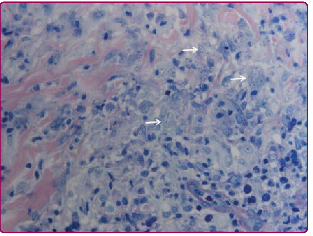
- Small, oval amastigotes (2-4 µm) within macrophages/histiocytes
- Pale blue cytoplasm, dark-staining nucleus (blue-purple), and a dot-like kinetoplast (bright red with Giemsa; identifiable with Leishman's)
- Found extracellularly and intracellularly (~70% of cases of cutaneous leishmaniasis)
- Giemsa/Leishman stain used with oil-immersion microscopy on air-dried, methanol-fixed smears
The histologic hallmark of cutaneous leishmaniasis is the presence of numerous extracellular and intracellular amastigotes (Leishman-Donovan bodies) within histiocytes. The organisms can also be highlighted by Wright and Feulgen stains. - Fitzpatrick's Dermatology
Histopathology Images
Fig A (H&E, low power): Cutaneous leishmaniasis showing tuberculoid granulomatous dermatitis with multinucleated giant cells and lymphoplasmacytic infiltrate:
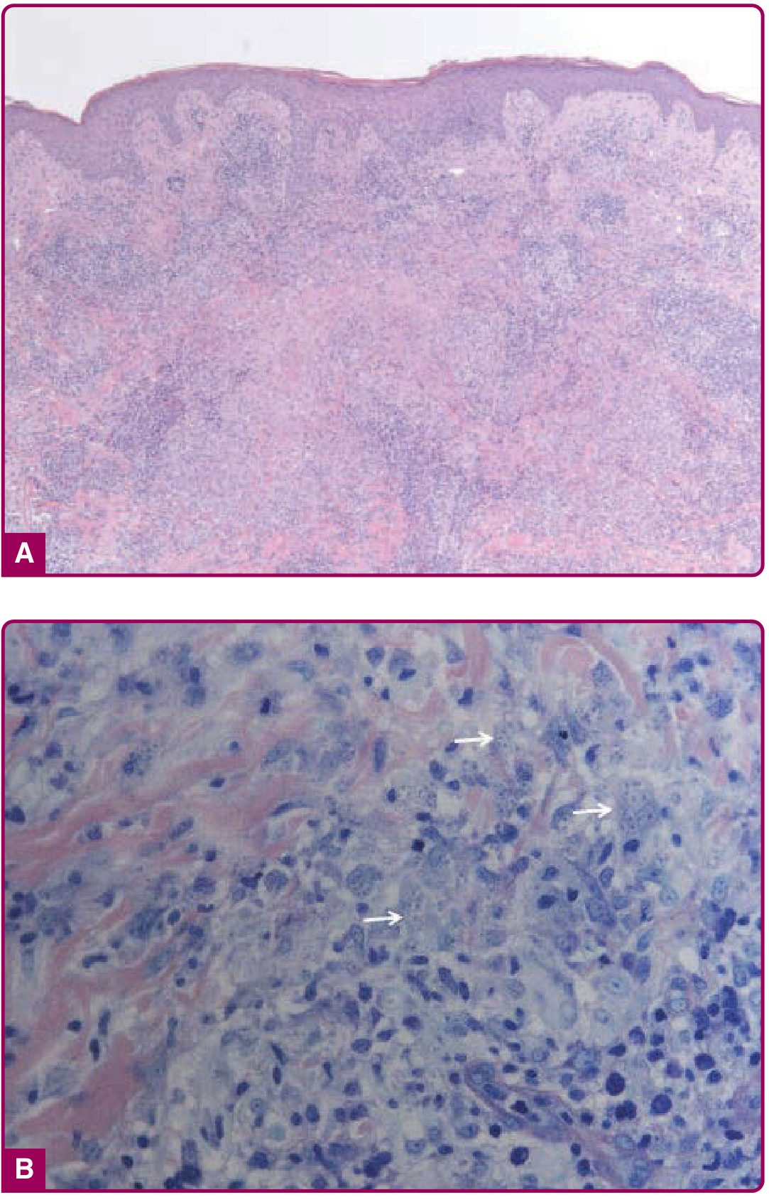
Fig B (Giemsa stain, high power): Parasitized macrophages with amastigotes (Leishman-Donovan bodies, white arrows):
Common Staining Problems & Troubleshooting
| Problem | Cause | Fix |
|---|---|---|
| Excessively blue | Thick film, prolonged staining, alkaline buffer | Shorten stain time, lower pH buffer |
| Excessively pink | Understaining, prolonged washing, acidic buffer | Prolong staining, raise pH buffer |
| Precipitate on film | Dirty slides, drying during staining, poor filtration | Use clean slides, filter stain, wash properly |
| Poorly stained RBCs | Understaining or excessive washing | Prolong staining, reduce washing |
Differential Diagnosis on Leishman-Stained Smears (Parasitized Macrophages)
When LD bodies are seen, consider distinguishing from:
- Histoplasmosis - yeasts with a surrounding halo
- Donovanosis (granuloma inguinale) - safety-pin appearance of Donovan bodies
- Rhinoscleroma - Mikulicz cells
- Trypanosomiasis, toxoplasmosis, blastomycosis
Summary: Leishman's stain is a simple, methanol-based Romanowsky stain used for blood films and parasite detection. Its most historically significant application - and the origin of its name - is the identification of Leishmania amastigotes (Leishman-Donovan bodies) in tissue and aspirate smears.
This is a shared conversation. Sign in to Orris to start your own chat.