
Submandibular pleomorphic adenoma case presentation
pleomorphic adenoma salivary gland histology
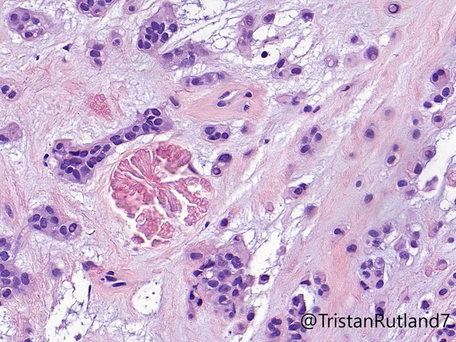
This histopathology image depicts a pleomorphic adenoma of ceruminous gland origin, a rare benign salivary-type mixed tumor arising in the external auditory canal. Acquired via standard bright-field light microscopy, the slide shows Hematoxylin and Eosin staining with widely scattered ductal epithelial structures embedded in a chondromyxoid stroma. The tumor exhibits biphasic cellularity: duct-forming epithelial cells arranged in cords and nests, and intervening myoepithelial/plasmacytoid cells with scant cytoplasm. The stroma is variably myxoid and chondroid, imparting a cartilaginous appearance. In this case, tyrosine-rich crystals are present within the stroma, a distinctive crystalline feature occasionally reported in salivary/glandular myoepithelial lesions. The lesion demonstrates benign histology without significant pleomorphism, mitotic activity, or perineural invasion, supporting a diagnosis of a benign ceruminous pleomorphic adenoma. Clinically, the finding is relevant for differential diagnosis of external auditory canal masses and informs surgical management; complete excision is curative with low recurrence when margins are negative. This image is useful for educational purposes in pathology, ENT, and oral medicine, illustrating the classic histopathological hallmarks: epithelial/myoepithelial biphasic proliferation, chondromyxoid stroma, ductal differentiation, and crystalline deposits, consistent with ceruminous gland-origin pleomorphic adenoma and histologic mimicry to salivary gland pleomorphic adenoma. The image supports diagnostic education in pathology, otolaryngology, and histology.
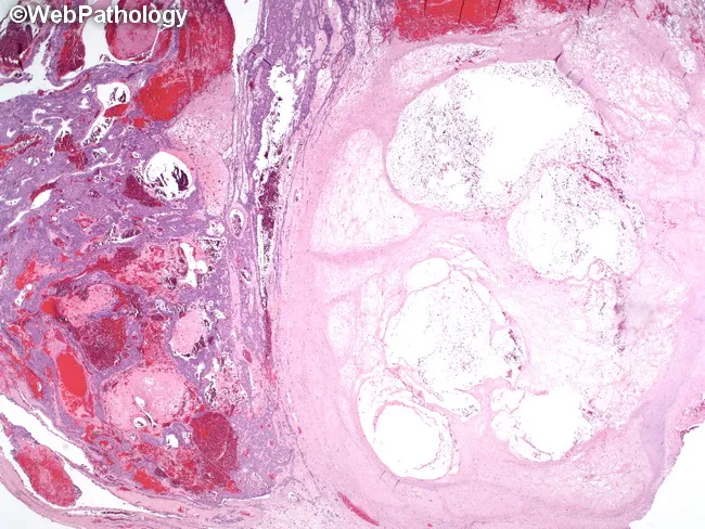
This is a histopathology slide of a salivary gland lesion, specifically a pleomorphic adenoma, showing post‑biopsy changes. The left half of the tumor demonstrates prominent hemorrhage with extravasated red cells and congested stroma, while the right half contains an infarcted nodule with coagulative features and ghosted cellular outlines. The lesion is composed of biphasic components characteristic of pleomorphic adenoma: duct‑forming epithelial cells arranged in cords within a myoepithelial-rich stroma that ranges from myxoid to chondroid, with areas of hyalinization. The infarcted region displays coagulative necrosis with preservation of overall architecture but loss of viable cytoplasm in many cells. The hemorrhagic component reflects biopsy‑related vascular injury following fine‑needle aspiration biopsy (FNA), a known iatrogenic change. The contrast between hemorrhagic zones and infarcted nodules can mimic malignant necrosis if not interpreted in context. Clinically, this image highlights the need to correlate cytology/fine needle aspiration findings with histology to avoid overcalling malignancy in post‑procedural salivary gland lesions. Diagnostic significance: FNA‑induced hemorrhage and infarction within pleomorphic adenoma; typical histology of benign salivary gland tumor preserved elsewhere; no overt atypia or invasive growth detected. Potential use cases: education, differential diagnosis in salivary gland tumors, radiology-pathology correlation, biopsy artifact recognition, pathology review.
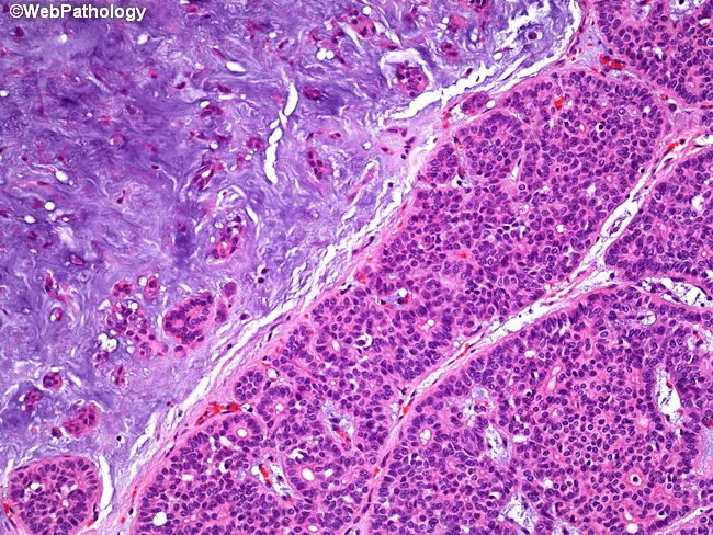
Imaging modality: light microscopy of hematoxylin and eosin stained paraffin-embedded salivary gland tissue from the parotid region. Anatomy: parotid gland tissue showing a classic biphasic neoplasm with epithelial elements intimately admixed with mesenchymal-like stroma. Description: Epithelial components form acinar and tubular structures, composed of duct‑forming cells and myoepithelial-like cells, set within a markedly hypocellular myxoid to chondromyxoid stroma. The epithelial nests are hypercellular in places and juxtaposed to expansive, basophilic stroma; the interface produces a biphasic appearance. The stroma often shows loose, lobulated myxoid material with mild chondroid differentiation, creating the distinctive mixed morphology. Overall, this pattern is typical of pleomorphic adenoma, a benign salivary gland tumor with epithelial–mesenchymal differentiation and typically a well circumscribed capsule. Diagnostic significance: the combination of acinar/tubular epithelial differentiation within a myxoid/chondromyxoid stroma strongly supports pleomorphic adenoma; differentiates from malignant salivary neoplasms. Differential considerations: mucoepidermoid carcinoma, adenoid cystic carcinoma, basal cell adenoma. Clinical correlation: presents as a slow-growing, painless parotid mass in adults; treatment usually involves complete surgical excision with facial nerve preservation and careful follow-up for recurrence. Potential applications: educational histology exemplar, pathology teaching repository, and radiology-pathology correlation studies. This image exemplifies classic pleomorphic adenoma histology suitable for teaching and diagnostic training in pathology education.
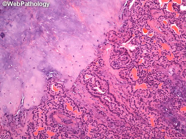
Histology, Light microscopy, Hematoxylin and Eosin stained section of a salivary gland mass. The epithelial component demonstrates ductal/acinar differentiation arranged in cords and small nests, with cuboidal to columnar cells and inconspicuous cytoplasm. Surrounding this proliferative epithelium is a abundant, hypocellular stromal background composed of chondromyxoid matrix with cartilaginous-like areas and occasional myxoid pools. The composite tissue exhibits a classic biphasic pattern typical of pleomorphic adenoma: epithelial/ductal elements embedded within a variably myxoid to chondroid stroma, often with myoepithelial cells contributing to the matrix. The lesion is circumscribed, showing well-defined limits without overt invasion into adjacent parotid tissue at this magnification. These features are diagnostic of a benign mixed tumor arising from salivary gland parenchyma. Clinically, this entity presents as a slow-growing, painless mass in the parotid region and has a low malignant potential. Differential considerations include other salivary gland neoplasms such as basal cell adenoma, mucoepidermoid carcinoma, and chondroid syringoma. Diagnostic significance lies in confirming a pleomorphic adenoma, guiding surgical planning toward complete excision with margin control to minimize recurrence risk. The image is optimal for teaching and research in head-and-neck pathology and salivary gland tumor histology. This image is valuable for medical education, pathology review, and diagnostic practice.
submandibular gland tumor swelling clinical presentation
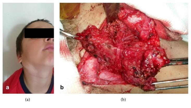
This composite educational material consists of a clinical photograph (a) and an intraoperative photograph (b) documenting a parotid gland tumor, identified as a plexiform neurofibroma. Panel A shows a pediatric patient with significant facial asymmetry due to a longitudinal swelling in the right parotid and submandibular region. The mass is approximately 3 cm in diameter, causing outward displacement of the mandibular angle. Panel B displays the surgical exploration during a superficial parotidectomy. The image shows a large, hemorrhagic, and multinodular mass with a gelatinous appearance. Surgical instruments are seen retracting tissues to expose the tumor's intimate relationship with the facial nerve trunk. The intraoperative view demonstrates displacement of the facial nerve's temporofacial and cervicofacial branches due to chronic compression. Key educational concepts include the clinical presentation of parotid masses, surgical anatomy of the facial nerve, and the characteristic 'bag of worms' appearance of plexiform neurofibromas in the cervicofacial region.
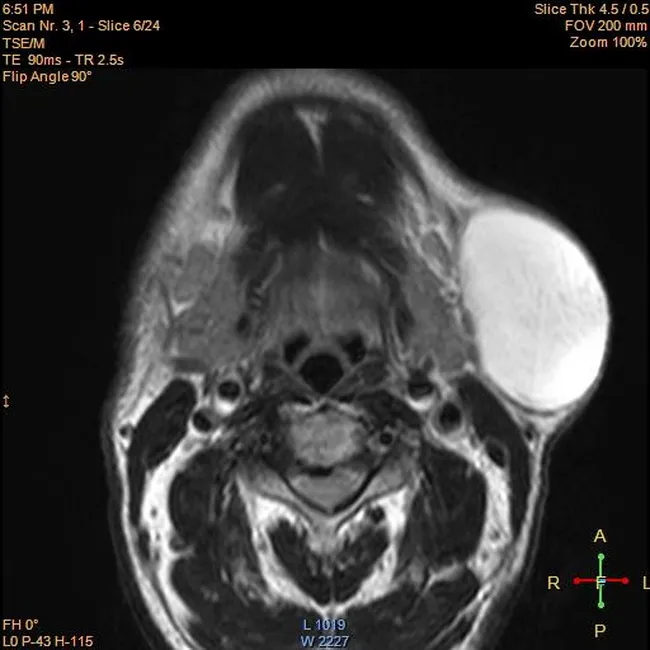
Imaging modality: Magnetic Resonance Imaging (MRI). This axial T1-weighted neck image demonstrates a well-defined oval lesion within the left submandibular soft tissues. The mass shows high signal intensity on T1 and remains bright on T2-weighted sequences, consistent with a fat-containing lesion. A thin capsule and sharp margins are evident; there is no invasion of adjacent bone or deep soft tissues. There is no edema, diffusion restriction, or surrounding inflammatory change. The radiologic appearance is classic for a lipoma, a benign adipose tumor composed of mature fat cells. The lesion is nonadherent to surrounding structures, and there is no surrounding lymphadenopathy. The clinical presentation is a painless, slowly enlarging swelling in a 60-year-old male. Differential considerations include other fat-containing lesions such as lipoblastoma in younger patients or liposarcoma in older adults, but the homogeneous high T1 signal with no suspicious septations or nodularity favors lipoma. Post-contrast imaging, if performed, would typically show minimal to no enhancement. This imaging feature set informs preoperative planning by delineating lesion size, extent, and relationship to the submandibular gland, mandible, and facial artery branches, aiding surgical approach and reducing biopsy need while histopathology remains definitive. Correlation with surgical and histopathology findings confirms benign diagnosis.
Submandibular Pleomorphic Adenoma - Case Presentation
Case Vignette
Clinical Presentation
Symptoms
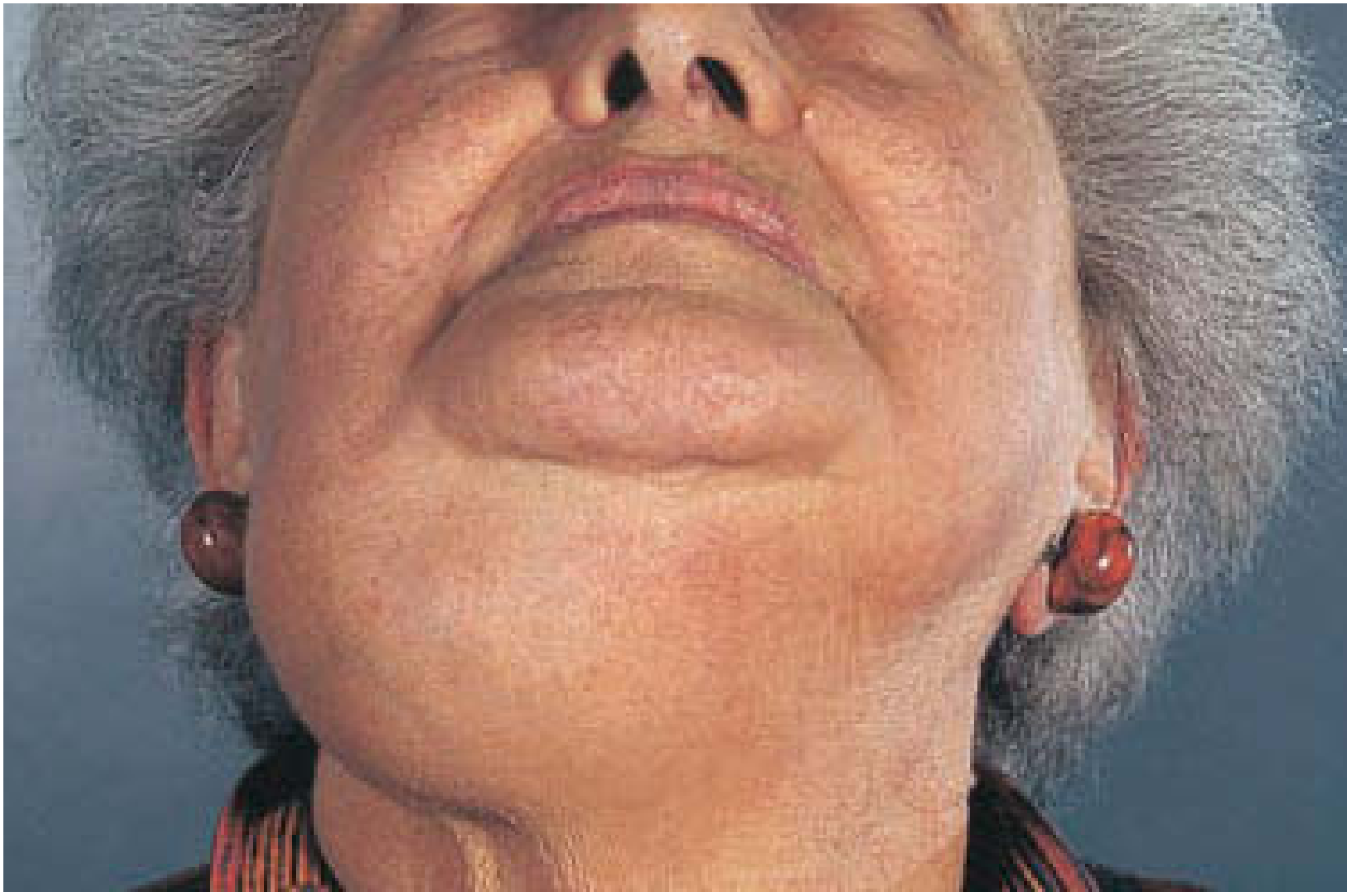
- Slow-growing, painless swelling in the submandibular triangle
- No pain (pain would suggest infection, sialadenitis, or malignant change)
- No facial nerve symptoms (weakness = red flag for malignancy)
- No skin ulceration (ulceration = red flag for malignancy)
Clinical Examination Findings
- Well-defined, firm to rubbery (firmness varies - soft if predominantly myxoid, hard if chondroid/collagenous), solitary, mobile mass in the submandibular triangle
- Overlying skin is normal, non-tender
- No facial nerve palsy (marginal mandibular branch tested by asking patient to show lower teeth)
- Bimanual palpation of the floor of the mouth localizes the tumor more accurately
- Sometimes difficult to distinguish from a tail-of-parotid tumor or submandibular lymph node
Background and Epidemiology
- Pleomorphic adenoma (PSA; benign mixed tumor) is the most common salivary gland neoplasm, accounting for ~50% of all salivary gland tumors
- Annual incidence: 2.4-3.05 per 100,000
- Distribution: >80% parotid gland, ~10-15% submandibular gland, ~5% minor salivary glands
- In the submandibular gland, 50% of all tumors are pleomorphic adenomas, but ~50% of submandibular gland tumors are malignant (compared to only 25% in the parotid); thus the index of suspicion for malignancy is higher
- "The larger the gland, the more likely the tumor is benign" - Sabiston Textbook of Surgery
Differential Diagnosis
| Condition | Distinguishing Features |
|---|---|
| Submandibular sialadenitis | Tender, painful, often with calculus on imaging |
| Submandibular lymphadenopathy | Regional head/neck infection/malignancy; multiple nodes |
| Warthin's tumor | Almost exclusive to parotid, older males, smokers |
| Mucoepidermoid carcinoma | More common in submandibular gland; firm, possible nerve involvement |
| Adenoid cystic carcinoma | Perineural invasion, pain, facial nerve dysfunction |
| Submandibular lipoma/cyst | Soft, fluctuant, not in gland |
Investigations
Imaging
- Ultrasound: First-line - shows well-defined, hypoechoic, lobulated mass; can localize it within the gland vs. adjacent structures; guides FNA
- CT scan: Delineates extent, relationship to adjacent structures, bony involvement, and cervical nodes
- MRI: Best for soft tissue characterization; pleomorphic adenoma typically shows intermediate T1, high T2 signal; calcification (if present on CT) is characteristic
Cytology / Histology
- FNAC (Fine Needle Aspiration Cytology): Commonly performed; sensitivity ~80% for distinguishing benign vs. malignant. Results classified using the Milan System for Reporting Salivary Gland Cytopathology
- Core needle biopsy: More tissue, higher diagnostic accuracy than FNAC; useful when FNA is inconclusive
- Note: Incisional/open biopsy is avoided due to risk of tumor spillage and implantation recurrence
Pathology
Macroscopic Features
- Well-demarcated, round or ovoid with broad-based surface bosselations (lobulations)
- Variably encapsulated (capsule may be thick/fibrotic OR attenuated/incomplete)
- Cut surface: solid, blue-grey (chondromyxoid) or white/tan; may show cystic areas and calcification
- Protuberant peri-capsular nodules can occur, attached by a pedicle - simple enucleation risks leaving these behind, causing recurrence
Microscopic Features
- Biphasic tumor: ductal epithelial cells + myoepithelial cells embedded in a chondromyxoid matrix
- Cellular diversity: oval, epithelioid, spindle, plasmacytoid, and clear cells
- Stromal components range from myxoid to chondroid to hyalinized
- Squamous metaplasia may be present (does not imply malignancy)
- Capsule: thin (<20 micron) in at least one area, incomplete in ~50%
Immunohistochemistry
- Luminal cells: CK7 (strong and diffuse)
- Myoepithelial cells: p63, S-100, SOX10, SMA
Management
Definitive Treatment
- Performed as transcervical submandibular gland excision (submandibularectomy)
- Excision should be extracapsular - the gland and tumor are removed entirely; simple enucleation is unacceptable (risks capsular breach, tumor spillage, and multifocal recurrence)
- A cuff of surrounding normal tissue is included where possible
Surgical Steps (Submandibular Gland Excision - Sabiston)
- Transcervical incision, 2 fingerbreadths below the mandible
- Raise subplatysmal flaps
- Protect the marginal mandibular branch of the facial nerve (Hayes-Martin maneuver: divide the facial vein at the inferior gland border, raise it with the gland fascia - the marginal mandibular nerve runs superficial to this vein)
- Divide the facial artery at the inferior aspect
- Dissect the superior gland free; divide the inferior border off the anterior belly of digastric
- Retract the mylohyoid medially-superiorly to identify the lingual nerve, submandibular ganglion, and submandibular duct
- Identify the hypoglossal nerve with medial-inferior retraction of mylohyoid
- Divide the facial artery posteriorly; free the gland completely
Structures at Risk
- Marginal mandibular nerve (branch of facial nerve) - most commonly injured; higher risk in submandibular gland excision for pleomorphic adenoma due to extracapsular dissection requirement
- Lingual nerve
- Hypoglossal nerve
- Facial artery and vein
Malignant Transformation (Carcinoma ex Pleomorphic Adenoma)
- Sudden increase in size
- Pain or tenderness
- Facial nerve palsy (marginal mandibular nerve involvement)
- Skin fixation or ulceration
- Cervical lymphadenopathy
- Older patient age
- Male sex
- Long duration of tumor (left untreated for years)
- Multiple recurrences
- Deep lobe parotid location
Prognosis
- Excellent with complete excision
- Recurrence is uncommon after proper extracapsular excision
- Recurrence is typically multifocal when it does occur (from capsular rupture and spillage during surgery)
- Long-term follow-up is recommended
Key Teaching Points
- Submandibular gland tumor = 50% benign (pleomorphic adenoma), 50% malignant - always investigate thoroughly
- Pleomorphic adenoma is diagnosed by FNA/core biopsy but definitively treated by surgical excision
- Enucleation is contraindicated - always extracapsular excision
- Capsule is incomplete in ~50% of cases - risk of spillage and recurrence
- Marginal mandibular nerve injury is the key surgical complication in submandibular gland excision