
Hemodynamic disorders: hemorrhage. Etiopathogenesis and morphology
Reading File
Finding Sources
Finding Sources
Searching PubMed
"hemorrhage"[MeSH Terms] AND "pathology"
Reading File
Reading File
Reading File
Hemorrhage: etiopathogenesis and morphology
Definition
Hemorrhage is the extravasation of blood from vessels. It results from vessel damage and may be made worse by defects in clotting. - Robbins & Kumar Basic Pathology, Ch. 3
Etiopathogenesis
Blood escapes from a vessel when the wall is breached or when hemostasis fails. Hemostasis depends on three things working together: the vessel wall, platelets and coagulation factors.
1. Vessel wall damage (rupture or erosion)
- Trauma: a laceration, incision or contusion. Small vessels bleed after blunt injury.
- Atherosclerosis: weakens the wall. Aneurysm rupture and dissection can cause massive bleeding (for example a retroperitoneal hematoma from a dissecting aortic aneurysm).
- Inflammatory erosion: examples are peptic ulcer, tuberculous cavity erosion and vasculitis.
- Neoplastic erosion: a tumor invades the wall.
- Chronic passive congestion: capillaries in chronically congested tissue can bleed.
- If the affected vessel is a large artery or vein, the hemorrhage may be massive.
2. Hemorrhagic diatheses
- These are disorders in which bleeding follows a seemingly trivial injury, or occurs spontaneously.
- Causes are inherited or acquired defects in one of the following:
- Vessel wall: vitamin C deficiency (scurvy), vasculitis, amyloid, Ehlers-Danlos syndrome, infections such as meningococcemia.
- Platelets: thrombocytopenia (ITP, TTP, marrow failure, drugs) or platelet dysfunction (aspirin, uremia, von Willebrand disease).
- Coagulation factors: hemophilia A and B, vitamin K deficiency, liver disease, DIC, anticoagulant therapy.
- Pattern of bleeding in a diathesis:
- Platelet and vascular defects usually cause mucocutaneous bleeding: petechiae, purpura and mucosal bleeding.
- Coagulation factor defects cause deep bleeding into muscle and joints.
3. Delayed or concealed bleeding
- A subcapsular hematoma of the liver or spleen can enlarge and later rupture into the peritoneal cavity.
- Trauma can create a false aneurysm that ruptures later. - P C Dikshit, Textbook of Forensic Medicine and Toxicology
Factors that determine clinical impact (Robbins):
- Volume lost.
- Rate of bleeding. Slow loss is compensated by vascular bed adjustment and fluid shifts from other compartments. Rapid loss is not.
- Site. A small bleed in the brainstem can be fatal, while the same volume in the pleural cavity is of little consequence.
Sealing mechanisms can limit blood loss. Transient vasospasm follows the injury, and a cut musculoelastic artery may retract and seal. A crush injury combined with arterial retraction can also limit bleeding.
Morphology
By location
| Term | Description |
|---|---|
| External hemorrhage | Blood escapes through the skin or a natural orifice. Names include epistaxis, hemoptysis, hematemesis, melena, hematochezia, menorrhagia and metrorrhagia. |
| Hematoma | Blood accumulates within a tissue as a mass lesion. It ranges from a trivial bruise to a fatal retroperitoneal or intracerebral hematoma. |
| Body cavity hemorrhage | Hemothorax, hemopericardium, hemoperitoneum, hemarthrosis. Hemopericardium can cause tamponade. |
By size (skin, mucous membranes, serosa)
- Petechiae: minute, 1 to 2 mm. They occur in skin, mucous membranes or serosal surfaces.
- Causes: thrombocytopenia, defective platelet function, loss of vascular support (scurvy).
- Purpura: 3 to 5 mm.
- Causes: the same disorders as petechiae, plus trauma, vasculitis and increased vascular fragility.
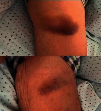
- Ecchymoses: 1 to 2 cm or larger subcutaneous hematomas, or "bruises".
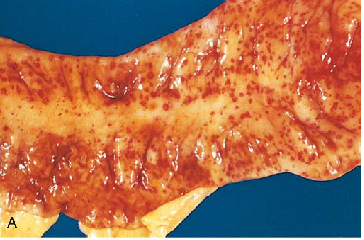
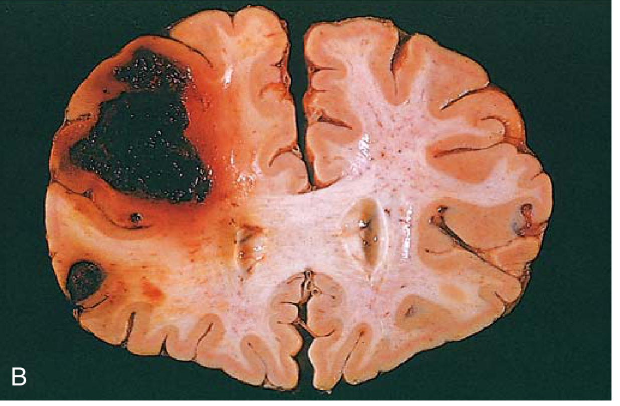
Robbins Fig. 3.4: (A) punctate petechial hemorrhages of the colonic mucosa in thrombocytopenia; (B) fatal intracerebral hemorrhage.
Evolution of a bruise (microscopic and biochemical)
Extravasated red cells are phagocytosed and degraded by macrophages. The colors change as follows:
- Hemoglobin: red-blue.
- Bilirubin: blue-green.
- Hemosiderin: golden-brown. Iron-laden macrophages (siderophages) are seen on microscopy and stain with Prussian blue.
Large hemorrhages can break down enough hemoglobin to cause jaundice. Persistent hemosiderin staining remains after old hemorrhage.
Clinical consequences
- Acute massive loss (roughly more than 20% of blood volume) causes hypovolemic shock. Hemodynamic compensation is overwhelmed, and hypoperfusion can lead to organ injury, especially of the kidney, heart and brain.
- Chronic or recurrent external loss (for example GI or menstrual) causes iron deficiency anemia. Blood lost into tissue keeps its iron, which is recycled.
- Critical sites: intracranial bleeding raises pressure and risks herniation. Hemopericardium causes tamponade. Hemothorax compromises ventilation.
Currency check
A quick PubMed search of recent systematic reviews (last 2 years) found only site-specific work. Examples are hemorrhagic progression of traumatic brain contusions and bleeding in treated head and neck cancer. None contradicts these basic pathology concepts.
If you want, I can go deeper on hemorrhagic diatheses (platelet versus coagulation disorders), on the lab workup (PT, aPTT, platelet count, bleeding time), or on the classes of hemorrhagic shock.
Compare platelet vs coagulation factor bleeding disorders in a table
Finding Sources
Reading File
Platelet (primary hemostasis) vs coagulation factor (secondary hemostasis) bleeding disorders
| Feature | Platelet / primary hemostasis defects | Coagulation factor / secondary hemostasis defects |
|---|---|---|
| Basic defect | Too few platelets (thrombocytopenia), or platelets that don't work (adhesion or aggregation defect). Von Willebrand disease is grouped here. | Deficiency or dysfunction of clotting factors, so the fibrin clot does not form or stabilize. |
| Typical causes | ITP, TTP/HUS, marrow failure, drug-induced thrombocytopenia, DIC (consumption), aspirin and NSAIDs, uremia, von Willebrand disease, Glanzmann thrombasthenia, Bernard-Soulier syndrome | Hemophilia A (factor VIII) and B (factor IX), vitamin K deficiency, liver disease, warfarin and heparin, DIC (consumption), factor deficiencies (II, V, VII, X, XI) |
| Inheritance | Mostly acquired. Inherited forms include von Willebrand disease, Glanzmann thrombasthenia and Bernard-Soulier syndrome. | Inherited forms are often X-linked recessive (hemophilia A and B). Acquired forms are common (liver disease, vitamin K deficiency, anticoagulants). |
| Site of bleeding | Skin and mucous membranes | Deep tissues: muscle, joints, soft tissue |
| Characteristic lesions | Petechiae (1 to 2 mm), purpura (3 mm or larger), epistaxis, gum bleeding, GI bleeding, menorrhagia | Large ecchymoses, deep hematomas, hemarthrosis after minor trauma |
| Timing after injury | Immediate bleeding. Platelets normally seal capillary defects at once, so bleeding starts right after the injury and tends to stop with pressure. | Often delayed or recurrent. The initial platelet plug forms, but without a stable fibrin clot the bleeding resumes later. |
| Response to local pressure | Usually controlled | Often poorly controlled |
| Feared complication | Intracerebral hemorrhage at very low platelet counts | Intracranial hemorrhage, joint destruction (hemophilic arthropathy) |
| Platelet count | Low in thrombocytopenia, normal in platelet function disorders | Normal (except in DIC) |
| Bleeding time / PFA-100 | Prolonged | Normal |
| PT | Normal | Prolonged if the extrinsic or common pathway is involved (factor VII, X, V, II, fibrinogen; warfarin; liver disease; vitamin K deficiency) |
| aPTT | Normal (prolonged in von Willebrand disease because factor VIII is reduced) | Prolonged if the intrinsic or common pathway is involved (hemophilia A and B, heparin) |
| Mixing study | Not applicable | Corrects with a factor deficiency and does not correct with an inhibitor |
Key points
- Mnemonic: platelet defects cause bleeding "on the surface" (skin, mucosa). Coagulation defects cause bleeding "in the depths" (joints, muscle).
- Overlap exists. Von Willebrand disease affects both limbs: vWF is needed for platelet adhesion and also carries and protects factor VIII. It can give a mixed picture.
- DIC causes both patterns. Platelets and factors are consumed, so PT, aPTT and the platelet count are all abnormal.
- Vascular disorders are a third group, not shown in the table. Scurvy, vasculitis and amyloid give palpable purpura and ecchymoses with a normal platelet count and normal coagulation tests.
- Mild bleeding tendencies: the most common causes are inherited vWF defects, aspirin and uremia.
Sources
- Robbins, Cotran & Kumar Pathologic Basis of Disease, "Hemorrhagic Disorders" (the bleeding patterns, hemarthrosis in hemophilia, intracerebral hemorrhage in thrombocytopenia, vWF, aspirin and uremia as common mild causes).
- Frameworks for Internal Medicine, case on fever and skin rash (platelet disorders present with petechiae and purpura, coagulation disorders with ecchymoses or hemarthrosis, and von Willebrand disease gives a mixed picture).
- The lab rows (bleeding time, PT, aPTT, mixing study), the delayed-bleeding row and the causes lists come from standard hematology knowledge, not directly from the retrieved passages.
I can add a short differential table for specific disorders (ITP vs TTP vs DIC vs hemophilia vs vWD) if you want one.
Shock. Etiology and classification. Pathogenesis of different types of shock (compensation)
Finding Sources
Finding Sources
Searching Images
shock pathophysiology compensatory mechanisms stages diagram
Searching PubMed
"shock"[MeSH Terms] AND "classification"
Reading File
Reading File
Reading File
Shock: etiology, classification and pathogenesis
Definition
Shock is circulatory failure that impairs tissue perfusion and causes cellular hypoxia. Early injury is reversible, but prolonged shock leads to irreversible tissue injury and death. - Robbins, Cotran & Kumar Pathologic Basis of Disease, Ch. 4
Etiology and classification
Robbins groups shock into three major categories, plus two less common ones. Physiology and critical care texts add a fourth category, obstructive shock. Together these map onto the four hemodynamic types: hypovolemic, cardiogenic, obstructive and distributive.
| Type | Primary problem | Causes | Hemodynamic profile |
|---|---|---|---|
| Hypovolemic | Low blood or plasma volume, so low preload | Hemorrhage, vomiting, diarrhea, burns, trauma, third-space loss | Low CO, high SVR, low CVP and PCWP |
| Cardiogenic | Pump failure | MI, ventricular rupture, arrhythmia, cardiomyopathy, acute valve failure | Low CO, high SVR, high CVP and PCWP |
| Obstructive (Robbins lists these under cardiogenic) | Extrinsic compression or outflow obstruction | Cardiac tamponade, massive pulmonary embolism, tension pneumothorax | Low CO, high SVR, high CVP. PCWP is low in PE and tamponade is variable. |
| Distributive: septic | Cytokine-mediated vasodilation, endothelial injury, microvascular thrombosis | Overwhelming bacterial or fungal infection; also viral infection | Usually high CO early with low SVR, then myocardial depression |
| Distributive: anaphylactic | IgE-mediated mast cell degranulation, with systemic vasodilation and increased vascular permeability | Drugs, foods, venoms | Low SVR, low preload |
| Distributive: neurogenic | Loss of sympathetic tone | Spinal cord injury, high spinal or epidural anesthesia | Low SVR, venous pooling, often bradycardia |
The terms sepsis, septic shock and SIRS are defined in Robbins as follows:
- Sepsis: life-threatening organ dysfunction caused by a dysregulated host response to infection.
- Septic shock: a subset of sepsis with profound circulatory, cellular and metabolic abnormalities and higher mortality.
- SIRS: a sepsis-like state triggered by nonmicrobial insults such as burns, trauma or pancreatitis.
Stages and compensation
Shock evolves through three stages. They were described most clearly in hypovolemic shock but apply to other forms too. - Robbins, Cotran & Kumar Pathologic Basis of Disease, Ch. 4, "Stages of Shock"
1. Nonprogressive (compensated) stage
Reflex mechanisms maintain cardiac output, blood pressure and vital organ perfusion. These mechanisms are:
- Baroreceptor reflexes and generalized sympathetic stimulation.
- Catecholamine release (adrenaline, noradrenaline).
- ADH release.
- Renin-angiotensin-aldosterone activation.
The net effects are:
- Tachycardia.
- Peripheral vasoconstriction. In the skin this causes the cool, pale "shocky" appearance.
- Renal fluid conservation.
- Preferential perfusion of the heart and brain. Coronary and cerebral vessels respond poorly to sympathetic signals, so their caliber and flow stay near normal.
Further compensatory mechanisms known from physiology texts:
- Capillary fluid shift. Falling hydrostatic pressure draws interstitial fluid into the vessels.
- Venoconstriction. This mobilizes blood from the venous reservoir.
- Respiratory compensation. Hyperventilation offsets metabolic acidosis.
2. Progressive stage
- Compensation fails and widespread tissue hypoxia develops.
- Aerobic respiration is replaced by anaerobic glycolysis, with excess lactic acid and metabolic acidosis.
- Acidosis blunts the vasomotor response. Arterioles dilate and blood pools in the microcirculation.
- Pooling reduces cardiac output further, and anoxic endothelial injury can trigger DIC.
- Vital organs begin to fail.
3. Irreversible stage
- Cell injury is so severe that survival is impossible even if the hemodynamic defect is corrected.
- Lysosomal enzymes leak from cells.
- Myocardial contractility falls, partly because of increased nitric oxide synthesis.
- Ischemic bowel lets intestinal flora enter the blood, so bacteremic shock may be superimposed.
- Renal failure follows ischemic kidney injury, and death ensues.
Pathogenesis by type
Hypovolemic shock
- Lost volume reduces venous return, preload and cardiac output.
- All the compensatory mechanisms above are fully engaged, so this form shows the stages most clearly.
- Rapid loss of up to about 20% of blood volume may have little effect in a healthy adult. Greater losses cause shock. - Robbins, Cotran & Kumar Pathologic Basis of Disease, Ch. 4
Cardiogenic shock
- The pump fails (infarction, arrhythmia, tamponade, outflow obstruction), so cardiac output falls despite adequate volume.
- The sympathetic and RAAS responses raise afterload and heart rate, which increases myocardial oxygen demand. This worsens ischemia and creates a vicious cycle.
- Venous congestion produces pulmonary edema.
Septic shock
This is the most complex form (Robbins, Fig. 4.20). The main elements are:
- Innate immune activation. PAMPs (endotoxin, enterotoxins, M protein) and DAMPs bind TLRs and other receptors.
- NF-kB activation increases TNF, IL-1, IL-12, IL-18, IFN-gamma and HMGB1.
- Reactive oxygen species, prostaglandins and PAF are also produced.
- Complement is activated (C3a, C5a).
- Endothelial activation and injury. This causes:
- Vasodilation (nitric oxide, prostaglandins), increased vascular permeability and edema.
- Pooling of blood and relative hypovolemia.
- Coagulation activation. Microbial products activate factor XII and tissue factor. The result is microvascular thrombosis and DIC.
- Counter-regulatory immunosuppression. Anti-inflammatory cytokines, lymphocyte apoptosis and anergy can occur, so patients may oscillate between hyperinflammatory and immunosuppressed states.
- Metabolic and cellular abnormalities. These include insulin resistance and hyperglycemia, adrenal insufficiency, and mitochondrial dysfunction that causes cytopathic hypoxia.
- Myocardial depression.
- Skin findings. Early septic shock can cause cutaneous vasodilation, so the skin may be warm and flushed rather than cool.
- Outcome. Organ failure follows. Mortality is about 40% despite treatment. Gram-positive bacteria are the most common trigger, followed by gram-negative bacteria and fungi.
Neurogenic and anaphylactic shock
- Acute vasodilation causes hypotension and tissue hypoperfusion. - Robbins, Cotran & Kumar Pathologic Basis of Disease, Ch. 4
- In anaphylaxis, the mediators are released from mast cells (histamine, leukotrienes and others).
- In neurogenic shock, loss of sympathetic vascular tone leaves the compensation incomplete. Bradycardia may occur instead of tachycardia.
Morphology
Changes are those of hypoxic injury from hypoperfusion plus microvascular thrombosis. - Robbins, Cotran & Kumar Pathologic Basis of Disease, Ch. 4
- Organs most involved: brain, heart, kidneys, adrenals and GI tract.
- Fibrin thrombi: seen in any tissue, most readily in glomeruli.
- Adrenal cortex: cell lipid depletion, which reflects the stress response.
- Lungs: resistant to hypoxia in hypovolemic shock, but sepsis or trauma can cause diffuse alveolar damage ("shock lung").
- Kidney: acute tubular necrosis.
- Heart: subendocardial ischemic necrosis and contraction band necrosis.
- Recovery: except for neurons and cardiomyocytes, which are lost, tissues can recover completely if the patient survives.
Currency check
A PubMed search of systematic reviews from the last 2 years found only prognostic and diagnostic work: shock index in trauma and ACS, and point-of-care ultrasound in shock. Nothing there contradicts the pathogenesis described above.
I can go deeper on septic shock mediators, the hemodynamic differentiation table, or hemorrhagic shock classes if you want.
This is a shared conversation. Sign in to Orris to start your own chat.