
Treatment of rheumatoid arthritis
"rheumatoid arthritis"[MeSH Terms] AND "guideline"[pt]
rheumatoid arthritis hand deformities X-ray
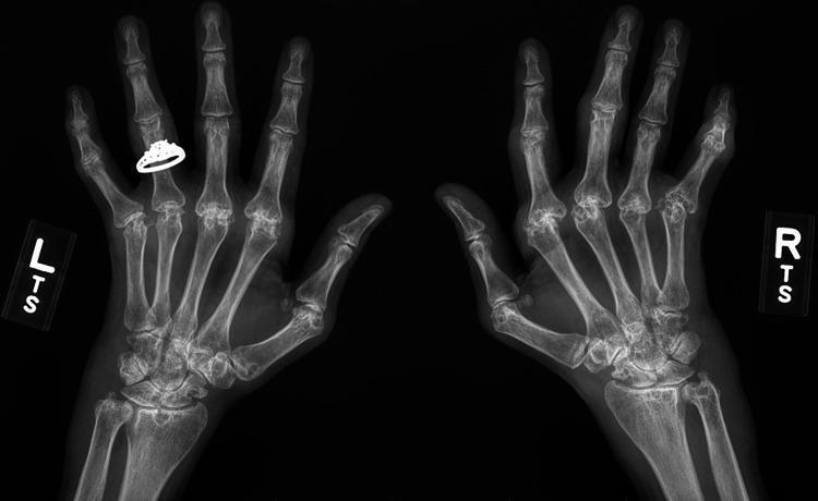
This diagnostic image is a posteroanterior (PA) bilateral hand and wrist X-ray demonstrating advanced erosive arthritis consistent with long-standing rheumatoid arthritis. Key findings include symmetric involvement of the metacarpophalangeal (MCP) joints and carpal bones. There is significant joint space narrowing, periarticular osteopenia, and marginal bone erosions particularly visible at the second and third MCP joints. Structural deformities are evident, characterized by ulnar deviation of the digits at the MCP joints and carpal crowding with secondary osteoarthritic changes in the wrists. A radiopaque ring is visible on the fourth digit of the left hand. The image serves as a clinical example of progression in treatment-resistant inflammatory arthritis, illustrating the irreversible joint destruction and malalignment that occurs in chronic seropositive rheumatoid arthritis.
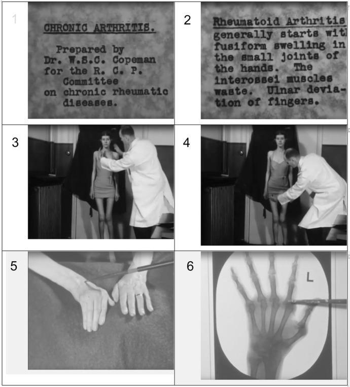
This composite educational material consists of six stills from a historical medical film titled 'Chronic Arthritis' (1938), depicting clinical and diagnostic aspects of Rheumatoid Arthritis (RA). Panels 1 and 2 provide textual context, identifying RA features such as fusiform swelling of small hand joints, interossei muscle wasting, and ulnar deviation of the fingers. Panels 3 and 4 show a clinician in a white coat performing a physical examination on a standing patient, focusing on the shoulder/clavicular region and the knee joint, respectively, to assess range of motion and deformities. Panel 5 is a clinical photograph showing a close-up of a patient's hands with a pointer highlighting fusiform swelling and classic RA hand deformities. Panel 6 displays a diagnostic X-ray of the left hand, with a pointer indicating potential periarticular osteopenia or joint space narrowing in the metacarpophalangeal (MCP) or proximal interphalangeal (PIP) joints. This visual sequence is intended for medical education, demonstrating the transition from clinical signs to radiographic confirmation in rheumatology.
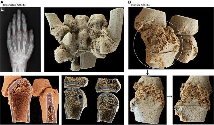
This composite educational image provides a comparative visual analysis of bone pathology in Rheumatoid Arthritis (RA) and Psoriatic Arthritis (PsA) using conventional radiography and high-resolution CT-based cinematic rendering. Panel A focuses on RA, showing a standard hand X-ray with highlighted carpal and metacarpophalangeal joint involvement, paired with 3D renderings that demonstrate significant marginal erosions, carpal bone deformities, and trabecular demineralization characteristic of chronic inflammation and joint destruction. Panel B illustrates PsA, utilizing photorealistic cinematic renderings to showcase the distinct dual processes of bone erosion and pathological new bone formation (proliferation). The high-resolution cross-sectional views reveal the 'inner life' of the bone, highlighting pitted surfaces and cortical irregularities. This visual comparison serves as a diagnostic educational tool for identifying the predominately destructive nature of RA versus the mixed erosive-proliferative features of PsA in the small joints of the hand. It is intended for rheumatology and radiology education to enhance the understanding of structural remodeling in inflammatory arthritides.
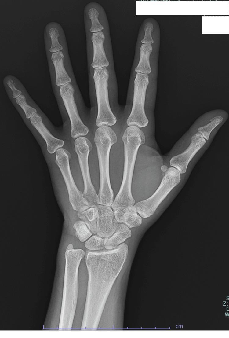
A posterior-anterior (PA) diagnostic X-ray of the left hand. The imaging demonstrates the distal radius and ulna, carpal bones, metacarpals, and phalanges in normal anatomical alignment. The joint spaces, including the radiocarpal, intercarpal, metacarpophalangeal (MCP), and proximal/distal interphalangeal (PIP/DIP) joints, appear well-preserved with no evidence of joint space narrowing, erosions, or subchondral sclerosis. The bone mineralization and cortical thickness are within normal limits. There are no periarticular osteopenia, marginal erosions, or joint deformities, such as ulnar drift or boutonnière deformity, which are typical of advanced rheumatoid arthritis. The surrounding soft tissues are unremarkable with no signs of swelling or calcification. This image serves as a baseline comparison for inflammatory arthropathies or to rule out structural bone disease in patients presenting with arthralgia or morning stiffness.
Treatment of Rheumatoid Arthritis (RA)
Goals and Overall Strategy
1. Non-pharmacologic measures
- Patient education, smoking cessation (smoking worsens disease severity and reduces DMARD response)
- Physical and occupational therapy to preserve joint function and muscle strength
- Splinting and assistive devices for affected joints
2. Bridging therapy (symptom control while DMARDs take effect)
- NSAIDs: control pain/stiffness but do not alter disease progression; watch for GI and cardiovascular risk
- Glucocorticoids (e.g., low-dose prednisone): rapid anti-inflammatory bridge during DMARD initiation, or for flares. Long-term use raises risk of osteoporosis (glucocorticoid-induced), so bisphosphonates, denosumab, or teriparatide should be considered for prevention in higher-risk patients - Harrison's, Ch. 370.
3. Conventional synthetic DMARDs (csDMARDs) - first line
| Drug | Notes |
|---|---|
| Methotrexate | DMARD of choice; anchor drug for combination therapy; weekly oral/SC dosing with folic acid co-therapy to reduce toxicity; onset ~6-12 weeks |
| Leflunomide | Pyrimidine synthesis inhibitor; efficacy similar to methotrexate, used alone or combined |
| Sulfasalazine | Reduces joint inflammation and radiographic progression |
| Hydroxychloroquine | Used for mild/early disease or as an adjunct; not a true disease-modifying agent since it doesn't slow radiographic progression; dosed ≤5 mg/kg to limit retinal toxicity |
4. Biologic DMARDs (added when csDMARD response is inadequate)
- TNF inhibitors (etanercept, infliximab, adalimumab, certolizumab, golimumab) - first biologics approved (1999), often combined with methotrexate for superior efficacy over monotherapy
- Abatacept (T-cell costimulation blocker, blocks CD28-CD80/86 interaction) - the AGREE trial in MTX-naive early erosive RA showed higher DAS28-CRP remission (41.4% vs 23.3%) and less radiographic progression than methotrexate alone; also showed a better safety profile than infliximab (fewer serious infections, adverse events) - Rheumatology, 2-Volume Set (2022, Elsevier), p. 627.
- Rituximab (anti-CD20, B-cell depletion) - typically for TNF-inhibitor failures
- Tocilizumab/sarilumab (IL-6 receptor blockers) - can be used as monotherapy if MTX is not tolerated
- Biosimilars - now widely available with comparable efficacy/safety to reference biologics
5. Targeted synthetic DMARDs
- JAK inhibitors (tofacitinib, baricitinib, upadacitinib) - oral small molecules for csDMARD/biologic failures; carry a boxed warning for cardiovascular events, malignancy, and thrombosis in at-risk populations (per FDA safety review), so are generally reserved after TNF-inhibitor failure in higher-risk patients
6. Surgical management
Monitoring and safety
Recent guideline evidence
- GUIPCAR 2025 Spanish guidelines (PMID: 41535165) and the 2024 Chinese RA guidelines (PMID: 39802551) reaffirm methotrexate as first-line therapy with early treat-to-target escalation to biologic/targeted synthetic DMARDs.
- The Canadian Rheumatology Association Living Guidelines (PMID: 37527865) and a 2023 systematic review of core RA recommendations (PMID: 37291382) similarly emphasize methotrexate, hydroxychloroquine, and sulfasalazine as core conventional agents, consistent with the textbook material above - no major contradictions found, but these are worth reviewing directly if you need the latest dosing/sequencing nuances.
Methotrexate dosage and usage
methotrexate rheumatoid arthritis dosing
Methotrexate: Dosage and Usage in Rheumatoid Arthritis
Mechanism (brief)
Place in therapy
Dosage
- Route/schedule: Given as a single weekly dose (occasionally split into 3 doses 12 hours apart over 24 hours in some regimens) - never taken daily.
- Starting dose: Typically 7.5-15 mg/week orally, titrated ("ramped up") based on response and tolerability.
- Dose range: 5-25 mg/week. High-dose therapy (20-25 mg/week) is more effective than low-dose (5-15 mg/week), but dosing is escalated gradually while monitoring for dose-dependent toxicity - Goldman-Cecil Medicine, Ch. 28, p. 2806.
- Route options: Oral absorption is dose-dependent and variable between individuals. Subcutaneous injection improves bioavailability, reduces GI side effects, and is often effective when oral therapy has a suboptimal response or is poorly tolerated.
- Renal adjustment: Methotrexate is cleared by glomerular filtration and active tubular secretion, so the dose must be reduced in renal impairment (including the elderly), and previously stable patients can develop toxicity if renal function worsens.
Folic acid co-therapy
Combination use
- Oral triple therapy: methotrexate + hydroxychloroquine + sulfasalazine, used when methotrexate monotherapy is insufficient.
- Biologic/targeted synthetic combinations: methotrexate is commonly continued alongside a TNF inhibitor or JAK inhibitor, since combination therapy generally outperforms biologic monotherapy.
- It is also FDA-approved for polyarticular juvenile idiopathic arthritis and psoriatic arthritis, where it treats peripheral arthritis/psoriasis but not enthesitis, dactylitis, or axial disease.
Toxicity and monitoring
| Toxicity | Monitoring/Management |
|---|---|
| Hepatotoxicity | CBC and AST/ALT at baseline, then every 8-12 weeks once dose is stable (monthly for the first 3 months and after dose changes per some protocols) |
| Bone marrow suppression | Same CBC monitoring; dose reduction or folic acid usually resolves mild cytopenias |
| Pneumonitis | Rare but can be fatal - if it occurs, stop methotrexate and do not restart |
| Oral ulcers, GI upset | Often improved with folic acid or dose reduction |
| Renal function | Monitor periodically since reduced clearance precipitates toxicity |
| Infection risk | Immunosuppressive effect increases susceptibility |
Contraindications
- Absolute contraindication in pregnancy (teratogenic, can cause fetal death) and breastfeeding - Goldman-Cecil Medicine; Harrison's.
- Avoid in significant renal impairment or pre-existing liver disease; avoid alcohol while on therapy.
- Use caution in diabetic or obese patients (higher hepatotoxicity risk, notably documented in psoriasis populations).
Recent evidence context
A 65 year old female present with rheumatoid arthritis with slightly obese
rheumatoid arthritis elderly onset treatment
Case Considerations: 65-Year-Old Obese Female with Rheumatoid Arthritis
1. Confirm the diagnosis and rule out mimics
2. Baseline workup before starting therapy
- CBC, renal function (eGFR - critical in this age group), LFTs, albumin
- Hepatitis B/C screening, TB screening (if biologics being considered)
- Lipid panel, blood pressure, fasting glucose (obesity + RA both independently raise cardiovascular risk, and this compounds)
- Baseline DEXA scan - postmenopausal women have elevated osteoporosis/fracture risk, which is further increased by RA-related inflammation and any glucocorticoid use
3. DMARD therapy - methotrexate with age-adjusted caution
- Start at a lower initial dose (e.g., 7.5-10 mg/week) rather than the higher end of range, since renal clearance of methotrexate declines with age and toxicity risk rises accordingly.
- Titrate slowly while monitoring CBC and AST/ALT every 8-12 weeks once stable (more frequently at initiation and after dose changes).
- Folic acid co-therapy (1-4 mg/day or 5 mg/week) as usual to reduce GI/hepatic toxicity.
- Renal function should be rechecked regularly - previously stable elderly patients can develop toxicity if GFR falls - Goldman-Cecil Medicine, Ch. 243.
4. Bridging glucocorticoids - use cautiously
5. NSAIDs - minimize use
6. Obesity-specific implications
- Obesity is independently associated with higher RA disease activity and lower rates of achieving remission, and with reduced response to TNF-inhibitor biologics specifically (pharmacokinetic dilution and inflammatory adipokine effects) - Rheumatology, 2-Volume Set (2022, Elsevier); Firestein & Kelley's Textbook of Rheumatology.
- Weight management (diet, supervised exercise/physical therapy) should be part of the plan - it improves both disease activity and cardiovascular risk profile, and may improve response to therapy.
- Cardiovascular risk from RA and obesity are additive - lipid and blood pressure monitoring should be routine, and statin therapy considered per standard CV risk calculators adjusted for RA (many guidelines apply a multiplier to standard risk scores in RA patients).
7. If escalation to biologic/targeted therapy is needed
- TNF inhibitors are generally reasonable, though obesity may blunt response - watch disease activity scores closely and consider dose/interval adjustment or an alternative mechanism (e.g., abatacept, IL-6 inhibitor) if response is inadequate.
- JAK inhibitors (tofacitinib, baricitinib, upadacitinib) warrant more caution here: the ORAL Surveillance trial and subsequent FDA boxed warning identified increased risk of major cardiovascular events, malignancy, venous thromboembolism, and death specifically in patients ≥65 years old (and current/past smokers). Given her age, JAK inhibitors would typically be reserved for after TNF-inhibitor failure, with a frank discussion of this risk, rather than used as an early-line biologic choice.
Monitoring summary for this patient
| Domain | Frequency |
|---|---|
| CBC, AST/ALT | Every 8-12 weeks (methotrexate) |
| Renal function | Periodically, more often if declining |
| Bone density | Baseline, then per osteoporosis risk protocol |
| Lipids/BP/glucose | At baseline and periodically, especially with IL-6 inhibitors or JAK inhibitors |
| Disease activity (DAS28/CDAI) | Every visit, treat-to-target escalation every ~3 months if not at target |