

Patient with age 30 years old blackish on neck with no pain no irritation and no swelling , what should prescribe for removing Blackesh
acanthosis nigricans neck hyperpigmentation velvety
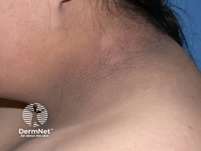
Clinical photography of the neck demonstrates acanthosis nigricans: hyperpigmented, velvety plaques within intertriginous regions of the cervical skin. The image shows a brown-to-dark brown, slightly pigmented area along the posterior and lateral neck folds, with smooth but thickened dermal-epidermal junction and papillomatosis consistent with acanthosis nigricans. The texture appears velvety on close inspection, and the surrounding skin remains comparatively lighter. There is no visible mucosal involvement or ulceration. The pattern is typically bilateral and symmetric, favoring the neck and adjacent flexural areas; extension to axillae or inguinal folds may be observed in some patients. This dermatologic finding is commonly associated with insulin resistance, obesity, type 2 diabetes mellitus, endocrine disorders, or, in rare adults, paraneoplastic syndromes. Clinically, acanthosis nigricans serves as a cutaneous sign prompting assessment of metabolic health, including fasting glucose, HbA1c, lipid profile, and weight management. The image is intended for educational purposes to illustrate morphological features such as hyperpigmentation, velvety texture, and intertriginous distribution; it aids differential diagnosis including frictional hyperpigmentation and postinflammatory changes. Potential uses include dermatology training, primary care screening, and research on metabolic disease correlations. Limitations: single-view photograph cannot assess texture depth or extent; clinical history crucial for confirming etiologies and guiding management decisions.
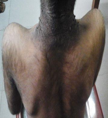
A clinical photograph capturing the posterior neck, upper back, and proximal arms of a patient, demonstrating classic manifestations of acanthosis nigricans and generalized hypertrichosis. The posterior neck displays prominent hyperpigmentation and velvety skin thickening, characteristic of acanthosis nigricans. The texture in this region appears coarse and verrucous with a cobblestone-like morphology. Concurrent with these skin changes is generalized hypertrichosis, marked by an abnormal density of fine, dark hair covering the shoulders, upper back, and posterior arms. These findings are clinically significant as they often serve as cutaneous markers for underlying metabolic or endocrine disorders, such as severe insulin resistance or lipodystrophy syndromes. The image is a valuable educational resource for dermatology and endocrinology, illustrating the co-occurrence of paraneoplastic or metabolic skin signs.
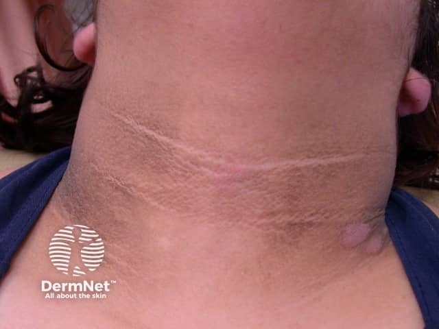
Clinical photography of acanthosis nigricans affecting the anterior neck. Modality: Clinical photography; Technique: close‑up, high‑resolution frontal view of the cervical skin with even illumination. Observed features include diffuse hyperpigmentation and velvety, slightly thickened plaques aligned with neck flexures, most prominent in the anterior cervical region and along the nape-to-thyroid area. Surface texture is smooth to slightly papillomatous with mild pleating; surrounding skin is comparatively lighter. No ulceration, edema, or exudate. The presentation is symmetric and localized to the neck in this image, consistent with classic acanthosis nigricans rather than post‑inflammatory hyperpigmentation alone. Pathophysiology reflects epidermal hyperplasia with papillomatosis and increased melanin deposition in the stratum basale, secondary to insulin resistance–driven growth factor signaling in susceptible individuals. Clinically, acanthosis nigricans serves as a cutaneous marker for metabolic syndrome, obesity, type 2 diabetes mellitus, and less commonly paraneoplastic processes. Differential considerations include frictional dermatitis, dermatitis neglecta, or other causes of hyperpigmented neck plaques. Diagnostic significance lies in prompting metabolic evaluation and search for underlying endocrine or oncologic disease. Useful in dermatology education, primary care triage, and research on cutaneous signs of systemic disease. Correlation with body mass index and fasting glucose improves diagnostic confidence and guides therapeutic lifestyle interventions and monitoring thereafter.
"acanthosis nigricans" AND treatment
Diagnosis: Acanthosis Nigricans (AN)
Step 1 - Rule Out Underlying Cause (Mandatory)
| Investigation | Reason |
|---|---|
| Fasting blood glucose + HbA1c | Insulin resistance / prediabetes / T2DM (most common cause in a 30-year-old) |
| Fasting insulin + HOMA-IR | Confirm insulin resistance |
| Lipid profile | Metabolic syndrome screen |
| BMI + waist circumference | Obesity-related AN |
| Thyroid function (TSH) | Hypothyroidism-associated AN |
| Androgens / LH:FSH (if female) | Rule out PCOS |
| Review medications | Steroids, OCP, niacin, insulin can all cause AN |
In a 30-year-old with no symptoms, insulin resistance is the most likely driver. The skin change is a metabolic warning sign.
Step 2 - Treat the Root Cause
- Weight reduction - even 5-10% body weight loss can visibly reduce AN
- Metformin - if insulin resistance or prediabetes is confirmed; also directly reduces AN severity
- Dietary changes - low glycaemic index diet, reduce refined carbohydrates
- Exercise - improves insulin sensitivity
Step 3 - Topical Treatments for Cosmetic Improvement
| Agent | How to Use | Evidence |
|---|---|---|
| Topical Tretinoin 0.025-0.05% cream | Apply nightly, thin layer to affected area | Good evidence; reduces hyperkeratosis |
| Ammonium Lactate 12% lotion (lactic acid) | Apply BD | Softens/lightens the thickened skin |
| Salicylic acid 6% cream/lotion | Apply once daily | Keratolytic; reduces thickness |
| Glycolic acid peels (20-30%) | Dermatologist-performed, 3-6 sessions | Useful for cosmetic improvement |
| Urea 10-20% cream | Apply BD | Moisturises and softens skin |
| Calcipotriol 0.005% ointment | Apply OD/BD | Evidence from case series |
Sample Prescription
1. Tab. Metformin 500 mg BD with meals (if insulin resistance confirmed)
2. Tretinoin 0.025% cream - apply to neck every night (thin layer)
3. Ammonium Lactate 12% lotion - apply morning
4. Sunscreen SPF 30+ on neck during daytime
- This is not dirt - it cannot be washed off
- Minimum 8-12 weeks to see improvement with topicals
- Metabolic workup is mandatory - skin will not clear unless underlying cause is addressed
- Avoid friction/rubbing the neck skin
Red Flags to Rule Out
- Rapid onset AN + weight loss + abdominal symptoms = may rarely signal paraneoplastic AN (gastric/other adenocarcinoma) - though this is rare at 30
- Extensive mucosal involvement = also warrants malignancy workup
How to use
What would you like instructions on?
How to Apply Tretinoin Cream for Acanthosis Nigricans (Neck)
Strength to Use
- Start with 0.025% (lowest strength) - the neck skin is sensitive
- Can step up to 0.05% after 4-6 weeks if tolerated well
Step-by-Step Application
Frequency Schedule (Start Low, Go Slow)
| Week | Frequency |
|---|---|
| Week 1-2 | Every other night (alternate nights) |
| Week 3-4 | Every night if no irritation |
| Week 5 onwards | Every night (maintenance) |
Important Instructions for Patient
- Do NOT apply near eyes, nostrils, or mouth
- Always use sunscreen SPF 30+ on the neck in the morning - tretinoin makes skin sun-sensitive; skipping sunscreen can worsen darkening
- Expect peeling, redness, or mild irritation in the first 2-4 weeks - this is normal (called "retinoid reaction")
- Do NOT stop because of mild peeling - reduce frequency to alternate nights and continue
- Avoid shaving over the area or wearing tight collars while on treatment
- Results take 8-12 weeks minimum - do not expect quick lightening
When to Stop / Seek Review
- Severe burning, blistering, or swelling - stop and review
- No improvement at all after 3 months - consider stepping up to 0.05% or adding glycolic acid peel