
Explain stomach anatomy
Reading File
Now let me search the medical library for stomach anatomy content in parallel:
Finding Sources
Finding Sources
Excellent results. Let me now read the most comprehensive anatomy sections in parallel:
Reading File
Reading File
Reading File
Now let me read more of the Sleisenger anatomy section and fetch the key images in parallel:
Reading File
Reading File
Loading Image
Now let me fetch a few more key images - the regions diagram and histology diagram:
Loading Image
Loading Image
Reading File
I now have comprehensive content from multiple authoritative textbooks. Let me compile the full answer.
Stomach Anatomy
The stomach is a J-shaped muscular dilation of the alimentary canal, continuous with the esophagus proximally and the duodenum distally. It serves as the primary food reservoir and begins protein digestion. Capacity ranges from ~30 mL in a neonate to 1.5-2 L in an adult.
Gross Anatomy and Regions
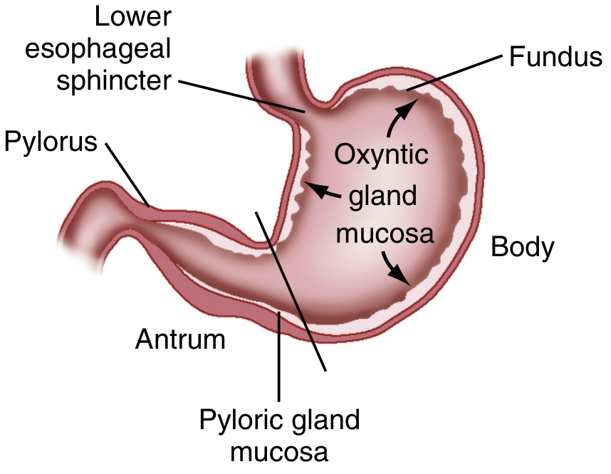
The stomach is divided into 4 anatomic regions:
-
Cardia - A small, ill-defined zone immediately adjacent to the esophagogastric junction. The esophagogastric junction lies at the level of T10, 1-2 cm below the diaphragmatic hiatus.
-
Fundus - The dome-shaped superior portion projecting upward above the cardia. It contacts the left hemidiaphragm superiorly and the spleen laterally.
-
Body (Corpus) - The largest region, located below and continuous with the fundus. The incisura angularis (a fixed sharp indentation two-thirds down the lesser curvature) marks the caudal limit of the body.
-
Antrum and Pylorus - The antrum extends from its indistinct border with the body to the pyloric junction. The pylorus is a tubular structure containing the palpable pyloric sphincter (circular muscle), located ~2 cm to the right of midline at L1. It controls gastric emptying into the duodenum.
Curvatures:
- Lesser curvature - right upper border; the incisura angularis is its most notable landmark
- Greater curvature - left lower border; may descend below the umbilicus when distended
Relations:
- Posterior: pancreas, transverse colon, diaphragm, spleen, left kidney and adrenal gland
- Anterior: liver (right lobe overlies the lesser curvature), anterior abdominal wall (left lower aspect)
- The posterior stomach wall forms the anterior wall of the omental bursa (lesser peritoneal sac)
Peritoneal Attachments and Ligaments
The stomach is entirely invested in peritoneum (except a small bare area at the esophagogastric junction). This peritoneum forms several named ligaments:
| Ligament | From | To |
|---|---|---|
| Gastrohepatic (lesser omentum) | Lesser curvature | Liver |
| Gastrocolic ligament | Greater curvature/fundus | Transverse colon |
| Gastrosplenic ligament | Greater curvature/fundus | Spleen |
| Gastrophrenic ligament | Fundus | Diaphragm |
| Greater omentum | Greater curvature | Hangs down (covers small intestine) |
Blood Supply
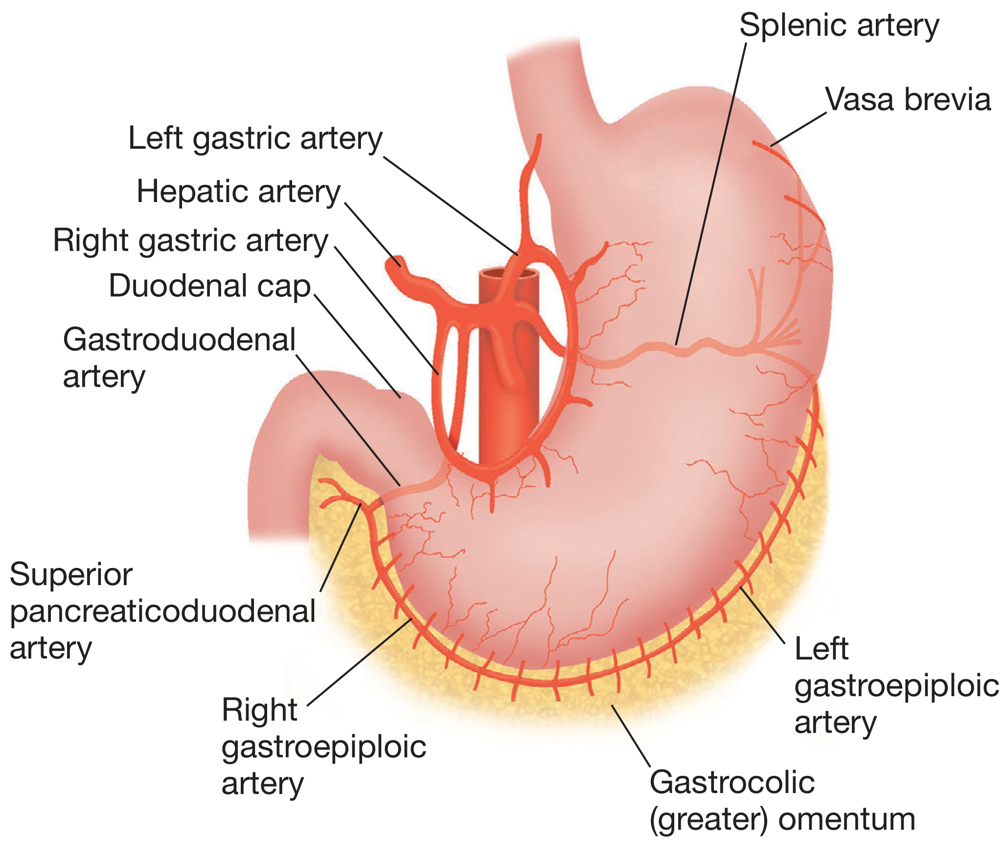
All gastric arteries derive from the celiac axis:
Lesser curvature arcade:
- Left gastric artery (direct celiac branch) - supplies the upper lesser curvature; gives branches toward the cardia
- Right gastric artery (from common hepatic artery) - anastomoses with the left gastric artery
Greater curvature arcade:
- Left gastroepiploic artery (from splenic artery) - supplies the upper greater curvature
- Right gastroepiploic artery (from gastroduodenal artery, itself a branch of common hepatic) - supplies the lower greater curvature; these two often anastomose to complete the arcade (though this arcade is sometimes variably incomplete)
Fundus:
- Vasa brevia (short gastric arteries) - multiple small branches arising near the terminal splenic artery; supply the fundus and upper left greater curvature
Venous drainage mirrors the arterial supply and drains into the portal system - lesser curvature veins into the portal vein, greater curvature veins into the splenic vein. The left gastric vein (coronary vein) is clinically important as it dilates markedly in portal hypertension, forming esophageal varices.
Lymphatic Drainage
Lymphatic channels anastomose freely within the gastric wall and ultimately drain to the celiac nodes via four regional pathways:
- Inferior gastric region → subpyloric and omental nodes → hepatic nodes → celiac nodes
- Splenic/superior greater curvature → pancreaticosplenic nodes → celiac nodes
- Superior gastric/lesser curvature → left and right gastric nodes → celiac nodes
- Pyloric/hepatic lesser curvature → suprapyloric nodes → hepatic nodes → celiac nodes
This extensive interconnection is why gastric cancer can spread early and widely.
Innervation
Parasympathetic (vagal):
- The right and left vagus nerves form the esophageal plexus, then coalesce into the posterior vagal trunk (predominantly right vagus) and anterior vagal trunk (predominantly left vagus) near the gastric cardia
- Each trunk gives off hepatic and celiac branches, then continues as the nerve of Latarjet (anterior and posterior) running along the lesser curvature
- The nerves of Latarjet supply the gastric body and end as the "crow's foot" at the antrum, stimulating motility and acid secretion
Sympathetic:
- Preganglionic fibers arise from T6-T8 spinal nerves, synapse in the bilateral celiac ganglia
- Postganglionic fibers travel along gastric vessels
- Includes afferent pain fibers and motor fibers to the pyloric sphincter
The embryological rotation of the stomach explains why the right vagus ends up innervating the posterior wall and the left vagus innervates the anterior wall.
Microscopic Anatomy (Histology)
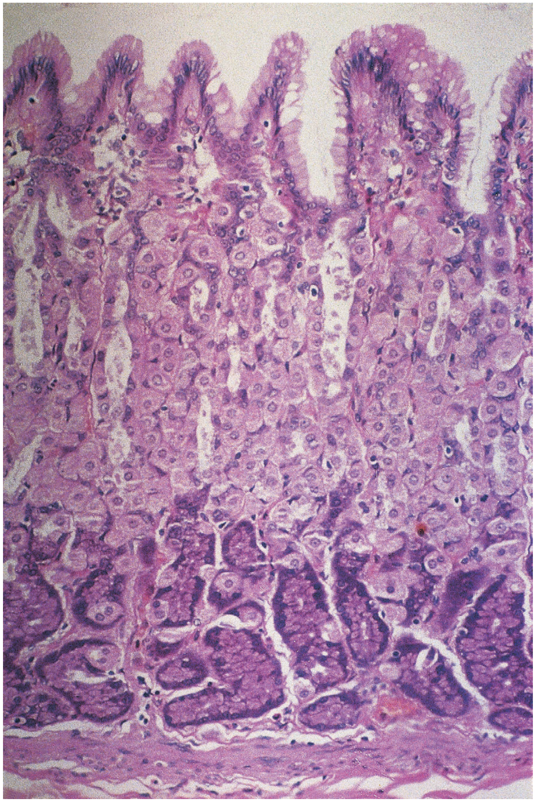
The gastric wall has the standard 4-layer GI structure (mucosa, submucosa, muscularis, serosa) with stomach-specific features:
Mucosal Layers and Gastric Glands
The mucosa is lined by a simple columnar epithelium that abruptly transitions from squamous esophageal epithelium at the cardia. Surface cells produce a highly viscous neutral mucus protecting the wall from self-digestion.
Glands are classified by region:
| Region | Gland Type | Key Cell Types | Secretions |
|---|---|---|---|
| Body & Fundus | Gastric glands proper (oxyntic glands) | Parietal cells, Chief cells, Mucous neck cells | HCl + intrinsic factor (parietal); pepsinogen (chief); mucus (neck) |
| Cardia | Cardiac glands | Mucous cells only | Mucus, lysozyme |
| Antrum/Pylorus | Pyloric glands | Mucous columnar cells, G cells | Neutral-to-slightly-acid mucus; gastrin (G cells) |
Parietal cells - Large, eosinophilic, triangular cells found in the body; they secrete H⁺ via the H⁺/K⁺-ATPase proton pump (the target of PPIs) against a concentration gradient of 1,000,000:1. They also secrete intrinsic factor, essential for vitamin B12 absorption.
Chief cells - Basophilic columnar cells at the base of body glands; produce pepsinogen I and II, which are activated to pepsin by the acidic environment. The pepsinogen I:II ratio falls with gastric atrophy.
Enteroendocrine cells:
- G cells (antrum) - produce gastrin, which stimulates acid secretion
- ECL cells (body) - produce histamine, a key local driver of acid secretion
- D cells (throughout) - produce somatostatin, which has a negative regulatory role
Muscular Layer
The stomach has 3 muscle layers (unlike the rest of the GI tract which has 2):
- Outer longitudinal layer - especially thick; runs along curvatures
- Middle circular layer - thickens at the pylorus to form the pyloric sphincter
- Inner oblique layer - unique to the stomach; helps with mechanical churning
The incisura angularis functionally separates the stomach into an upper digestive sac (fundus + body) and a lower pyloric canal with emptying function.
Embryology (Brief)
The stomach is recognizable in the 4th week of gestation as a dilation of the distal foregut. The dorsal aspect grows faster than the ventral, forming the greater curvature. The stomach then rotates 90 degrees around its longitudinal axis, placing the greater curvature to the left and lesser curvature to the right - this explains the vagal arrangement described above.
Sources:
- Sleisenger and Fordtran's Gastrointestinal and Liver Disease, Chapter 49 (Embryology and Anatomy of the Stomach)
- Bailey and Love's Short Practice of Surgery, 28th ed., Chapter 67 (Anatomy of the Stomach and Duodenum; Microscopic Anatomy)
- Color Atlas of Human Anatomy Vol. 2, Internal Organs (Microscopic Anatomy of the Stomach)
Explain Types of circulation
Reading File
Searching sources...
Finding Sources
Finding Sources
Finding Sources
Finding Sources
Reading File
Reading File
Finding Sources
Reading File
Finding Sources
Finding Sources
Reading File
Reading File
Loading Image
Loading Image
Finding Sources
Reading File
Reading File
I now have rich content from multiple authoritative physiology and anatomy textbooks. Let me compile the comprehensive answer.
Types of Circulation
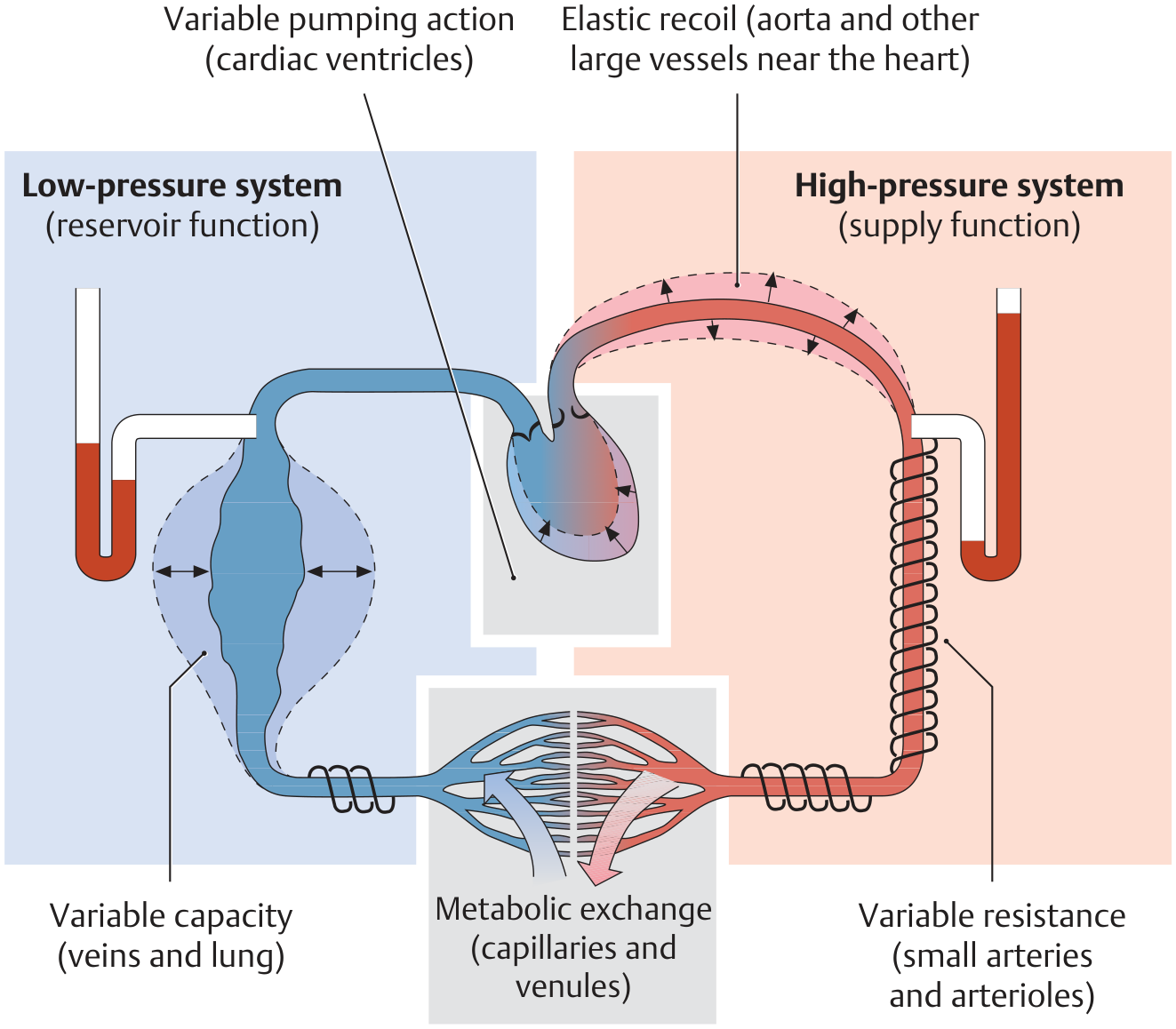
The circulatory system is a closed vascular system kept in motion by the heart's pumping action. Functionally it is divided into two main circuits - pulmonary and systemic - plus several specialized sub-circuits. Blood flows along a pressure gradient: mean arterial pressure ~100 mmHg in the high-pressure arterial system, falling to under 20 mmHg in the venous low-pressure system.
1. Pulmonary Circulation (Lesser Circulation)
Path: Right ventricle → Pulmonary arteries → Lung capillaries → Pulmonary veins → Left atrium
This is the short, low-pressure loop dedicated entirely to gas exchange.
- Deoxygenated venous blood from the entire body returns via the superior and inferior vena cava to the right atrium, then enters the right ventricle
- The right ventricle pumps blood through the pulmonary trunk and arteries to the lungs
- Pulmonary arteries travel alongside the bronchi all the way down to the respiratory bronchioles, keeping perfusion and ventilation in close proximity
- Gas exchange occurs across the enormous alveolar capillary surface (~50-70 m²)
- Oxygenated blood returns via pulmonary veins to the left atrium
Key feature - low pressure: Mean pulmonary arterial pressure is normally ≤15 mmHg (vs ~100 mmHg in the aorta). The entire pulmonary circuit is a low-pressure system; capillary pressures do not rise above ~10 mmHg.
Unique regulation: Unlike every other vascular bed, the pulmonary circulation responds to hypoxia with vasoconstriction (hypoxic pulmonary vasoconstriction). This shunts blood away from poorly ventilated alveoli toward well-ventilated zones, optimizing ventilation-perfusion matching.
2. Systemic Circulation (Greater Circulation)
Path: Left ventricle → Aorta → Arteries → Arterioles → Capillaries → Venules → Veins → Vena cava → Right atrium
This is the long, high-pressure loop that supplies every organ and tissue in the body.
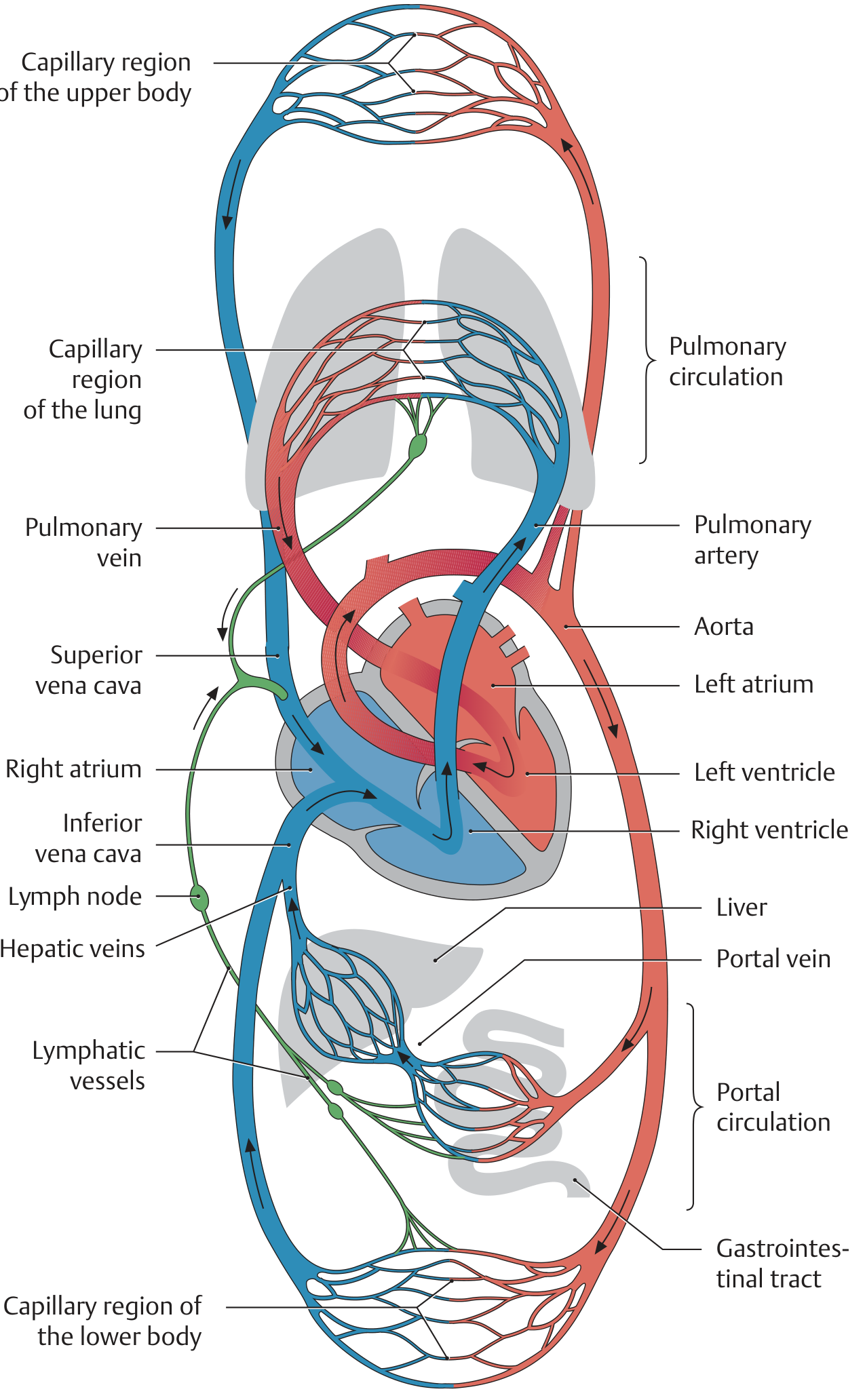
- Oxygenated blood from the left atrium enters the left ventricle, which pumps it into the aorta at ~120 mmHg systolic
- Blood distributes through arteries → arterioles → capillaries across all organs
- Arterioles are the principal site of vascular resistance - the steepest pressure drop in the systemic circulation occurs here (not at capillaries), because although individual capillary resistance is high, there are so many capillaries in parallel that aggregate arteriolar resistance dominates
- Exchange of O₂, CO₂, nutrients, and waste occurs in capillaries
- Deoxygenated blood returns through venules → veins → superior and inferior vena cava → right atrium
- Veins are capacitance vessels - they can hold ~80% of total blood volume and serve as the primary blood reservoir
Pressure Profile Summary
| Segment | Mean Pressure |
|---|---|
| Aorta (systemic arterial) | ~100 mmHg |
| Arterioles | ~35 mmHg (steepest drop) |
| Capillaries | ~25 mmHg |
| Veins | <20 mmHg |
| Pulmonary artery | ~15 mmHg |
| Pulmonary capillaries | <10 mmHg |
3. Portal Circulation
Path: Capillaries of gut/spleen/pancreas → Portal vein → Liver sinusoids → Hepatic veins → Inferior vena cava
The portal circulation is a special part of the systemic circuit with two successive capillary beds (a portal system).
- Nutrient-rich venous blood from the stomach, small and large intestine, pancreas, and spleen is collected by the portal vein rather than draining directly back to the heart
- It is carried first to the liver's sinusoidal capillary bed, where absorbed nutrients are metabolized, detoxified, and processed
- Blood then exits via hepatic veins into the inferior vena cava
- Clinical significance: In portal hypertension (e.g., from cirrhosis), pressure backs up into the portal system, causing dilation of portosystemic anastomoses (esophageal varices, hemorrhoids, caput medusae)
4. Coronary Circulation
Path: Aortic root → Left and right coronary arteries → Myocardial capillaries → Cardiac veins → Coronary sinus → Right atrium
The coronary circulation supplies the heart muscle itself.
- The coronary arteries arise from the root of the aorta just above the aortic valve
- Controlled almost entirely by local metabolites (primarily hypoxia and adenosine); sympathetic innervation plays only a minor role
- When myocardial O₂ demand rises, local hypoxia triggers vasodilation of coronary arterioles (active hyperemia), increasing coronary blood flow to match demand
Unique feature - systolic compression: During systole, myocardial contraction physically compresses the intramural coronary vessels, briefly reducing or occluding blood flow. When systole ends, reactive hyperemia restores O₂ delivery. This means the left coronary artery primarily fills during diastole, which is why tachycardia (shortened diastole) can precipitate ischemia in diseased coronary arteries.
5. Cerebral Circulation
- The brain receives ~750 mL/min (~15% of cardiac output) despite being only ~2% of body mass
- Regulated almost entirely by local metabolites, with CO₂ (or H⁺) being the most potent cerebrovascular vasodilator
- Rising PCO₂ → increased H⁺ → vasodilation of cerebral arterioles → increased cerebral blood flow (to wash out excess CO₂)
- Blood-brain barrier means many circulating vasoactive substances cannot affect the cerebral vasculature
- Exhibits tight autoregulation - cerebral blood flow is kept constant across a wide range of perfusion pressures (~50-150 mmHg)
6. Renal Circulation
- Receives ~20-25% of cardiac output (~1,200 mL/min) - disproportionately large relative to the kidney's metabolic needs
- This high flow serves filtration and excretion, not just O₂ delivery
- Blood flow is tightly autoregulated independent of sympathetic innervation, using a combination of myogenic response and tubuloglomerular feedback
- The renal vasculature has a unique double-capillary arrangement: glomerular capillaries (for filtration) → efferent arteriole → peritubular capillaries / vasa recta (for reabsorption and concentration)
7. Skeletal Muscle Circulation
- Skeletal muscle receives ~15-20% of cardiac output at rest, but this can increase to >80% during maximal exercise
- At rest: controlled primarily by sympathetic α₁ adrenergic vasoconstriction
- During exercise: controlled by local metabolites (lactate, adenosine, K⁺); sympathetic β₂ receptor activation by epinephrine (adrenal) causes additional vasodilation
- Because of the enormous mass of skeletal muscle, the degree of arteriolar vasoconstriction in this tissue is a major determinant of total peripheral resistance
8. Cutaneous Circulation
- Skin blood flow is regulated primarily by sympathetic innervation for thermoregulation, not metabolic needs
- When body temperature rises (e.g., during exercise), sympathetic inhibition → cutaneous vasodilation → warm blood shunted to the skin surface for heat dissipation
- Local metabolites have relatively little effect on skin blood flow
9. Fetal Circulation
Fetal circulation is fundamentally different because the lungs are non-functional and the placenta serves as the gas-exchange organ. Three shunts divert blood away from the lungs and liver:
| Shunt | Location | Function |
|---|---|---|
| Ductus venosus | Umbilical vein → IVC | Bypasses liver; allows well-oxygenated placental blood to enter systemic circulation |
| Foramen ovale | Right atrium → Left atrium | Bypasses pulmonary circulation |
| Ductus arteriosus | Pulmonary artery → Aorta | Bypasses the high-resistance collapsed lungs |
The umbilical vein carries oxygenated blood (~80% saturated) from the placenta. After birth, rising O₂ levels and falling pulmonary resistance cause these shunts to close (functional closure within hours, anatomical closure over weeks to months).
Summary Overview
| Circulation | Pump | Direction | Purpose |
|---|---|---|---|
| Pulmonary | Right ventricle | Heart → Lungs → Heart | Gas exchange (O₂/CO₂) |
| Systemic | Left ventricle | Heart → Body → Heart | O₂/nutrient delivery, waste removal |
| Portal | None (passive) | Gut capillaries → Liver → IVC | Hepatic first-pass metabolism |
| Coronary | Left ventricle | Aortic root → Myocardium → RA | Heart muscle perfusion |
| Cerebral | Left ventricle | Carotids/vertebrals → Brain → IJV | Brain perfusion |
| Renal | Left ventricle | Renal arteries → Kidneys → IVC | Filtration and excretion |
| Fetal | Both ventricles (in parallel) | Placenta → Body → Placenta | Placental gas exchange, liver bypass |
Sources:
- General Anatomy and Musculoskeletal System - THIEME Atlas of Anatomy, Section 6.1 (Overview of the Human Cardiovascular System)
- Costanzo Physiology, 7th ed. (Coronary, Cerebral, Renal, Skeletal Muscle, Skin Circulations)
- Medical Physiology (Boron & Boulpaep), Chapter 19 (Pulmonary Circulation pressure profiles)
- Murray & Nadel's Textbook of Respiratory Medicine (Pulmonary Circulation anatomy)
- Ganong's Review of Medical Physiology, 26th ed. (Fetal Circulation)
Explain in detail intestine both small and large
Reading File
Searching sources...
Finding Sources
Finding Sources
Finding Sources
Finding Sources
Finding Sources
Reading File
Reading File
Reading File
Reading File
Reading File
Reading File
Reading File
Reading File
Reading File
Now let me fetch the key images in parallel:
Loading Image
Loading Image
Reading File
Loading Image
Loading Image
I now have comprehensive content from multiple authoritative surgical and physiology textbooks. Let me compile the full detailed answer.
The Intestines: Small and Large
PART 1 — THE SMALL INTESTINE
Overview
The small intestine spans from the pylorus (gastric outlet) to the ileocecal valve, measuring approximately 5 to 7 meters (16-23 feet) in an adult - comprising more than 60% of the total length of the alimentary tract. It is divided into three distinct regions: the duodenum, jejunum, and ileum. It is the principal site of digestion and nutrient absorption, and is also the largest endocrine organ in the human body.
Embryology
The small intestine derives from the distal foregut, midgut, and adjacent splanchnic mesenchyme. Between weeks 7-10 of gestation, a large portion of the midgut herniates through the umbilicus and rotates 270 degrees counterclockwise around the axis of the superior mesenteric artery (SMA) before returning to the abdominal cavity. Failure of any step in this rotation produces malrotation of the intestine. During weeks 5-6, the duodenal lumen temporarily obliterates due to mucosal proliferation, then recanalizes - failure of this produces duodenal atresia.
1. Duodenum
The duodenum is the first and shortest segment (~20-30 cm), beginning at the pylorus and ending at the ligament of Treitz (the suspensory muscle of the duodenum). It is largely retroperitoneal (fixed) and forms a C-shaped loop around the head of the pancreas.
It is divided into four parts:
| Part | Length | Key Features |
|---|---|---|
| 1st (Bulb/Cap) | ~5 cm | Attached to pylorus; hepatoduodenal ligament attaches here; smooth mucosa. Site of ~90% of duodenal ulcers (posterior wall erodes into gastroduodenal artery) |
| 2nd (Descending) | ~10 cm | Contains Kerckring folds (plicae circulares); receives bile and pancreatic juice at the ampulla of Vater (7-10 cm from pylorus) via the sphincter of Oddi; minor papilla (duct of Santorini) opens proximally |
| 3rd (Transverse/Horizontal) | ~10 cm | Crosses the aorta and IVC; the SMA crosses anteriorly - can compress it in SMA syndrome |
| 4th (Ascending) | ~5 cm | Curves upward to the ligament of Treitz at L2; marks start of jejunum |
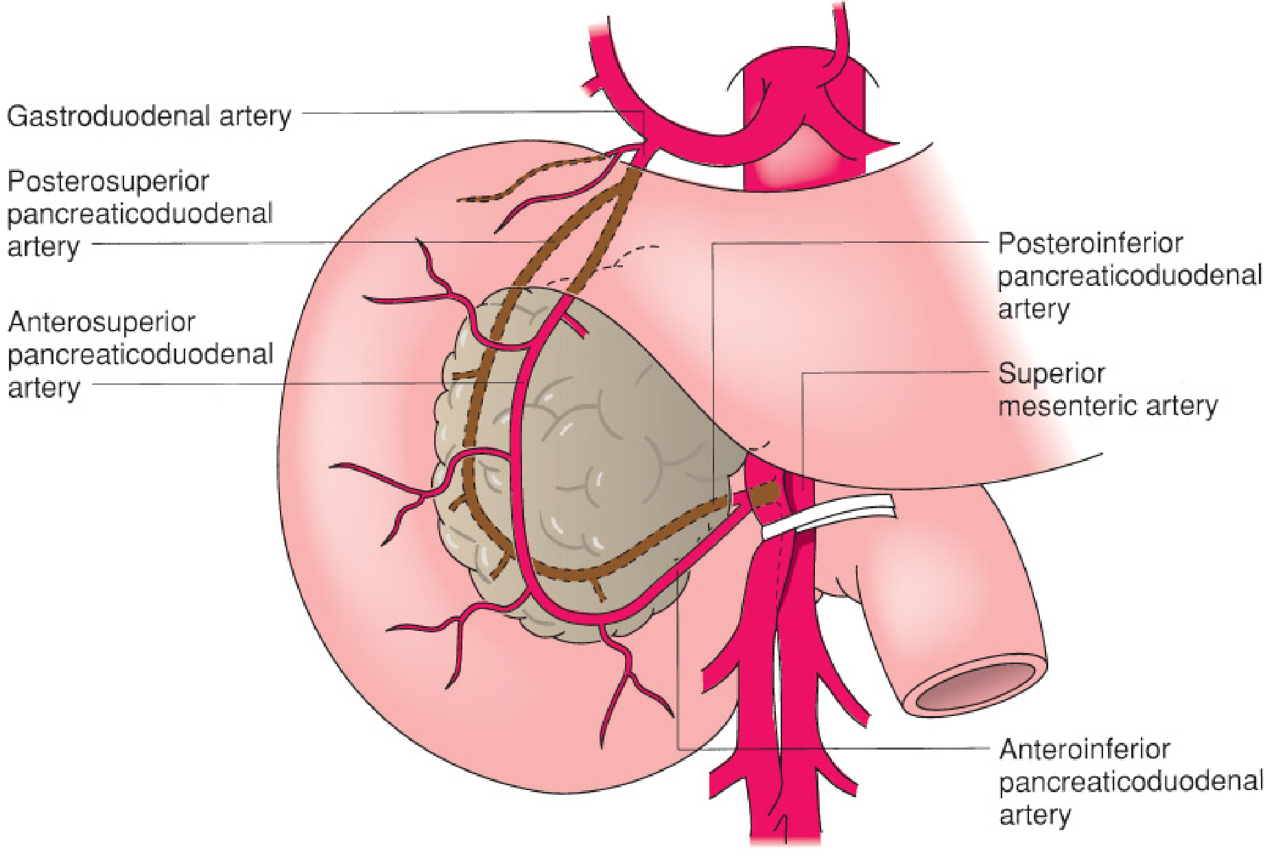
Vascular supply: Dual supply reflecting its embryological origin - the celiac trunk (via gastroduodenal → superior pancreaticoduodenal arteries) supplies the foregut-derived proximal duodenum; the SMA (via inferior pancreaticoduodenal arteries) supplies the midgut-derived distal duodenum. These form anterior and posterior pancreaticoduodenal arcades.
2. Jejunum
- Begins at the ligament of Treitz (L2); constitutes the proximal 40% of the intraperitoneal small intestine
- Widest part of the small intestine; located primarily in the left upper quadrant
- Mucosa has prominent, tall, closely-spaced plicae circulares (Kerckring folds) - these circular folds maximize absorptive surface area and are visible on barium studies
- Vasa recta (arterial branches from SMA) in the jejunum are long and straight, with only 1-2 arterial arcades in the mesentery
- Wall is thicker and more vascular than the ileum; appears "redder" at surgery
- Site of absorption of most carbohydrates, proteins, water-soluble vitamins, and iron
3. Ileum
- Constitutes the distal 60% of the intraperitoneal small intestine
- Located primarily in the right lower quadrant; terminates at the ileocecal valve
- Plicae circulares become shorter, fewer, and more sparse distally; ileum appears thinner-walled
- Vasa recta are shorter with multiple, more complex mesenteric arcades (more arborization)
- Contains Peyer's patches (large lymphoid follicles in the submucosa) - most abundant in ileum, most prominent in children, they represent the gut's major organized lymphoid tissue
- Specific absorptive functions: bile acid reabsorption (enterohepatic circulation) and vitamin B12 absorption (via intrinsic factor-B12 complex binding to cubilin receptors) occur exclusively in the terminal ileum
- The ileocecal valve is a sphincter-like junction that slows transit and partially prevents colonic contents from refluxing into the ileum (though studies show 70-90% are anatomically incompetent)
Microscopic Anatomy (Histology)
The small intestinal wall has 4 layers - from lumen outward:
1. Mucosa
The innermost layer, responsible for absorption and secretion. Consists of:
- Epithelial layer
- Lamina propria
- Muscularis mucosae (thin smooth muscle sheet)
The structural unit is the crypt-villus axis:
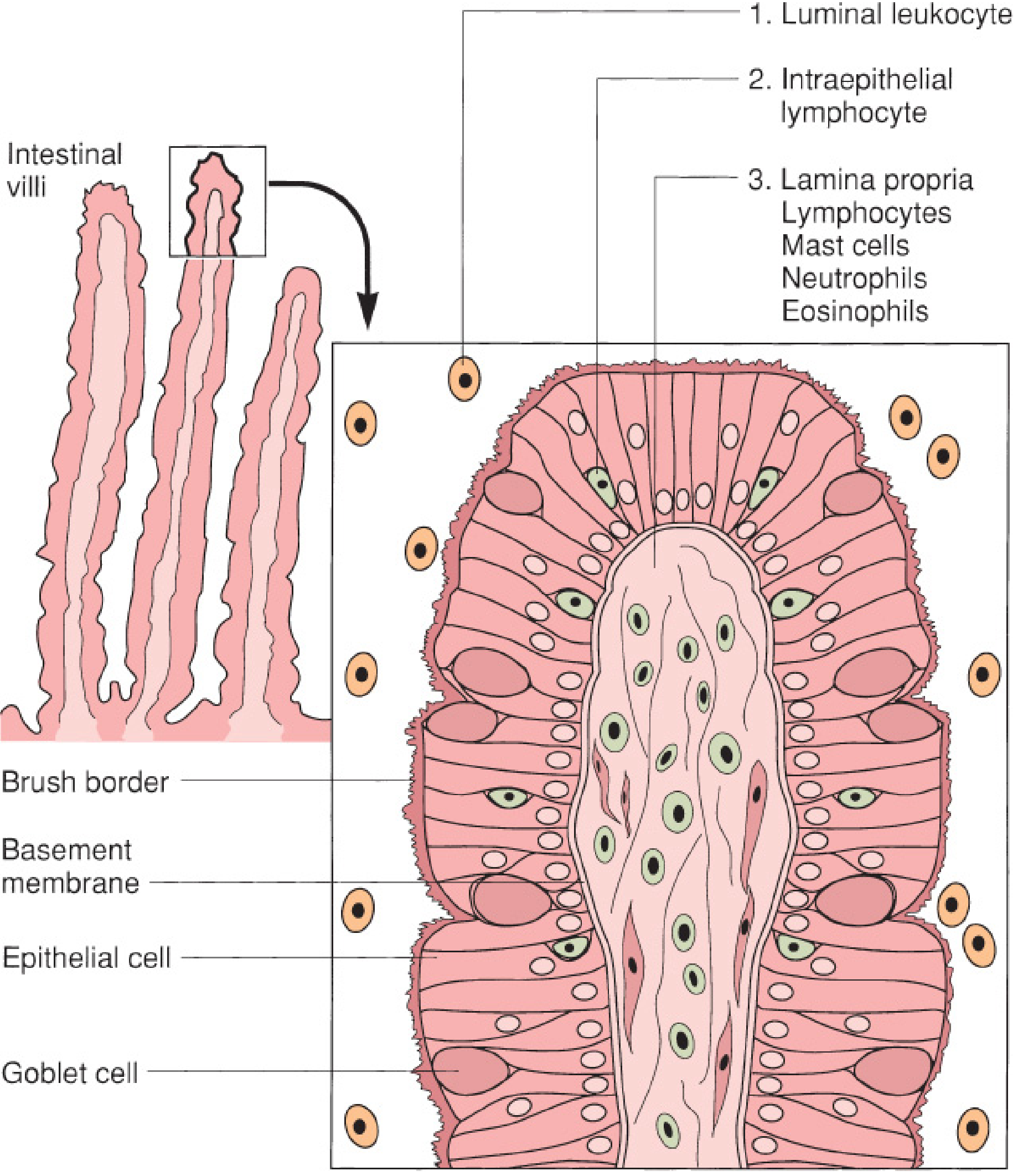
Villi - finger-like projections projecting into the lumen; each contains:
- A central lacteal (lymphatic vessel) for fat absorption
- A small artery, vein, and capillary network
- 90% columnar absorptive enterocytes with microvilli (brush border) coated by the glycocalyx - glycoprotein filaments essential for final digestion and absorption
Crypts of Lieberkühn - glands at the base of villi containing stem cells that produce 4 main cell types:
| Cell Type | Location | Function |
|---|---|---|
| Absorptive enterocytes | Villi (majority) | Nutrient and water absorption via brush border |
| Goblet cells | Villi and crypts | Secrete mucus to protect epithelium and assist transit |
| Paneth cells | Crypt base | Secrete antimicrobial peptides (defensins, lysozyme); maintain stem cell niche |
| Enteroendocrine cells | Crypts and villi | Secrete hormones (largest endocrine organ in the body); see table below |
The entire intestinal epithelium renews every ~5 days - stem cells divide in the crypt, migrate up the villus, and shed from the tip by apoptosis.
Mucosal specializations by region:
- Duodenum: deepest crypts; Brunner glands in submucosa secrete bicarbonate-rich mucus to neutralize gastric acid
- Jejunum: tallest villi, most numerous plicae - maximum absorptive surface area
- Ileum: shorter villi; Peyer's patches prominent in submucosa; cubilin receptors for B12-IF complex
2. Submucosa
- Dense connective tissue with blood vessels, lymphatics, and nerves
- Contains Meissner's plexus (submucosal nerve plexus - regulates secretion and blood flow)
- Strongest layer of the intestinal wall (important in surgical anastomoses)
- Brunner glands (duodenum only)
- Peyer's patches (ileum predominant)
3. Muscularis Propria
- Inner circular layer + outer longitudinal layer
- Auerbach's (myenteric) plexus lies between them - coordinates peristaltic contractions
4. Serosa
- Thin mesothelial layer overlying loose connective tissue
- Covers intraperitoneal bowel completely; covers only the anterior surface of retroperitoneal segments
Blood Supply of the Jejunum and Ileum
All blood comes from the Superior Mesenteric Artery (SMA):
- Main SMA branches form vascular arcades within the mesentery
- From the final arcade, vasa recta (straight terminal arteries) enter the intestinal wall without anastomosis - making individual segments vulnerable to ischemia if a vasa recta is interrupted
- Venous drainage via the Superior Mesenteric Vein (SMV) → portal system
- Lymphatics drain into mesenteric lymph nodes → cisterna chyli
Key Hormones of the Small Intestine
| Hormone | Cell | Site | Stimulus | Actions |
|---|---|---|---|---|
| Gastrin | G cell | Duodenum/antrum | Protein, vagal stimulation | Stimulates HCl, pepsinogen, gastric motility |
| Secretin | S cell | Duodenum | Acid/fat in duodenum | Stimulates pancreatic HCO₃⁻, inhibits gastric acid |
| CCK | I cell | Duodenum/proximal jejunum | Amino acids, fat | Gallbladder contraction, pancreatic enzyme secretion, sphincter of Oddi relaxation |
| GIP | K cell | Duodenum/jejunum | Glucose, fat | Stimulates insulin release (incretin effect) |
| GLP-1 | L cell | Ileum/colon | Mixed nutrients | Insulin secretion, slows gastric emptying |
| Motilin | M cell | Upper small intestine | Fasting | Initiates migrating motor complex |
PART 2 — THE LARGE INTESTINE
Overview
The large intestine extends from the ileocecal valve to the anus, measuring approximately 150 cm (5 feet). It is divided anatomically and functionally into:
- Colon (cecum, ascending, transverse, descending, sigmoid)
- Rectum
- Anal canal
Its primary functions are water and electrolyte absorption, fermentation of unabsorbed carbohydrates by gut flora, formation and storage of feces, and ultimately defecation.
Embryology
- Midgut (SMA territory) → cecum, ascending colon, proximal 2/3 transverse colon
- Hindgut (IMA territory) → distal 1/3 transverse colon, descending colon, sigmoid, rectum, upper anal canal
- Ectoderm → distal anal canal
- The dentate line marks the junction between endodermal hindgut mucosa and ectodermal anoderm
Gross Anatomy: The Colon
Distinguishing Features of the Colon (vs Small Bowel)
Three external features uniquely identify the colon:
- Teniae coli - three narrow bands of the outer longitudinal muscle that run along the length of the colon (they converge at the appendix proximally and merge into a complete longitudinal layer at the rectum distally)
- Haustra (sacculations) - pouches formed between the teniae coli due to the shorter teniae relative to the colon length
- Appendices epiploicae - small fat-filled peritoneal appendages along the teniae
Segments of the Colon
Cecum (~7.5-8.5 cm diameter)
- Widest and thinnest-walled portion of the colon
- Most vulnerable to perforation (Law of Laplace: wall tension ∝ radius)
- Least vulnerable to obstruction
- The vermiform appendix (typically 6-9 cm) hangs from the posteromedial cecum; the teniae coli converge at its base - a surgical landmark for appendectomy
Ascending Colon
- Extends from cecum to the hepatic flexure
- Fixed to the retroperitoneum on the right side
- Supplied by the SMA (ileocolic and right colic arteries)
Transverse Colon
- Extends from hepatic flexure to splenic flexure
- Most mobile segment; suspended by the transverse mesocolon
- Tethered by the gastrocolic ligament superiorly (greater omentum attaches to its anterior/superior edge)
- Characteristic triangular appearance on colonoscopy due to these attachments
- Receives blood from middle colic artery (SMA)
Splenic Flexure
- Highest and most posterior colonic flexure
- Attached to spleen by the lienocolic ligament - can be short and dense, making surgical mobilization challenging
- Also known as a watershed zone - junction of SMA and IMA territories, susceptible to ischemia
Descending Colon
- From splenic flexure to sigmoid
- Fixed to retroperitoneum on the left; relatively immobile
- Supplied by left colic artery (IMA)
Sigmoid Colon
- Narrowest and most mobile segment
- Located in left lower quadrant but can extend to right lower quadrant due to redundancy
- Most common site of volvulus (due to mobility) and diverticular disease
- Most vulnerable to obstruction (narrowest caliber)
- Supplied by sigmoid branches of IMA
Blood Supply of the Colon
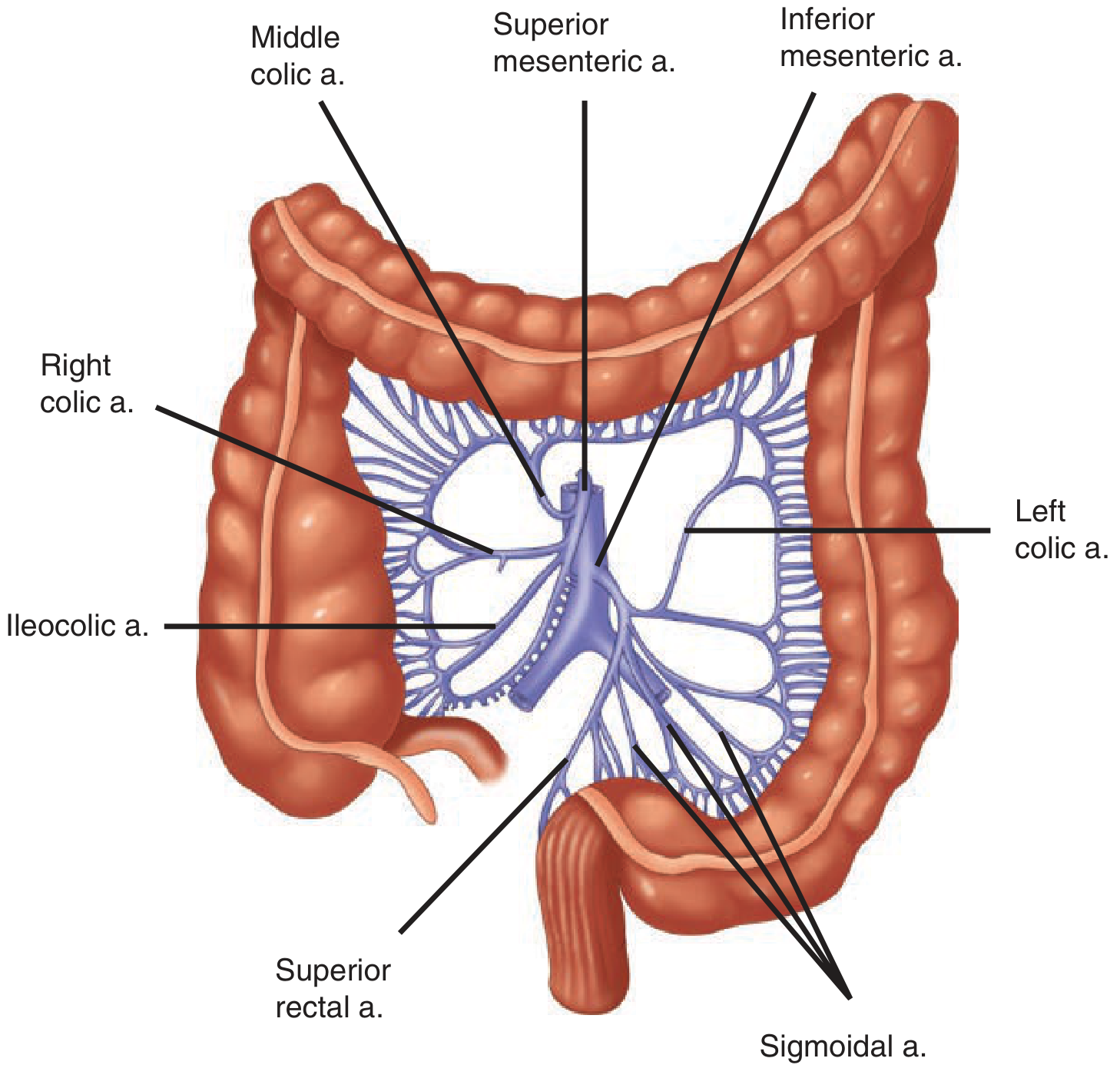
Superior Mesenteric Artery (SMA) supplies right colon:
- Ileocolic artery (absent in ~20%) → terminal ileum and proximal ascending colon
- Right colic artery → ascending colon
- Middle colic artery → transverse colon
Inferior Mesenteric Artery (IMA) supplies left colon:
- Left colic artery → descending colon
- Sigmoid arteries (2-6 branches) → sigmoid colon
- Superior rectal artery → proximal rectum
The terminal branches anastomose via the marginal artery of Drummond (present and complete in only 15-20% of people), and via the arc of Riolan (central anastomosis between middle and left colic). The splenic flexure, being at the SMA-IMA watershed, is most vulnerable to ischemia.
Venous drainage: Parallels arteries and drains into the portal system. The inferior mesenteric vein ascends retroperitoneally and joins the splenic vein posterior to the pancreas.
Lymphatic drainage: Runs along regional arteries through four nodal groups: epicolic → paracolic → intermediate → principal (origin of SMA/IMA) → para-aortic nodes.
Nerve supply:
- Sympathetic (inhibitory): T6-T12 and L1-L3
- Parasympathetic (stimulatory): Vagus nerve (right and transverse colon); sacral nerves S2-S4 via nervi erigentes (left colon and rectum)
The Rectum
- Approximately 12-15 cm in length; begins at the rectosigmoid junction (level of the sacral promontory, where teniae coli coalesce)
- Three submucosal folds called the valves of Houston extend into the rectal lumen
- No serosa on the mid and lower rectum (only the proximal 1/3 is covered by peritoneum)
Key fascial relationships:
- Posterior: Presacral fascia separates rectum from presacral venous plexus and pelvic nerves. At S4, Waldeyer's fascia (rectosacral fascia) attaches anteriorly to the anorectal junction
- Anterior: Denonvilliers' fascia separates rectum from prostate/seminal vesicles (in men) and vagina (in women)
Arterial supply: Superior rectal artery (IMA) + middle rectal arteries (internal iliac) + inferior rectal arteries (internal pudendal → internal iliac)
Venous drainage:
- Superior rectal vein → IMV → portal system
- Middle rectal vein → internal iliac vein
- Inferior rectal vein → internal pudendal vein → internal iliac vein
- The submucosal hemorrhoidal plexus drains into all three, creating portosystemic anastomoses
The Anal Canal
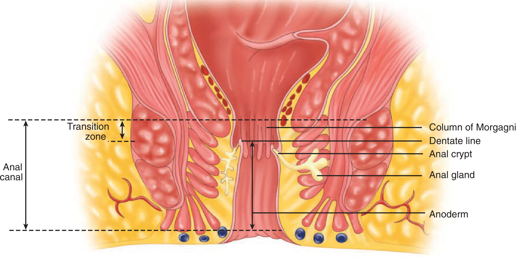
Anatomic anal canal (2 cm): dentate line to anal verge
Surgical anal canal (2-4 cm): anorectal junction to anal verge
The Dentate (Pectinate) Line is the critical anatomical landmark:
- Marks transition from columnar rectal mucosa (above) to squamous anoderm (below)
- Above: visceral innervation (sympathetic/parasympathetic) - insensate to pain; venous drainage to portal system
- Below: somatic innervation (pudendal nerve) - exquisitely pain-sensitive; venous drainage to systemic (iliac) veins
- This distinction explains why internal hemorrhoids (above dentate) are painless and external hemorrhoids (below) are painful
Columns of Morgagni - longitudinal mucosal folds surrounding the dentate line; the anal crypts between them can become infected → cryptoglandular abscess → fistula-in-ano
Sphincter Complex:
- Internal anal sphincter - involuntary; thickened smooth muscle from the inner circular rectal layer; maintains resting tone (~85% of resting anal pressure)
- External anal sphincter - voluntary striated muscle; three portions (subcutaneous, superficial, deep); supplied by the pudendal nerve (S2-S4)
- Puborectalis - part of the levator ani complex; forms a U-shaped sling around the anorectal junction; its tonic contraction creates the anorectal angle (~90°) which is critical for continence
Microscopic Anatomy of the Large Intestine
The colon lacks villi. Instead, the mucosa is flat with deep straight crypts of Lieberkühn packed with:
- Absorptive colonocytes - absorb water and electrolytes
- Goblet cells - very abundant; secrete mucus (more than in small bowel)
- Enteroendocrine cells
- No Paneth cells (except in the proximal cecum) and no Brunner glands
The muscularis propria has:
- Inner circular layer (thickens to form internal anal sphincter distally)
- Outer longitudinal layer condensed into three teniae coli in the colon → becomes complete circumferential layer in the rectum
Comparison: Small vs Large Intestine
| Feature | Small Intestine | Large Intestine |
|---|---|---|
| Length | 5-7 m | ~1.5 m |
| Diameter | 2.5-4 cm | 4-8.5 cm |
| Villi | Present (abundant) | Absent |
| Plicae circulares | Present | Absent |
| Teniae coli | Absent | Present (3) |
| Haustra | Absent | Present |
| Appendices epiploicae | Absent | Present |
| Peyer's patches | Present (ileum) | Absent |
| Brunner glands | Duodenum only | Absent |
| Main function | Digestion + absorption | Water absorption, fermentation, defecation |
| Blood supply | Entirely SMA | SMA (right) + IMA (left) |
| Mesentery | Long, mobile | Partly fixed retroperitoneal segments |
Sources:
- Mulholland and Greenfield's Surgery, 7th ed., Chapter 48 - Anatomy and Physiology of the Small Intestine
- Schwartz's Principles of Surgery, 11th ed., Chapter 29 - Colon, Rectum, and Anus (Embryology and Anatomy)
- Sleisenger and Fordtran's Gastrointestinal and Liver Disease - Colon and Rectum
Explain in detail about intestine, boat, small intestine, and large intestine
I want to make sure I cover exactly what you're looking for. The term "boat" in the context of intestines isn't a standard medical term I recognize - it might be a typo or autocorrect issue.
Asking for Preferences
What did you mean by 'boat' in the context of intestines? It doesn't appear to be a standard anatomical term.
"Boat" is likely a typo for bloat (intestinal distension). I'll cover the intestines in detail along with a dedicated section on intestinal bloat/distension.
Searching sources...
Finding Sources
Finding Sources
Finding Sources
Finding Sources
Finding Sources
Finding Sources
Reading File
Reading File
Reading File
Reading File
I now have excellent content. The previous session already covered the detailed small and large intestine anatomy extensively. This query adds "bloat" which I've now researched thoroughly. Let me deliver the complete answer.
The Intestines: A Complete Guide
Note on "boat": This appears to be a typo for bloat (intestinal gas and distension). All four topics are covered below: intestines in general, bloat, small intestine, and large intestine.
PART 1 — THE INTESTINES: OVERVIEW
The intestines form the largest part of the gastrointestinal (GI) tract, extending from the pyloric outlet of the stomach to the anus. In total they measure approximately 6.5-8.5 meters in an adult. They perform the core tasks of the digestive system:
- Digestion - enzymatic breakdown of food
- Absorption - uptake of nutrients, water, electrolytes, vitamins
- Secretion - hormones, enzymes, mucus, bicarbonate
- Immunity - gut-associated lymphoid tissue (GALT) is the body's largest immune organ
- Excretion - formation and elimination of feces
They are divided into two major segments:
| Feature | Small Intestine | Large Intestine |
|---|---|---|
| Length | 5-7 m | ~1.5 m |
| Diameter | 2.5-4 cm | 4-8.5 cm |
| Main function | Digestion and absorption | Water absorption, fermentation, defecation |
| Villi | Present | Absent |
| Teniae coli | Absent | Present |
| Haustra | Absent | Present |
| Blood supply | Entirely SMA | SMA (right half) + IMA (left half) |
The intestinal wall throughout has the same 4-layer architecture (from lumen outward):
- Mucosa (epithelium + lamina propria + muscularis mucosae)
- Submucosa (strongest layer; contains Meissner's plexus, vessels, lymphatics)
- Muscularis propria (inner circular + outer longitudinal; Auerbach's/myenteric plexus between them)
- Serosa (peritoneal covering)
PART 2 — INTESTINAL BLOAT
What Is Bloating?
Bloating is the subjective sensation of increased abdominal pressure or fullness, often described as a feeling of swelling or tightness in the abdomen. It is one of the most common GI complaints worldwide.
Abdominal distension is distinct - it is the objective, measurable increase in abdominal girth, often visible externally. CT and inductance plethysmography studies have confirmed real increases in anteroposterior diameter during episodes of distension.
Mechanisms of Bloating
Contrary to popular belief, bloating is not simply caused by too much gas. Research using CT imaging and argon gas washout techniques shows that the mean volume of intestinal gas in symptomatic bloaters (~176 mL) is actually slightly less than in healthy controls (~199 mL). The key findings are:
1. Impaired Gas Transit and Handling
- Gas transit studies consistently show that bloaters have impaired intestinal reflex control of gas propulsion
- Gas refluxes back into the stomach more frequently and moves in a "to-and-fro" pattern rather than forward
- This poor clearance, most often in the jejunum and proximal bowel, leads to gas entrapment
2. Visceral Hypersensitivity
- Patients with IBS and bloating have increased sensitivity of the intestines to normal volumes of gas and bowel contents - the issue is perception, not volume
- This explains why symptoms can be severe even without objective distension
3. Abdominophrenic Dyssynergia
- A crucial and underappreciated mechanism: abnormal diaphragm activity holds gas in place
- Normally the diaphragm ascends to push gas forward; in some bloaters, the diaphragm descends (instead of ascending) and the anterior abdominal wall relaxes - creating a "container" that traps gas
- CT studies confirm diaphragmatic descent and increased anteroposterior diameter during episodes
- This can be corrected with biofeedback training targeting diaphragm and abdominal wall coordination
4. Abnormal Gas Composition (Flatus)
Normal flatus is composed of: N₂, O₂, CO₂, H₂, and CH₄ - none of which have an odor. The unpleasant smell comes from trace sulfur-containing compounds (methanethiol, dimethyl sulfide) produced by bacterial fermentation. Normally fewer than 25 flatus episodes per day is considered normal.
5. Increased Gas Production
In a subset of patients, true excess gas production occurs from:
- Small intestinal bacterial overgrowth (SIBO) - bacteria fermenting food residues in the small bowel (normally sterile)
- Carbohydrate malabsorption (lactose, fructose, fructans, sorbitol) - unabsorbed sugars reach the colon and are fermented
- Aerophagia - excessive air swallowing
- Carbonated drinks, chewing gum (especially sorbitol-containing)
Common Causes of Bloating
| Category | Causes |
|---|---|
| Small bowel | Lactose/fructose malabsorption, SIBO, celiac disease, Crohn's disease, giardiasis, small bowel obstruction |
| Colonic | Constipation (slow transit), colonic stricture, Hirschsprung disease, diverticular disease, colon cancer |
| Gastric | Gastroparesis, gastric outlet obstruction |
| Drugs | Narcotics, anticholinergics, loperamide, calcium channel blockers |
| Metabolic | Hypothyroidism, hypokalemia, hypercalcemia, diabetes |
| Functional | IBS, functional bloating |
Special Bloat Syndromes
- Gas-bloat syndrome - occurs in 25-50% of patients after gastric fundoplication; inability to belch traps swallowed air in the stomach
- Magenblase syndrome - excessive accumulation of swallowed air causing marked postprandial epigastric fullness, often relieved by belching
Treatment of Bloating
Non-pharmacologic:
- Physical activity and upright posture - exercise increases intestinal gas clearance; lying supine worsens it
- Dietary modification - reduce fermentable carbohydrates (FODMAPs: lactose, fructose, fructans, galactans, polyols)
- Probiotics - some strains reduce IBS-associated bloating (variable results depending on species and dose)
- Biofeedback - for patients with abdominophrenic dyssynergia; trains correct diaphragm-abdominal wall coordination
- Treat constipation - reduces colonic bulking and fermentation
Pharmacologic:
- Rifaximin (non-absorbable antibiotic) - reduces SIBO and bacterial fermentation; modest benefit
- Prokinetics (neostigmine, pyridostigmine) - improve gas transit
- Smooth muscle relaxants / antispasmodics - superior to placebo for abdominal pain and distension in IBS
- Peppermint oil - antispasmodic effect via calcium channel blockade (menthol); benefit in IBS is uncertain
PART 3 — THE SMALL INTESTINE
Overview
- Spans from pylorus to ileocecal valve
- Length: 5-7 meters (16-23 feet) in an adult; 200-300 cm in a newborn
- Comprises >60% of total alimentary tract length
- Three segments: Duodenum → Jejunum → Ileum
- The largest endocrine organ in the human body
Embryology
Derived from the distal foregut, midgut, and splanchnic mesenchyme. Between weeks 7-10, the midgut herniates through the umbilicus and rotates 270° counterclockwise around the axis of the superior mesenteric artery before returning to the abdominal cavity. Failure of rotation → intestinal malrotation (risk of volvulus).
A. Duodenum (~20-30 cm)
Begins at the pylorus, ends at the ligament of Treitz (L2). C-shaped, largely retroperitoneal and fixed.
| Part | Length | Key Anatomy |
|---|---|---|
| 1st (Bulb/Cap) | ~5 cm | Smooth mucosa; hepatoduodenal ligament attaches here; ~90% of duodenal ulcers occur here (erodes into gastroduodenal artery posteriorly) |
| 2nd (Descending) | ~10 cm | Kerckring folds begin; Ampulla of Vater (bile + pancreatic juice entry); sphincter of Oddi; minor papilla (Santorini duct) |
| 3rd (Horizontal) | ~10 cm | Crosses midline over aorta and IVC; SMA crosses anteriorly (SMA syndrome if compressed) |
| 4th (Ascending) | ~5 cm | Rises to ligament of Treitz; becomes jejunum |
Blood supply: Dual - celiac trunk (superior pancreaticoduodenal arteries) + SMA (inferior pancreaticoduodenal arteries) forming anterior and posterior pancreaticoduodenal arcades.
B. Jejunum (proximal 40% of intraperitoneal small bowel)
- Located in left upper quadrant; widest segment
- Mucosa: tall, closely-packed plicae circulares (Kerckring folds) → maximum absorptive surface
- Vasa recta (arterial end-branches): long and straight, 1-2 mesenteric arcades
- Thick, vascular, "redder" wall
- Primary site of absorption: carbohydrates, proteins, water-soluble vitamins, iron, calcium
C. Ileum (distal 60% of intraperitoneal small bowel)
- Located in right lower quadrant; terminates at ileocecal valve
- Mucosa: plicae circulares become shorter and fewer distally
- Vasa recta: shorter with multiple complex arcades
- Contains Peyer's patches (organized lymphoid follicles in submucosa) - key immune surveillance
- Specific absorption: bile acid reabsorption (enterohepatic circulation) and vitamin B12 (via intrinsic factor + cubilin receptors) - both exclusive to terminal ileum
- Ileocecal valve slows transit and partially prevents colonic reflux
Microscopic Anatomy of the Small Intestine
The Crypt-Villus Unit
Villi - finger-like projections into the lumen. Each contains:
- Central lacteal (lymphatic, for fat absorption as chylomicrons)
- Arteriole, venule, capillary network
- 90% absorptive enterocytes with microvilli (forming the brush border) coated by the glycocalyx - essential for surface digestion
Crypts of Lieberkühn - tubular glands at the base. Stem cells here generate 4 cell types:
| Cell | Function |
|---|---|
| Absorptive enterocytes | Nutrient, water, and electrolyte absorption |
| Goblet cells | Mucus secretion (protection + lubrication) |
| Paneth cells (crypt base) | Antimicrobial peptides (defensins, lysozyme); maintain stem cell niche |
| Enteroendocrine cells | Hormone secretion (CCK, secretin, GLP-1, GIP, etc.) |
Cells migrate from crypt → villus tip in ~5 days, then shed by apoptosis - one of the fastest self-renewing tissues in the body.
Four Wall Layers
- Mucosa - villi + crypts + muscularis mucosae
- Submucosa - strongest layer; Meissner's plexus; Brunner glands (duodenum - secrete HCO₃⁻ mucus to neutralize acid); Peyer's patches (ileum)
- Muscularis propria - inner circular + outer longitudinal; Auerbach's plexus between them
- Serosa - complete peritoneal covering on intraperitoneal segments
Blood Supply
- Duodenum: Celiac + SMA pancreaticoduodenal arcades
- Jejunum + Ileum: Entirely from SMA → arterial arcades → vasa recta (no anastomosis within wall)
- Venous: SMV → portal vein
- Lymphatics: Mesenteric nodes → cisterna chyli (fat absorbed as chylomicrons travels via lacteals, NOT portal blood)
Key Hormones
| Hormone | Cell | Location | Stimulus | Effect |
|---|---|---|---|---|
| Secretin | S cell | Duodenum | Acid/fat | Pancreatic HCO₃⁻, inhibits acid |
| CCK | I cell | Duodenum/jejunum | Amino acids, fat | Gallbladder emptying, pancreatic enzymes, sphincter of Oddi relaxation |
| GIP | K cell | Duodenum/jejunum | Glucose, fat | Insulin release (incretin) |
| GLP-1 | L cell | Ileum/colon | Nutrients | Insulin, slows gastric emptying |
| Motilin | M cell | Upper small intestine | Fasting | Migrating motor complex (MMC) |
PART 4 — THE LARGE INTESTINE
Overview
- Extends from ileocecal valve to anus, approximately 150 cm (5 feet)
- Divided into: Colon → Rectum → Anal canal
- No villi - flat mucosa with deep crypts rich in goblet cells
- Functions: water and electrolyte absorption, fermentation of residues by gut microbiota, fecal formation and storage, defecation
Distinguishing Features of the Colon
Three external landmarks unique to the colon:
- Teniae coli - 3 condensed bands of the outer longitudinal muscle running lengthwise (converge at appendix; merge into complete layer at rectum)
- Haustra - sacculations between teniae coli
- Appendices epiploicae - fat-filled peritoneal pouches along teniae
Segments of the Colon
Cecum (~7.5-8.5 cm)
- Widest, thinnest-walled segment → most vulnerable to perforation (Laplace's law), least to obstruction
- The vermiform appendix (~6-9 cm) attaches posteromedially; teniae coli converge at its base
Ascending Colon
- Fixed retroperitoneally on the right
- Hepatic flexure marks transition to transverse colon
- SMA territory (ileocolic + right colic arteries)
Transverse Colon
- Most mobile; suspended by transverse mesocolon
- Greater omentum attaches to its anterior/superior edge
- SMA territory (middle colic artery)
Splenic Flexure
- Highest and most posterior flexure
- Watershed zone between SMA and IMA - most vulnerable to ischemia
- Lienocolic ligament to spleen makes mobilization surgically challenging
Descending Colon
- Fixed retroperitoneally on the left
- IMA territory (left colic artery)
Sigmoid Colon
- Narrowest → most vulnerable to obstruction
- Most mobile → most common site of volvulus
- Most common site of diverticular disease and diverticulitis
- IMA territory (sigmoid arteries)
Blood Supply of the Large Intestine
Venous: Parallels arteries → SMV/IMV → portal vein. The inferior mesenteric vein ascends retroperitoneally to join the splenic vein behind the pancreas.
Lymphatics: Epicolic → paracolic → intermediate → principal nodes at SMA/IMA origins → para-aortic nodes.
Nerve supply:
- Sympathetic (inhibitory): T6-T12, L1-L3
- Parasympathetic (stimulatory): Vagus nerve (right colon + transverse); Sacral nerves S2-S4 via nervi erigentes (left colon + rectum)
Rectum (12-15 cm)
- Begins at rectosigmoid junction (sacral promontory - where teniae coli coalesce)
- Three submucosal folds: valves of Houston (project into lumen)
- Upper 1/3: covered by peritoneum anteriorly; mid and lower rectum: no serosa
- Denonvilliers' fascia anteriorly separates it from prostate/seminal vesicles (men) or vagina (women)
- Waldeyer's fascia (rectosacral fascia) posteriorly attaches at S4 to anorectal junction
- Venous plexuses here are portosystemic anastomoses (source of hemorrhoids; important in portal hypertension)
Anal Canal and Sphincters
The Dentate (Pectinate) Line - the most important anatomical landmark in the anal canal:
| Feature | Above Dentate Line | Below Dentate Line |
|---|---|---|
| Mucosa | Columnar (rectal) | Squamous (anoderm) |
| Innervation | Visceral (autonomic) - insensate | Somatic (pudendal nerve) - pain-sensitive |
| Venous drainage | Portal (superior rectal v.) | Systemic (internal iliac v.) |
| Lymphatics | Internal iliac nodes | Inguinal nodes |
| Hemorrhoids | Internal (painless) | External (painful) |
Columns of Morgagni - longitudinal mucosal folds at the dentate line; anal crypts between them can become infected → cryptoglandular abscess → fistula-in-ano
Sphincter Complex:
- Internal anal sphincter (IAS) - involuntary smooth muscle; thickened continuation of inner circular rectal layer; provides ~85% of resting anal pressure
- External anal sphincter (EAS) - voluntary striated muscle (3 portions: subcutaneous, superficial, deep); innervated by pudendal nerve (S2-S4)
- Puborectalis - U-shaped sling of the levator ani around the anorectal junction; creates the anorectal angle (~90°) which is the primary mechanical barrier to incontinence; part of levator ani (puborectalis + iliococcygeus + pubococcygeus)
Microscopic Anatomy of the Large Intestine
- No villi - flat mucosal surface with deep straight crypts
- Crypts are packed with goblet cells (more abundant than anywhere in the small intestine) → large quantities of lubricating mucus
- Absorptive colonocytes - absorb Na⁺, Cl⁻, and water (colon absorbs ~1-2 L water/day)
- Enteroendocrine cells - produce peptide YY, GLP-1, GLP-2
- No Paneth cells (except proximal cecum), no Brunner glands
- Muscularis: inner circular + condensed outer longitudinal (3 teniae in colon → full circumferential in rectum → thickened internal sphincter distally)
Summary: The Three Segments Compared
| Feature | Jejunum | Ileum | Colon |
|---|---|---|---|
| Plicae circulares | Tall, prominent | Short, sparse | Absent |
| Villi | Tall | Shorter | Absent |
| Wall thickness | Thick | Thinner | Variable |
| Mesenteric arcades | 1-2, long vasa recta | Multiple, short vasa recta | N/A |
| Special structures | Brunner glands (duodenum) | Peyer's patches | Teniae coli, haustra, appendices epiploicae |
| Key absorption | Carbs, protein, fat, vitamins | Bile acids, B12 | Water, electrolytes |
| Blood supply | SMA | SMA | SMA (right) + IMA (left) |
Sources:
- Mulholland and Greenfield's Surgery, 7th ed., Chapter 48 - Anatomy and Physiology of the Small Intestine
- Schwartz's Principles of Surgery, 11th ed., Chapter 29 - Colon, Rectum, and Anus
- Sleisenger and Fordtran's Gastrointestinal and Liver Disease, Chapter 17 - Abdominal Bloating and Distension
- Yamada's Textbook of Gastroenterology, 7th ed., Chapter 34 - Pathophysiology of Bloating and Gas
This is a shared conversation. Sign in to Orris to start your own chat.