
Explain me anatomy of lungs for my bds exam 1st year
Reading File
Now let me search the library for lung anatomy content, following the medical library skill protocol.
Finding Sources
Finding Sources
Finding Sources
Finding Sources
Finding Sources
Finding Sources
Now let me read the key sections from Gray's Anatomy for Students and the Color Atlas in parallel:
Reading File
Reading File
Reading File
Reading File
I now have enough detailed content. Let me also fetch the key lung diagram images to include in the answer.
Loading Image
Loading Image
Excellent! I now have rich content and beautiful diagrams from Gray's Anatomy for Students and the Color Atlas of Human Anatomy. Let me now compose a comprehensive, exam-focused answer.
Anatomy of the Lungs - BDS 1st Year
(Sources: Gray's Anatomy for Students; Color Atlas of Human Anatomy Vol. 2)
Overview
The lungs are the primary organs of respiration. They are paired, spongy organs lying in the thoracic cavity, one on each side of the mediastinum, each enclosed in its own pleural cavity. Each lung is shaped like a half cone.
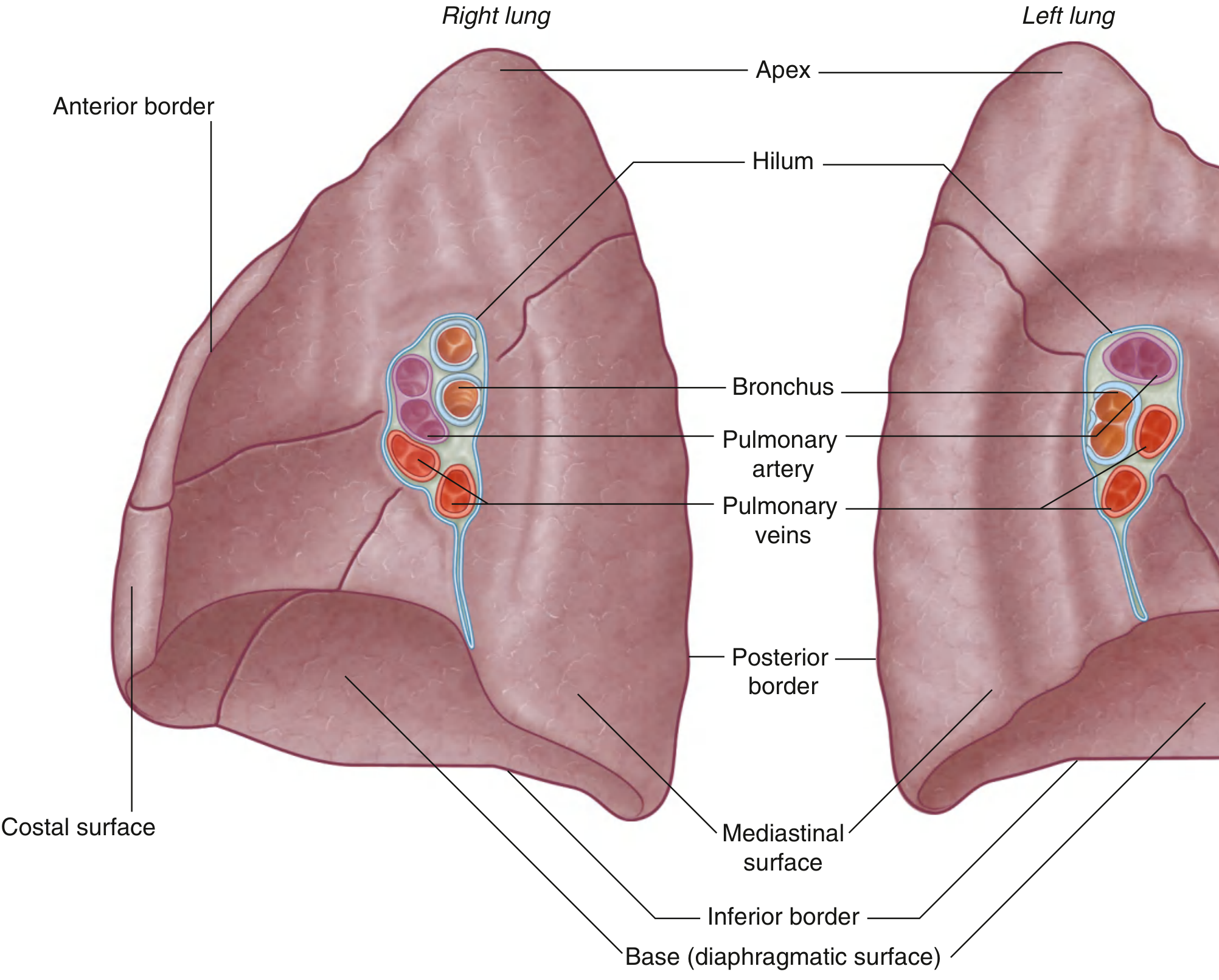
1. External Features (Surfaces & Borders)
Each lung has:
Surfaces
| Surface | Description |
|---|---|
| Costal surface | Convex; faces the thoracic wall/ribs |
| Mediastinal surface | Faces the mediastinum; contains the hilum and cardiac impression |
| Diaphragmatic surface (Base) | Concave; rests on the diaphragm |
Borders
- Anterior border - Sharp; where costal and mediastinal surfaces meet anteriorly
- Inferior border - Sharp; separates costal and diaphragmatic surfaces
- Posterior border - Rounded; in the paravertebral gutter
Apex
- The dome-like apex projects a few centimeters above the superior thoracic aperture (above the first rib, into the root of the neck)
- It is related to the subclavian artery and brachial plexus
2. Lobes and Fissures
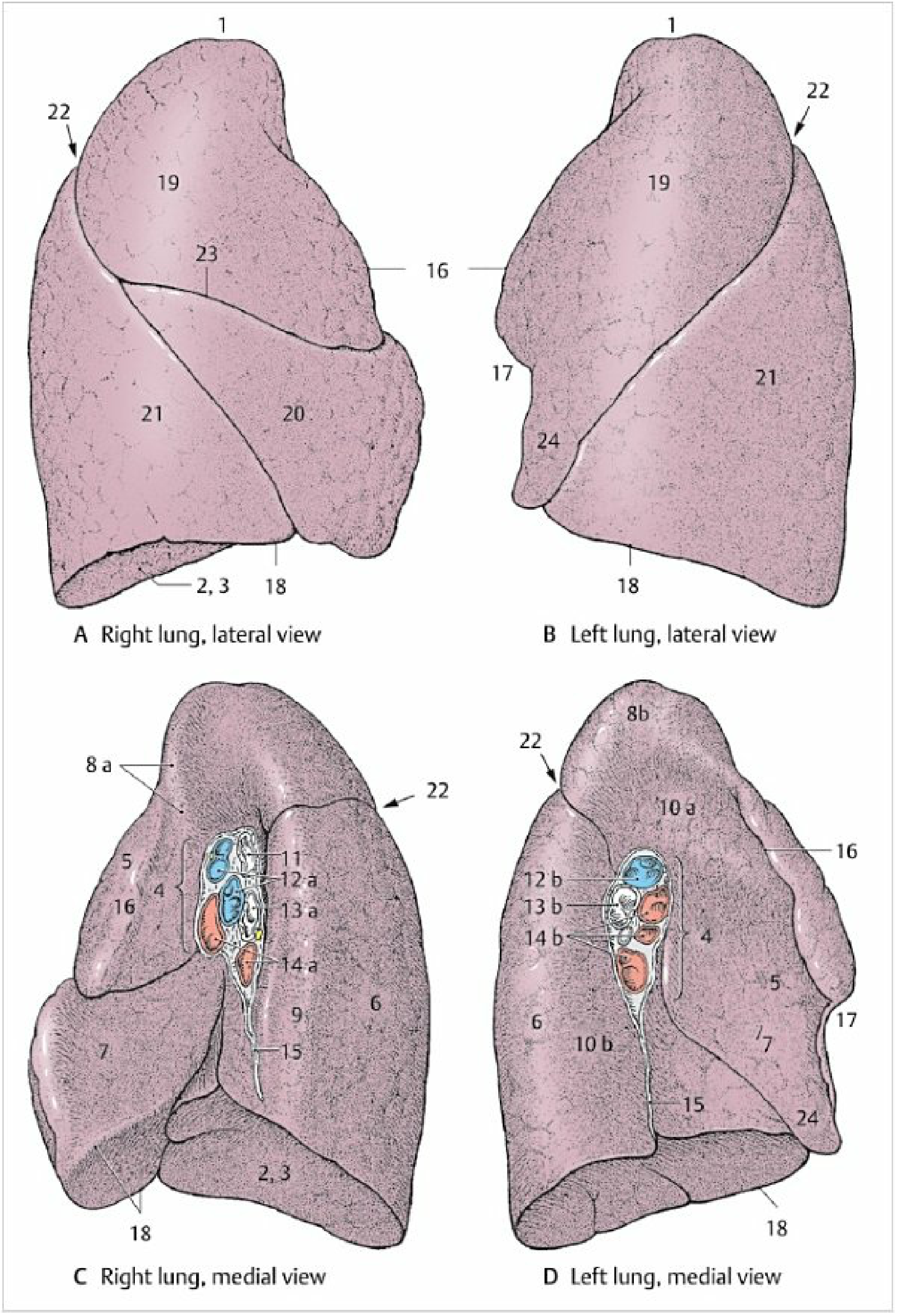
Right Lung (3 lobes, 2 fissures)
- Upper (Superior) lobe
- Middle lobe
- Lower (Inferior) lobe
- Oblique fissure - separates the lower lobe from the upper and middle lobes
- Horizontal fissure - separates the upper lobe from the middle lobe; runs along the 4th intercostal space anteriorly
Surface markings of fissures (right):
- Oblique fissure: begins at T4 spinous process, crosses 5th interspace laterally, follows rib 6 anteriorly
- Horizontal fissure: follows the 4th intercostal space from the sternum to where it meets the oblique fissure at rib 5
Left Lung (2 lobes, 1 fissure)
- Upper (Superior) lobe
- Lower (Inferior) lobe
- Oblique fissure only (slightly more oblique than the right)
- The left lung is smaller than the right due to the heart projecting into the left pleural cavity
- The lingula - a tongue-like projection of the upper lobe that extends over the cardiac impression
- Cardiac notch - an indentation on the anterior border of the left lung caused by the heart
Surface markings (left):
- Oblique fissure: begins between T3-T4 spinous processes, crosses 5th interspace laterally, follows rib 6 anteriorly
3. Hilum and Root of the Lung
The hilum is the site on the mediastinal surface where structures enter and leave the lung. The collection of all these structures forms the root of the lung.
Structures at the Hilum
- Main bronchus
- Pulmonary artery
- Two pulmonary veins (superior and inferior)
- Bronchial arteries and veins
- Lymphatic vessels
- Autonomic nerve plexus
Arrangement at the Hilum (important exam point)
General rule: Pulmonary artery is superior, pulmonary veins are inferior, bronchi are posterior
| Side | Arrangement (superior to inferior) |
|---|---|
| Right | Superior lobar bronchus (eparterial) → Pulmonary artery → Main bronchus → Pulmonary veins |
| Left | Pulmonary artery → Main bronchus → Pulmonary veins |
Key difference: On the right, the superior lobar bronchus lies above (eparterial to) the pulmonary artery. On the left, the bronchus lies below (hyparterial to) the pulmonary artery.
The root is surrounded by a pleural sleeve that extends downward as the pulmonary ligament.
4. Relations of Mediastinal Surface
Right Lung
- Heart (right atrium and ventricle)
- Inferior vena cava
- Superior vena cava
- Azygos vein (arches over the root)
- Esophagus
- Right subclavian artery and vein (related to apex)
Left Lung
- Heart (left ventricle predominantly)
- Aortic arch (groove above the hilum)
- Descending thoracic aorta (groove below and behind the hilum)
- Left subclavian artery
- Esophagus (lower part)
5. Bronchopulmonary Segments
Each lung is divided into bronchopulmonary segments - the surgical unit of the lung. Each segment:
- Has its own segmental bronchus and accompanying branch of pulmonary artery (centrally located)
- Is drained by pulmonary veins that run in the intersegmental connective tissue (at the periphery/borders of segments)
- Is a pyramidal/wedge-shaped unit with apex toward the hilum
- Can be resected independently (surgically)
| Lung | Lobe | Segments |
|---|---|---|
| Right | Upper | 3 (apical, posterior, anterior) |
| Middle | 2 (lateral, medial) | |
| Lower | 5 (superior + 4 basal) | |
| Left | Upper | 4 (apicoposterior, anterior, superior lingular, inferior lingular) |
| Lower | 4-5 (superior + 3-4 basal) |
- Right lung: 10 segments
- Left lung: 8-9 segments (some segments fused)
6. Bronchial Tree
- Trachea bifurcates at the sternal angle (T4-T5) into right and left main bronchi
- Right main bronchus: shorter, wider, more vertical - foreign bodies more commonly lodge here
- Left main bronchus: longer, narrower, more horizontal
- Main bronchi → Lobar bronchi → Segmental bronchi → Medium/small bronchi → Bronchioles (no cartilage) → Terminal bronchioles → Respiratory bronchioles → Alveolar ducts → Alveoli
The terminal bronchiole is the last purely conducting airway. Respiratory bronchioles onward form the acinus (functional unit of gas exchange).
7. Blood Supply
| Vessel | Function |
|---|---|
| Pulmonary arteries (right and left) | Carry deoxygenated blood from right ventricle for gas exchange |
| Pulmonary veins (2 per side, 4 total) | Return oxygenated blood to left atrium |
| Bronchial arteries (from descending aorta) | Nutritive supply to bronchial walls, connective tissue, pleura |
| Bronchial veins | Drain into azygos/hemiazygos veins |
8. Nerve Supply
- Pulmonary plexus (anterior and posterior) - at the root of each lung
- Sympathetic (T2-T5 via cardiac plexus): bronchodilation, vasoconstriction
- Parasympathetic (vagus nerve, CN X): bronchoconstriction, increased secretion, vasodilation
- Visceral afferents travel with vagus (stretch receptors in alveoli - Hering-Breuer reflex)
9. Lymphatic Drainage
- Drains toward the tracheobronchial lymph nodes (at bifurcation of trachea)
- Ultimately drains into the right lymphatic duct (from right lung) and thoracic duct (from left lung)
- The subpleural plexus and peribronchial plexus ultimately converge at the hilum
Quick Exam Mnemonics
| Point | Mnemonic / Key Fact |
|---|---|
| Right vs. Left main bronchus | "Right = Shorter, Wider, More vertical" - foreign body risk |
| Hilum arrangement | "ABVV" from back to front: Artery (superior), Bronchus (posterior), Veins (inferior) |
| Cardiac notch | Left lung only |
| Lingula | Left upper lobe only |
| Eparterial bronchus | Right upper lobe bronchus only |
| Horizontal fissure | Right lung only |
| Lobes | Right = 3 (upper, middle, lower); Left = 2 (upper, lower) |
All anatomical details sourced from Gray's Anatomy for Students and Color Atlas of Human Anatomy Vol. 2 (Internal Organs).
Now anatomy of heart from bdc
Finding Sources
Finding Sources
Reading File
Finding Sources
Reading File
Reading File
Reading File
Reading File
Reading File
Reading File
Reading File
Reading File
Now let me fetch the key heart diagrams:
Loading Image
Loading Image
Loading Image
Loading Image
This is a shared conversation. Sign in to Orris to start your own chat.