
Treatment of spider bite ??
spider bite treatment management 2024 clinical guidelines
spider bite wound necrosis brown recluse black widow
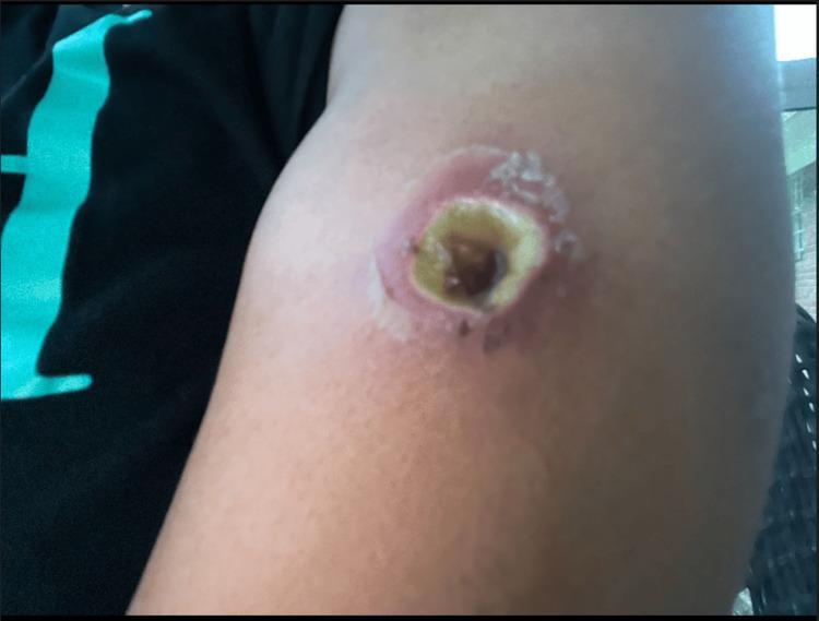
This clinical photograph displays a focal, well-demarcated skin lesion on the left upper extremity, characteristic of cutaneous loxoscelism (brown recluse spider bite). The wound exhibits a central necrotic core, appearing as a dark brown to black eschar, indicative of tissue infarction. This is immediately surrounded by a ring of yellowish, fibrinous slough and pale tissue, suggesting liquefactive necrosis or secondary infection. The periphery of the lesion shows a prominent concentric band of erythema, signaling intense localized inflammation and possible cellulitis. Evidence of epidermal sloughing and desquamation is visible at the margins of the inflammatory ring. Clinically, this sequence represents the 'red, white, and blue' sign often associated with recluse bites, where central necrosis (blue/black) is bordered by pallor (white) and extensive erythema (red). The image demonstrates the progression of dermal necrosis and the inflammatory response typical of venom-induced tissue damage.
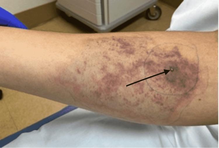
This clinical photograph displays a characteristic skin lesion resulting from a brown recluse spider (Loxosceles reclusa) bite on an extremity. The primary lesion consists of a central tense vesicle (indicated by a black arrow) resting upon an area of developing cutaneous necrosis, presenting as a darkened, dusky induration. Surrounding this central point is a large, irregularly shaped region of mixed ecchymosis and gravitational spreading of erythema. The discoloration demonstrates the classic 'red, white, and blue' sign associated with loxoscelism: peripheral erythema (red), intermediate blanching or ischemia (white), and central hemorrhagic necrosis (blue/purple). A pen-marked circle outlines the immediate inflammatory reaction to monitor progression. This image serves as an educational example of loxoscelism-induced skin changes, illustrating local tissue destruction and the typical inflammatory response involving vasospasm and thrombosis.
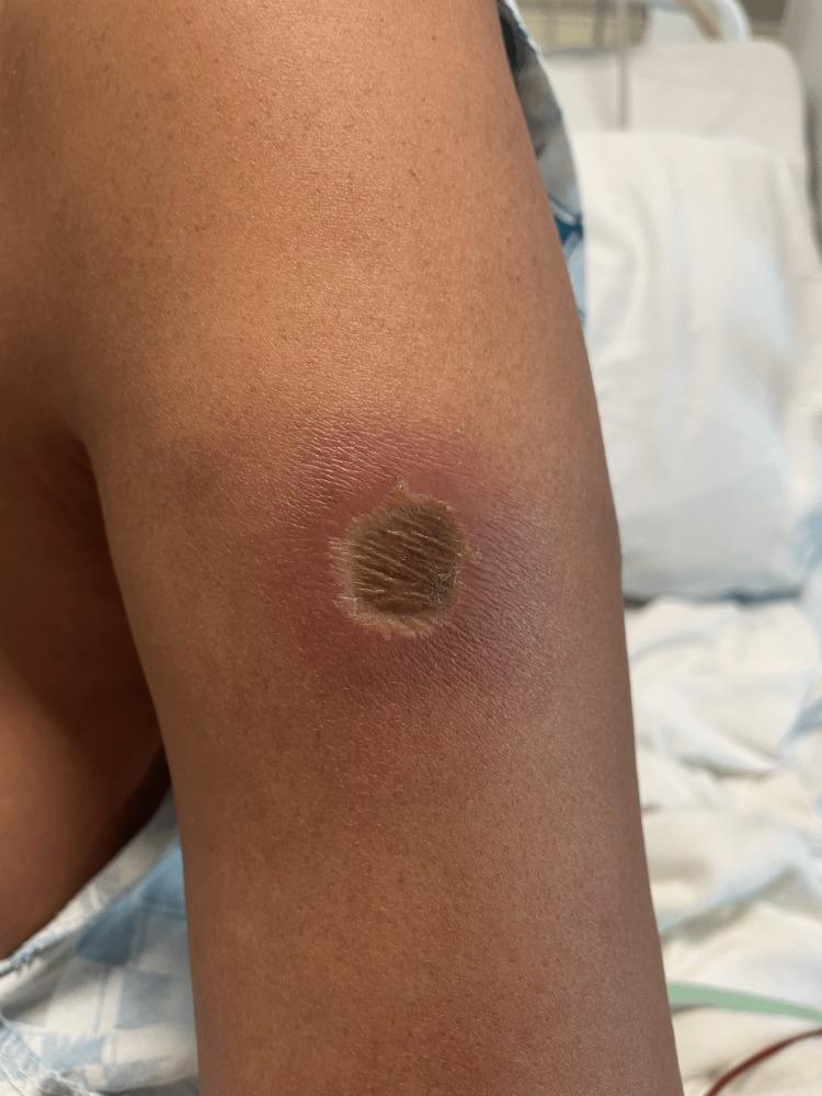
Clinical photograph of a skin lesion on the left upper extremity, characteristic of a brown recluse spider bite (Loxoscelism). The lesion exhibits a classic 'red, white, and blue' sign of envenomation. It features a central, depressed necrotic core that is dark brown and textured, indicating early dermal necrosis. Immediately surrounding the necrotic center is a pale, ischemic ring (central pallor). The outermost region consists of a well-defined area of erythema and inflammatory edema, which gradually fades into the surrounding normal skin. This appearance illustrates the progression of venom-induced tissue destruction, involving localized vasoconstriction and neutrophil-mediated inflammation leading to liquefactive necrosis. This image is an educational example of cutaneous arachnoidism and the diagnostic morphology used to identify necrotizing spider bites in a clinical setting.
Treatment of Spider Bites
1. General First Aid (All Spider Bites)
-
Clean the bite site thoroughly with soap and water
-
Apply a cold pack / ice wrapped in cloth for local pain relief
-
Elevate the affected limb; remove rings/jewelry to prevent constriction from swelling
-
Keep the patient calm and still
-
Update tetanus prophylaxis if not current within 5-10 years
-
Do NOT try to capture the spider live - a photograph is sufficient for identification
-
Transport to a medical facility if systemic symptoms develop
-
Tintinalli's Emergency Medicine, p. 1393
2. Brown Recluse Spider (Loxosceles reclusa) - Necrotic Arachnidism
Recognition
- Bite is often painless initially
- "Red, white, and blue" sign develops over 3-4 days: peripheral erythema (red), central blanching (white), ecchymosis/necrosis (blue)
- Hemorrhagic blister may appear at 24 hours
- Systemic effects (rare, mainly in children): hemolysis, nausea, fever, rhabdomyolysis, thrombocytopenia, renal failure - occurring 24-72 hours post-bite
Treatment
| Measure | Details |
|---|---|
| Wound care | Clean, keep dressed; monitor progression |
| Analgesia | Acetaminophen or NSAIDs for pain |
| Antibiotics | Only if secondary infection is present (not prophylactically) |
| Dapsone | Previously used to limit necrosis - no longer recommended (no validated evidence) |
| Surgical debridement | Avoid in the acute phase - early debridement can spread venom effects and worsen necrosis |
| Skin grafting | Reserved for large cosmetically significant necrotic defects (weeks later) |
| Systemic loxoscelism | Hospital admission; supportive care; monitor CBC, creatinine, coagulation; transfusion if needed |
| Antivenom | Available in Brazil; evidence for effectiveness is equivocal |
- Lab workup for possible systemic involvement: CBC, BUN/creatinine, LFTs, coagulation profile, urinalysis (hemoglobinuria), LDH
- Tintinalli's Emergency Medicine, p. 1394; Goldman-Cecil Medicine; Rosen's Emergency Medicine
3. Black Widow Spider (Latrodectus) - Latrodectism
Recognition
- Pinprick bite with characteristic target lesion (1-2 cm)
- Venom releases acetylcholine and norepinephrine -> massive neurotransmitter release
- Syndrome: localized pain -> spreads to entire extremity -> severe muscle cramps of trunk, back, abdomen
- Hypertension, tachycardia, diaphoresis, nausea, malaise
Treatment
| Step | Details |
|---|---|
| Wound care | Cleanse with soap and water; assess tetanus status |
| Analgesia | IV opioids (for severe pain) |
| Benzodiazepines | IV (e.g., diazepam or lorazepam) for muscle cramps - first-line |
| Calcium gluconate | NOT recommended - lacks supporting evidence |
| Antivenom (Latrodectus) | Equine IgG (Merck); 1 vial (2.5 mL) diluted in 10-50 mL NS, given IV over 15-30 min |
-
Whole equine IgG - risk of anaphylaxis and serum sickness
-
Skin testing does NOT reliably exclude anaphylaxis - not recommended
-
Short supply in the United States
-
Latrodectism is generally not life-threatening - weigh risk vs. benefit carefully
-
Consult Poison Control (USA: 1-800-222-1222) before use
-
Rosen's Emergency Medicine, p. 821
4. Funnel-Web Spider (Australia only)
- Extremely dangerous - can kill within 60 minutes
- Severe pain, parasympathetic storm, fasciculation, pulmonary edema, cerebral edema
- First aid: Pressure immobilization bandage (unique to this spider - venom acts rapidly)
- Specific antivenom is very effective - no deaths reported since its availability
- Urgent hospital transfer
5. Banana/Armed Spider (Phoneutria, South America)
- Intense pain, autonomic instability, priapism (common in boys)
- Antivenom available in Brazil - reserved for severe cases
- Mild-moderate cases: analgesia + local anesthetic agents
6. Tarantula Bites
- Painful bite with local erythema and edema only
- Major risk: barbed urticating hairs can penetrate eyes - ophthalmology consult if red eye/pain after exposure
- No antivenom; symptomatic treatment
7. Other/Unidentified Spider Bites
- Supportive and symptomatic treatment
- Ice/cold compress for pain
- Do NOT use pressure immobilization (except funnel-web - see above)
- No antivenoms available for most species
Quick Summary Table
| Spider | Key Feature | Main Treatment |
|---|---|---|
| Brown recluse (Loxosceles) | Painless bite, necrotic ulcer | Wound care, avoid early surgery, no dapsone |
| Black widow (Latrodectus) | Severe muscle cramps, hypertension | Opioids + benzodiazepines, antivenom if severe |
| Funnel-web (Australia) | Rapid systemic collapse | Pressure bandage + specific antivenom URGENTLY |
| Banana spider (Phoneutria) | Pain + priapism | Analgesia, antivenom for severe cases |
| Tarantula | Eye hair injury | Ophthalmology consult |
| Unknown/most spiders | Local pain only | Ice, analgesia, supportive care |
In India house hold in Neemuch MP
common household spiders India Madhya Pradesh venomous species bites
venomous spiders found in houses India Rajasthan Madhya Pradesh redback widow wolf spider
black widow spider India household
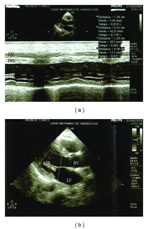
This clinical diagnostic image displays two panels of transthoracic echocardiography from a patient following Black Widow Spider (BWS) envenomation, illustrating myocarditis-like cardiac dysfunction and recovery. Panel (a) features a M-mode echocardiogram in the parasternal long-axis view. It identifies the Right Ventricle (RV), Interventricular Septum (IVS), Left Ventricle (LV), and Posterior Wall (PW). The M-mode tracing is used to assess ventricular wall motion and dimensions, with quantitative measurements of distance and slope (velocity) visible on the right. This panel corresponds to an initial Left Ventricular Ejection Fraction (LVEF) of 48% with noted hypokinesis. Panel (b) shows a 2D echocardiogram with color Doppler overlay in a similar view. Key cardiac chambers are labeled (RV, LV), and a color Doppler flow signal is identified as MR (Mitral Regurgitation) near the mitral valve. This follow-up image demonstrates clinical improvement, showing a reduction in mitral regurgitation to grade I and a recovering LVEF of 50%. The color scale on the right provides a reference for blood flow velocity and direction.
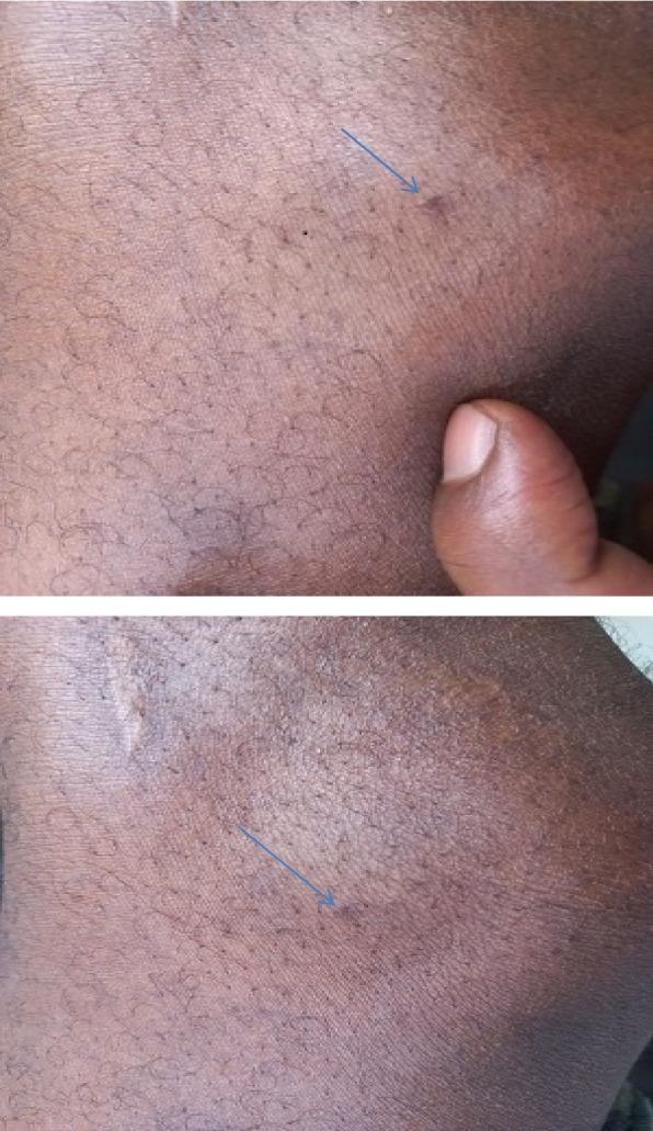
This clinical photograph shows a close-up of a skin lesion on the lateral aspect of the left knee of a patient with dark complexion. The image is split into two panels, both utilizing a blue arrow to indicate the primary site of interest. The lesion consists of a tiny, dark, circular puncture mark, consistent with a fang mark from a Latrodectus (black widow spider) bite. The surrounding skin exhibits minimal inflammatory response, with no significant erythema, edema, or induration visible in the immediate area. The skin texture is generally smooth with normal hair distribution and fine surface lines. A clinician's thumb is visible in the upper panel providing a scale for the lesion's minute size. This visual serves as a teaching tool for recognizing the subtle local dermatological findings often associated with latrodectism, where systemic symptoms (such as muscle pain and autonomic instability) typically outweigh visible skin trauma.
Spider Bites in Indian Households - Neemuch, MP
Spiders You Are Likely to Encounter at Home
| Spider | Local Name | Risk Level | Likely Location in Home |
|---|---|---|---|
| Wolf Spider (Lycosidae) | "Bhediya makdi" | Low-Moderate | Floors, under furniture, garden, shoes |
| Black Widow / Latrodectus (L. elegans / L. erythromaculatus) | "Kali makdi" | High | Dark corners, storerooms, under stones, toilets, boxes |
| Yellow Sac Spider (Cheiracanthium) | - | Low-Moderate | Walls, ceiling corners, inside folded clothes |
| Huntsman Spider | - | Low | Walls, behind cupboards |
| Jumping Spider | - | Very Low | Walls, windows |
| Common House Spider | - | Negligible | Corners, window frames |
Most important species in your region: The black widow (Latrodectus species) is present across India including Madhya Pradesh and Rajasthan. It is the most medically significant spider in Indian households. Wolf spiders are very common on floors and in shoes.
What to Do If Bitten at Home in Neemuch
Step 1 - First Aid (Do Immediately)
- Stay calm - panic increases heart rate and speeds venom spread
- Clean the bite with soap and water thoroughly
- Apply ice wrapped in cloth to the bite site for pain relief
- Elevate the bitten limb
- Remove rings / bangles / kadas from the affected hand/arm
- Try to photograph the spider (do NOT catch it)
- Note the time of the bite
Step 2 - When to Rush to Hospital
- Pain is spreading beyond the bite site (entire arm/leg/abdomen)
- Muscle cramps or rigidity, especially in the abdomen or back
- Excessive sweating
- Nausea, vomiting, headache
- Chest tightness or difficulty breathing
- High blood pressure / fast heart rate
- The person is a child, elderly, or pregnant
Treatment Based on Spider Type (India-Relevant)
Black Widow Bite (Latrodectus) - Most Dangerous in Your Area
- IV opioids (morphine/tramadol) for pain
- IV benzodiazepines (diazepam) for muscle cramps - this is the backbone of treatment
- IV fluids + monitoring of BP, heart rate
- Antivenom: In India, no widely available commercial Latrodectus antivenom exists. Management is primarily supportive (pain + muscle cramp control). If severe, contact National Poisons Information Centre (NPIC), AIIMS Delhi: 1800-11-6117 (toll free, 24/7)
- Admit and observe for 12-24 hours
Wolf Spider Bite - Common in Neemuch Homes
- Painful bite like a bee sting, local redness and swelling
- Not dangerous in otherwise healthy adults
- Treatment: Clean wound, ice pack, OTC paracetamol or ibuprofen for pain
- Watch for signs of infection (increasing redness, pus, warmth) over next 48 hours
- Tetanus booster if not updated in 5 years
Yellow Sac Spider / Unknown House Spider Bite
- Mild local pain, redness, and possible small blister
- Treatment: Clean, ice, analgesics (paracetamol/ibuprofen)
- Antibiotics only if infected - do NOT self-prescribe prophylactically
- Usually heals in 3-7 days without specific treatment
Practical Home Prevention (Neemuch Context)
- Shake out shoes before wearing, especially in the morning
- Check stored clothes, bags, and boxes before putting hands inside
- Seal gaps and cracks in walls, especially in storerooms and bathrooms
- Clear clutter from under beds, behind furniture, and in unused corners
- Use a torch at night when working in dark areas (garage, godown, outdoor toilets)
- Mosquito/pest control (general pest control reduces spider food sources)
- Teach children not to poke at webs or dark corners
Emergency Contacts - Neemuch, MP
| Resource | Contact |
|---|---|
| Neemuch District Hospital | 07423-222444 |
| National Poisons Information Centre (AIIMS Delhi) | 1800-11-6117 (24/7, toll-free) |
| MP Medical Helpline | 104 |
| National Emergency | 112 |