
Expansile medullary glioma with mass effects on cm cistern ,adjacent cerebellar hemispheres & pontomedullary junction understanding with images & treatment plan
medullary glioma MRI brainstem expansile mass
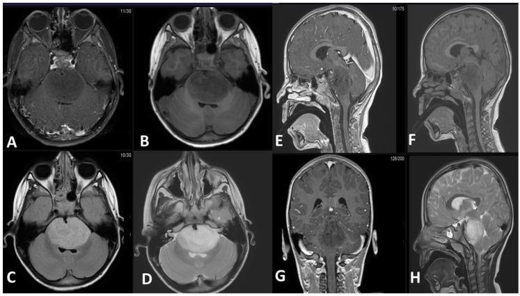
This diagnostic imaging panel consists of eight MRI slices (axial, sagittal, and coronal planes) demonstrating an expansile intra-axial mass in the brainstem, characteristic of a Diffuse Midline Glioma (DMG). The mass is primarily centered in the pons and exhibits a classic radiological profile: hypointense signal on T1-weighted pre-contrast (B, F) and post-contrast (A, E, G) sequences, indicating a lack of significant enhancement. T2-weighted (D, H) and FLAIR (C) sequences show the lesion as uniformly hyperintense. Anatomically, the tumor causes significant expansion of the pons, leading to the obliteration of the prepontine cistern and encasement of the basilar artery. Posteriorly, the mass compresses the fourth ventricle, though no proximal obstructive hydrocephalus is currently evident. Sagittal and coronal views (E, F, G, H) illustrate the longitudinal extent of the lesion, stretching from the pontomesencephalic junction superiorly to the pontomedullary junction inferiorly, with characteristically ill-defined, infiltrative borders. This series serves as a pedagogical example of the typical imaging features and local mass effect of pontine gliomas in neuro-oncology.
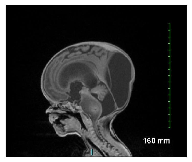
This sagittal T1-weighted fetal MRI scan demonstrates significant intracranial pathology in a developing fetus. The primary finding is a large, expansile, and poorly demarcated mass involving the brainstem, centered in the pons. The mass appears heterogeneously signal-intense and significantly expands the diameter of the pons, extending superiorly towards the midbrain and inferiorly toward the medulla. Secondary to this mass, there is evidence of severe obstructive hydrocephalus, characterized by marked dilation of the lateral ventricles and the third ventricle, with associated thinning of the overlying cerebral mantle and macrocephaly. The posterior fossa shows displacement of the cerebellar structures. This visual presentation is highly characteristic of a congenital brainstem glioma, specifically a diffuse intrinsic pontine glioma (DIPG) or similar anaplastic neoplasm, illustrating the typical obstructive complications and mass effect associated with neonatal neuro-oncology.
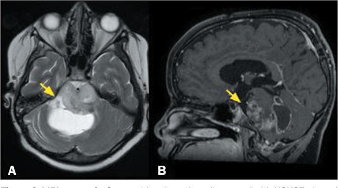
This diagnostic imaging composite displays two magnetic resonance imaging (MRI) sequences of the brain in a pediatric patient, illustrating a diffuse midline glioma affecting the brainstem. Image A is an axial T2-weighted sequence showing an expansile, infiltrative lesion located in the pons. The lesion demonstrates a heterogeneous signal intensity, with hyperintense focal areas (yellow arrow) indicative of cystic components or internal necrosis. The mass effect is evident as it expands the pontine contours. Image B is a sagittal, fat-saturated, T1-weighted sequence with gadolinium contrast enhancement. It reveals the solid portions of the tumor exhibit irregular, heterogeneous enhancement (yellow arrow), suggesting varying vascularity and blood-brain barrier disruption within the mass. The clinical presentation and imaging features are characteristic of a high-grade pediatric-type diffuse midline glioma, specifically the H3K27-altered molecular subtype. This case highlights the typical neuroradiological appearance of aggressive midline gliomas in children, focusing on expansile growth, signal heterogeneity, and variable contrast uptake.
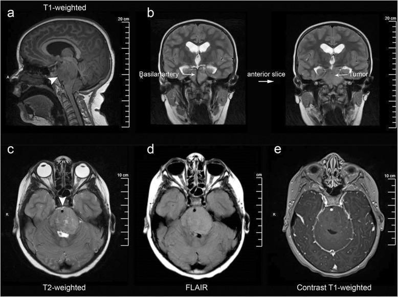
Multi-modal MRI series of the brain demonstrating a Diffuse Intrinsic Pontine Glioma (DIPG). (a) T1-weighted sagittal view shows an infiltrative, expansile mass centered within the pons. (b) Coronal T2-weighted images reveal the exophytic tumor portion extending into the prepontine and suprasellar cisterns, notably encasing the basilar artery. (c-e) Axial sequences highlight characteristic signal patterns: the lesion is hyperintense on T2-weighted (c) and Fluid-Attenuated Inversion Recovery (FLAIR) (d) sequences, indicating significant vasogenic edema or tumor infiltration. (e) Post-gadolinium T1-weighted axial sequence shows a conspicuous absence of enhancement, a typical feature of DIPG indicating an intact blood-brain barrier within the tumor. Associated secondary findings include obstructive hydrocephalus, evidenced by dilated lateral ventricles in the coronal views. This imaging series illustrates the classic neuroradiological criteria for pediatric brainstem gliomas, emphasizing anatomical distortion of the brainstem, cisternal extension, and specific signal characteristics across varying MRI sequences.
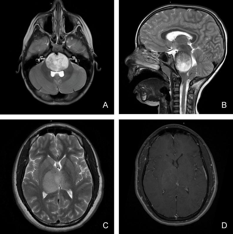
This composite of four magnetic resonance imaging (MRI) scans illustrates the diagnostic features of pediatric diffuse midline glioma in different anatomical locations. Panels A (axial) and B (sagittal) display T2-weighted sequences showing a large, hyperintense, and expansile mass involving the brainstem (pons), characteristic of a diffuse intrinsic pontine glioma (DIPG). The lesion demonstrates an infiltrative growth pattern, causing significant local mass effect and compression of adjacent structures. Panels C and D focus on a thalamic presentation. Panel C is an axial T2-weighted image showing a homogenous, hyperintense lesion within the right thalamus with poorly defined margins extending into the surrounding white matter tracts. Panel D shows the corresponding axial T1-weighted post-gadolinium sequence, revealing minimal to heterogeneous contrast enhancement within the thalamic mass. These images highlight key radiological hallmarks including the tumor's midline location, infiltrative nature, perilesional edema, and variable vascular permeability as indicated by enhancement patterns, which are critical for the clinical classification of these high-grade pediatric gliomas.
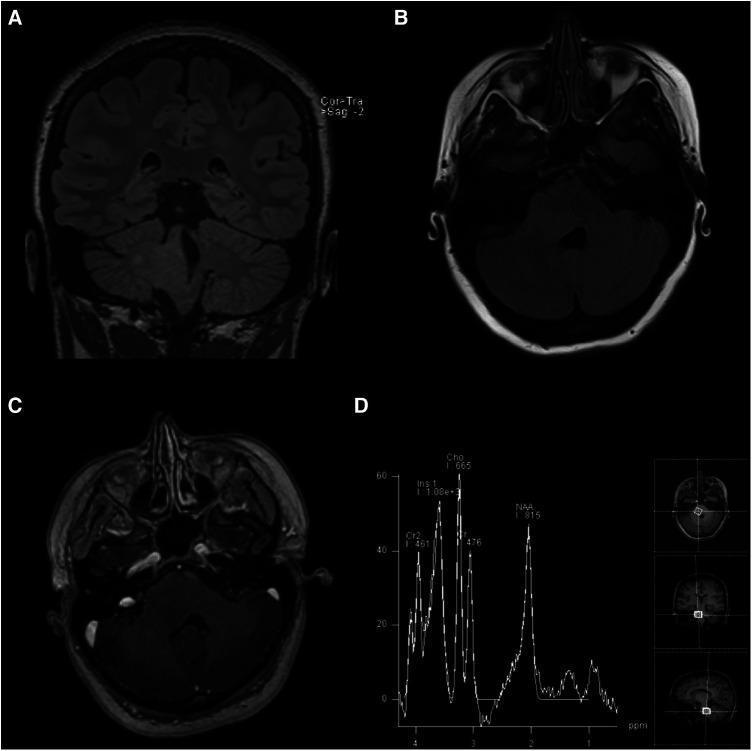
This composite of four images (A-D) presents a multi-modal MRI evaluation of a brainstem lesion. Panels (A) Coronal T2-weighted FLAIR and (B) Axial T2-weighted FLAIR demonstrate an ill-defined, hyperintense, and expansile mass within the right pons. The lesion extends into the right middle cerebellar peduncle and the right posterolateral medulla, causing partial effacement of the fourth ventricle. Panel (C) represents an axial post-gadolinium contrast T1-weighted image, which shows no contrast enhancement, indicating a preserved blood-brain barrier within the lesion. Panel (D) provides Magnetic Resonance Spectroscopy (MRS) data, showing a markedly increased choline (Cho) peak and a significantly decreased N-acetylaspartate (NAA) peak. This metabolic profile (high Cho:NAA ratio) is characteristic of increased cellular turnover and loss of neuronal integrity, typically seen in infiltrating gliomas such as Diffuse Intrinsic Pontine Glioma (DIPG). The findings are essential for neurosurgical and oncological education regarding the diagnostic imaging and metabolic characteristics of pediatric-type diffuse low-grade gliomas in adult patients.
medullary glioma brainstem treatment outcomes
diffuse midline glioma medulla brainstem H3K27M
diffuse midline glioma H3K27M ONC201 treatment 2024
PMID: 38704025
PMID: 39700833
Expansile Medullary Glioma: Anatomy, Imaging, Mass Effects & Treatment Plan
1. Overview and Classification
| Type | Behavior | Prognosis |
|---|---|---|
| Diffuse infiltrating (majority) | Asymmetric expansion, ill-defined margins, T2 hyperintense | Poor (median survival ~12-15 months) |
| Focal/nodular (dorsal exophytic) | Exophytic growth into cisterna magna, better defined | Better (may be pilocytic) |
- Adams and Victor's Principles of Neurology, 12th Edition - Glioma of the Brainstem
2. Anatomy of the Medulla and Relevant Structures
2.1 Cisternal Compression (Cisterna Magna)
- The cisterna magna is the largest subarachnoid cistern, dorsal to the medulla and inferior to the cerebellar vermis/tonsils
- An expanding dorsal or posterolateral medullary tumor pushes into this cistern, obliterating it
- This can obstruct CSF flow at the level of the fourth ventricular outflow foramina (Magendie and Luschka), producing obstructive hydrocephalus
- Compression of cerebellar tonsils downward can mimic or coexist with Chiari malformation
2.2 Adjacent Cerebellar Hemisphere Compression
- Lateral expansion compresses the cerebellar peduncles (inferior and middle) disrupting cerebellar input/output pathways
- Results in ipsilateral cerebellar ataxia, limb dysmetria, intention tremor, and wide-based gait
- Mass effect on the flocculus/nodulus disrupts vestibulo-cerebellar function (nystagmus, vertigo)
2.3 Pontomedullary Junction Involvement
- This junction is anatomically packed: cranial nerves VI, VII, VIII exit here; ascending/descending tracts cross
- Tumor spread to or pressure on this region produces a complex syndrome of lower pons + upper medullary signs
- Corticospinal and corticobulbar tracts pass through, so contralateral hemiparesis is common
- Localization in Clinical Neurology, 8e emphasizes the pontomedullary sulcus as the exit zone of CN VI-VIII
3. Clinical Presentation
Lower Cranial Nerve Palsies (most common initial symptom)
- CN IX/X (glossopharyngeal/vagus): dysphagia, dysphonia, hoarseness, absent gag reflex
- CN XII (hypoglossal): tongue deviation, dysarthria, atrophy
- CN XI (accessory): weakness of SCM and trapezius
- CN VI/VII if pontomedullary junction is involved: lateral gaze palsy, facial weakness
Long Tract Signs
- Contralateral hemiparesis (corticospinal tract involvement)
- Hemisensory loss
- Bilateral pyramidal signs if diffuse
Cerebellar Signs
- Ipsilateral limb ataxia, gait instability, dysmetria
- Nystagmus (especially horizontal or downbeat)
Autonomic and Vital Sign Abnormalities (medullary-specific)
- Hiccups, central respiratory irregularity
- Medullary satiety syndrome - anorexia, early satiety from involvement of the nucleus tractus solitarius and dorsal vagal complex
- Orthostatic hypotension (lateral tegmental pathways)
Raised ICP Features (late)
- Headache, vomiting, papilledema from hydrocephalus
4. Neuroimaging
MRI - The Gold Standard
| Sequence | Typical Appearance in DMG |
|---|---|
| T1 | Hypointense (darker than normal brainstem) |
| T2/FLAIR | Diffuse hyperintensity filling/expanding the medulla |
| T1 + Gadolinium | Usually NO enhancement (intact BBB); ring/nodular enhancement = higher grade |
| DWI | Variable; restricted diffusion may suggest cellularity |
| MR Spectroscopy | High Cho:NAA ratio (increased turnover, neuronal loss) |
Image Gallery
Mass Effect Summary on MRI
- Cisterna magna: Obliteration/effacement by posteriorly expanding tumor; hydrocephalus if fourth ventricular outflow is blocked
- Cerebellar hemispheres: Lateral tumor spread compresses/displaces cerebellar tissue; edema in adjacent cerebellar white matter
- Pontomedullary junction: Loss of normal sulcal anatomy; encasement of the basilar artery branches and AICA/PICA if exophytic
Tissue Diagnosis
5. Molecular Biology
- Global reduction in H3K27me3: Activates oncogenic transcription programs
- Polycomb repressive complex dysregulation: Widespread epigenetic reprogramming
- Drives aggressive, treatment-resistant behavior
- Diagnostic significance: Loss of H3K27me3 immunostaining on IHC is a surrogate marker
- ACVR1 mutations (seen especially in H3.1K27M tumors, pontine predominance)
- PDGFRA amplification
- TP53, PPM1D mutations
- EGFR amplification (rare)
6. Treatment Plan
6.1 Surgical Management
- High risk to vital nuclei (respiratory, cardiac, swallowing centers)
- Infiltrative margins preclude gross total resection
- No survival benefit from debulking
- Focal/dorsal exophytic pilocytic astrocytoma: Resection of the mural nodule via suboccipital craniotomy can be curative
- Hydrocephalus management: Endoscopic third ventriculostomy (ETV) or ventriculoperitoneal (VP) shunt for obstructive hydrocephalus from cisternal obliteration
- Stereotactic biopsy: For molecular diagnosis when imaging is atypical - increasingly feasible and low-morbidity in experienced centers
6.2 Radiation Therapy - Primary Treatment
| Regimen | Dose | Fractions | Context |
|---|---|---|---|
| Standard fractionation | 54 Gy | 30 fractions (1.8 Gy/fx) | Definitive primary treatment |
| Alternative standard | 59.4 Gy | 33 fractions | Some centers |
| Hypofractionation | 39 Gy | 13 fractions (3 Gy/fx) | Young children <3 yrs, palliative intent |
| Re-irradiation | 24 Gy | 12 fractions | Salvage at recurrence |
- Target volume includes GTV (T2/FLAIR abnormality) + 1-1.5 cm CTV margin
- Special attention to brainstem dose constraints: max point dose <54 Gy recommended for uninvolved brainstem
- Response: ~80% have temporary clinical improvement; median PFS ~6-9 months
6.3 Systemic Therapy
Standard Chemotherapy (Limited Benefit)
- Temozolomide: No established survival benefit in DMG; MGMT promoter methylation is rare
- Carboplatin + vincristine: Limited pediatric protocols; no Phase III data supporting routine use
- Bevacizumab: No survival benefit; may reduce steroid dependence transiently
Novel Targeted Therapies (Active Investigation)
- Mechanism: DRD2 antagonist + ClpP agonist leading to mitochondrial stress and selective tumor cell death
- Activity: Particularly active in H3K27M-mutant DMGs; regression seen in recurrent/progressive disease
- Clinical data (2025): Di Carlo et al., Eur J Cancer 2025 (PMID 39700833) - real-world compassionate use (174 patients, 14 countries): median OS from diagnosis 15.5 months overall; thalamic location and older age were favorable prognostic factors
- Current status: Phase III randomized trial ongoing for H3K27-altered DMG; FDA Breakthrough Therapy Designation
- ACVR1 inhibitors (e.g., LDN-212854): For ACVR1-mutant tumors (especially H3.1K27M)
- Panobinostat: HDAC inhibitor targeting the epigenetic dysregulation from H3K27M; clinical trials ongoing
- CAR-T cell therapy: GD2-targeting CAR-T cells; early-phase trials (GD2 is highly expressed on H3K27M tumors)
- Convection-enhanced delivery (CED): Direct intratumoral drug delivery bypassing BBB; clinical trials active for brainstem DMG
- Oncolytic viruses: Preliminary series show some prolongation of survival (per Adams and Victor's); mechanism (direct oncolytic vs. inflammatory) remains under study
6.4 Corticosteroids
- Dexamethasone (4-16 mg/day) for symptomatic vasogenic edema surrounding the tumor
- Not recommended as long-term maintenance - side effects substantial
- Particularly helpful when cisternal/cerebellar compression produces acute neurological deterioration
6.5 Supportive and Multidisciplinary Care
- Dysphagia management: Speech therapy, modified diet, nasogastric tube or PEG if severe
- Respiratory monitoring: SpO2 monitoring, overnight oximetry; NIV if hypoventilation occurs
- Physiotherapy: Ataxia/gait rehabilitation, fall prevention
- Palliative care integration: Early referral, goals-of-care discussions - prognosis is uniformly poor for diffuse type
- Endocrine assessment: HPA axis screening if hypothalamic extension occurs
- Audiological follow-up: CN VIII compression from cistern involvement
7. Prognosis
| Factor | Outcome Impact |
|---|---|
| Diffuse infiltrating type | Median OS 9-15 months |
| Focal/pilocytic type | Potentially curable with surgery |
| H3K27M present | WHO Grade 4; poor regardless of histology |
| H3.1K27M (vs H3.3K27M) | H3.1 slightly longer survival |
| Thalamic location | Better prognosis than brainstem |
| Medullary/pontine location | Worst prognosis |
| Adult vs. pediatric | Adults slightly longer OS (ONC201 data: 19.6 mo adults vs. 14.2 mo children) |
| No enhancement on MRI | Slightly better BBB integrity, less aggressive behavior |
8. Differential Diagnosis
- Diffuse Midline Glioma H3K27-altered (most likely in children/young adults)
- Pilocytic astrocytoma (focal, cystic, enhancing, better prognosis)
- ATRT (Atypical Teratoid/Rhabdoid Tumor) - infants, SMARCB1 loss
- Medulloblastoma - typically arises from roof of 4th ventricle, not intrinsic to medulla
- Ependymoma - typically 4th ventricular origin, may extend into cisterna magna
- Brainstem cavernous hemangioma - T1 hypointense hemosiderin rim, no mass effect pattern
- Multiple sclerosis plaque (usually no mass effect, enhances transiently)
- Metastasis - more common in adults, usually enhancing, may be multiple
- Brainstem encephalitis - diffuse T2 change but usually no mass effect, clinical course differs
Summary
- Cisternal obliteration → hydrocephalus risk, CSF flow obstruction
- Cerebellar compression → ipsilateral ataxia, dysmetria, nystagmus
- Pontomedullary compression → lower cranial nerve palsies, long tract signs, autonomic instability