
Azzoparde effect
Searching the Web
Azzopardi effect
The Azzopardi effect (also called the Azzopardi phenomenon) is a histopathological finding in which DNA from dying tumor cells leaches out and encrusts the walls of small blood vessels (venules), giving them a distinctive basophilic (blue) appearance on H&E-stained slides. It is most classically associated with small cell carcinoma of the lung.
Searching sources...
Reading File
Reading Web Page
https://pmc.ncbi.nlm.nih.gov/articles/PMC4774277
Finding Sources
I found it in Robbins Pathology. Let me pull the relevant section and image.
Reading File
I have the Robbins image showing the Azzopardi effect. Let me fetch it.
Loading Image
Here is a complete overview of the Azzopardi Effect (Azzopardi Phenomenon):
Azzopardi Effect
Definition
The Azzopardi effect is a histopathological finding in which DNA released from necrotic tumor cells encrusts the walls of small blood vessels (capillaries and venules), producing a characteristic deep basophilic (dark blue/purple) staining of the vessel wall on H&E-stained sections.
History and Eponym
Named after John G. Azzopardi, a Maltese-British pathologist, who in 1959 correctly identified the basophilic material as DNA - not calcium (as it had been incorrectly assumed before his work). He first described it after examining 100 cases of oat cell (small cell) carcinoma of the lung, finding the phenomenon in 32 of them.
Mechanism
- Small cell carcinoma consists of highly malignant, rapidly dividing cells with large hyperchromatic nuclei and scant cytoplasm.
- Rapid tumor growth outstrips blood supply, causing extensive necrosis.
- As tumor cells die and nuclei disintegrate, nuclear DNA is released into the surrounding tissue.
- This free DNA preferentially deposits on and encrusts the walls of nearby venules and capillaries - particularly in necrotic areas - due to physicochemical affinity.
- On H&E stain, DNA stains intensely hematoxyphilic (basophilic), giving vessels a smudged, deeply blue appearance throughout the full thickness of the wall.
Histological Appearance
On H&E sections you see:
- Deeply basophilic staining of vessel walls (full-thickness)
- Located within or adjacent to tumor, especially near areas of necrosis
- Coarsely granular, smudged material encrusting venules
Panel F in the image from Robbins below shows small cell carcinoma with the characteristic basophilic vessel wall encrustation (Azzopardi effect):
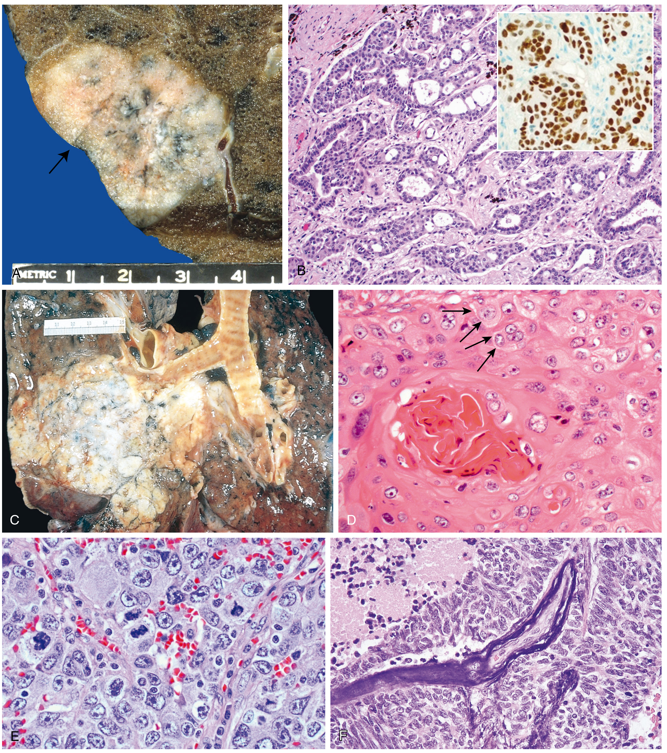
Fig. 15.34F from Robbins, Cotran & Kumar Pathologic Basis of Disease - Small cell carcinoma with deep basophilic staining of vascular walls (Azzopardi effect)
Tumors Associated with the Azzopardi Effect
The effect is not pathognomonic for small cell carcinoma - it can appear in any high-grade malignancy with rapid cell turnover and significant necrosis:
| Tumor | Frequency |
|---|---|
| Small cell carcinoma of the lung | Most classic - best described |
| Merkel cell carcinoma | Frequently seen |
| Medulloblastoma | Occasionally seen |
| Oral squamous cell carcinoma | Rarely |
| Polymorphous low-grade adenocarcinoma (salivary gland) | Rare |
Diagnostic Significance
- It is a marker of high-grade malignancy with brisk necrosis and rapid cell turnover.
- It is not specific for small cell carcinoma but should prompt the pathologist to consider it.
- Historically, it helped distinguish DNA deposition from calcium deposits (which were the earlier presumed cause).
- No direct therapeutic implications - it is a histological sign, not a treatable condition.
Key Points to Remember
- What: DNA from necrotic tumor cells encrusting venule walls - basophilic vessel staining on H&E
- Where: Predominantly in/near necrotic tumor tissue
- Classic tumor: Small cell carcinoma of the lung
- Who described it: John G. Azzopardi, 1959
- Previously confused with: Calcium deposition (dystrophic calcification)
- Diagnostic use: Indicator of high-grade tumor with necrosis; not specific
Source: Robbins, Cotran & Kumar Pathologic Basis of Disease, p. 671; PMC article on the Azzopardi phenomenon
This is a shared conversation. Sign in to Orris to start your own chat.