can myringostapediopexy that is done with short columella graft be called type 3 tympanoplasty
Reading File
Finding Sources
Finding Sources
Searching the Web
myringostapediopexy short columella type 3 tympanoplasty classification Wullstein
Reading File
Reading File
Loading Image
Loading Image
Now I have a very complete picture - including a subtle but important finding from the Shambaugh history chapter. Here is the full answer:
Yes - with an important nuance about how Wullstein himself labeled it
Short answer: In the standard modern (Wullstein) classification that most textbooks teach, myringostapediopexy using a short columella graft is classified as Type III tympanoplasty. However, the original Wullstein diagram actually labeled this configuration as "Type I+II" - which reveals an interesting historical wrinkle.
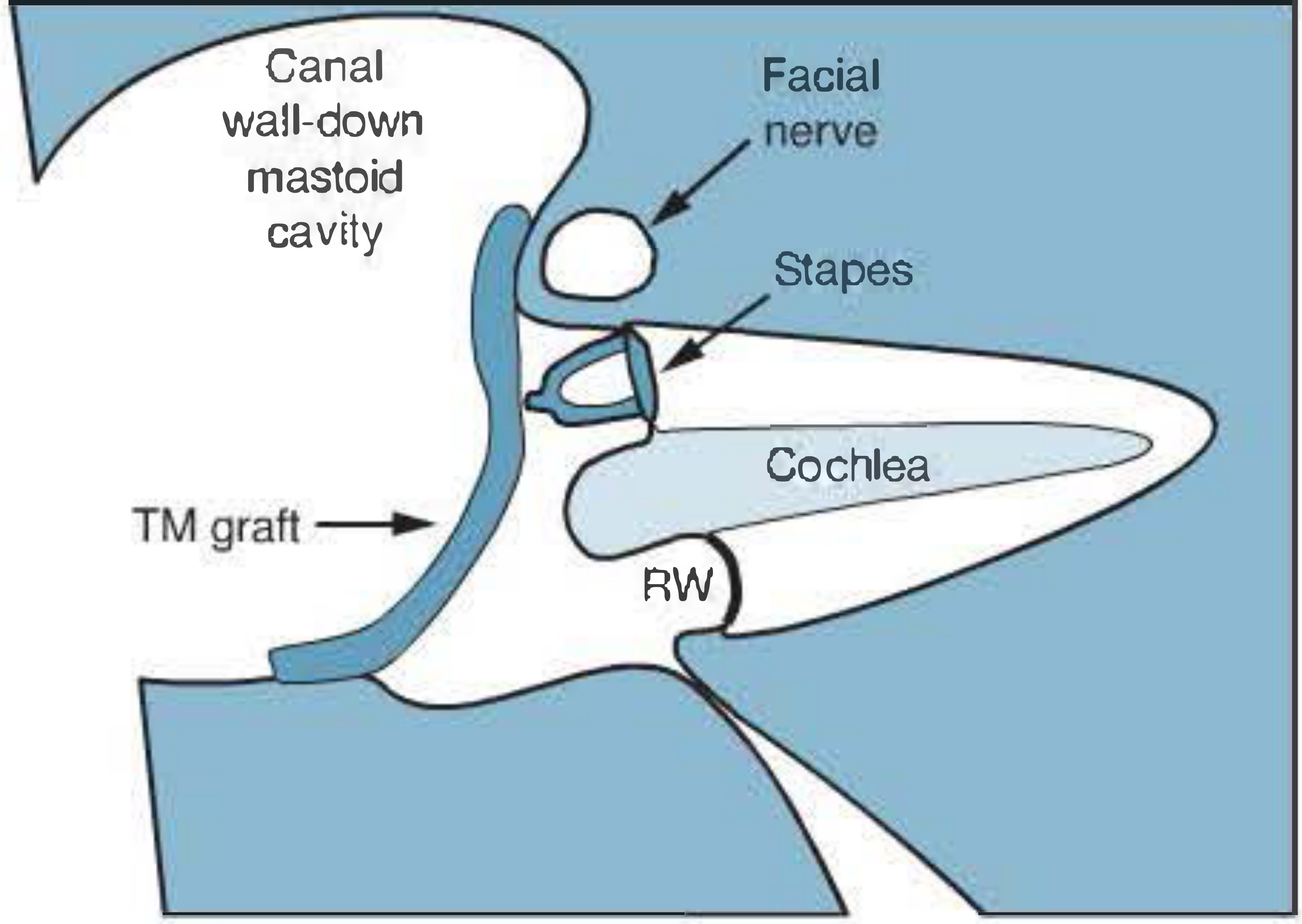
What is Myringostapediopexy?
It is the direct apposition of the tympanic membrane (or a graft) onto the stapes head (capitulum), using the intact stapes suprastructure as a "short columella." The malleus and incus are absent; sound is conducted directly from the new eardrum to the stapes.
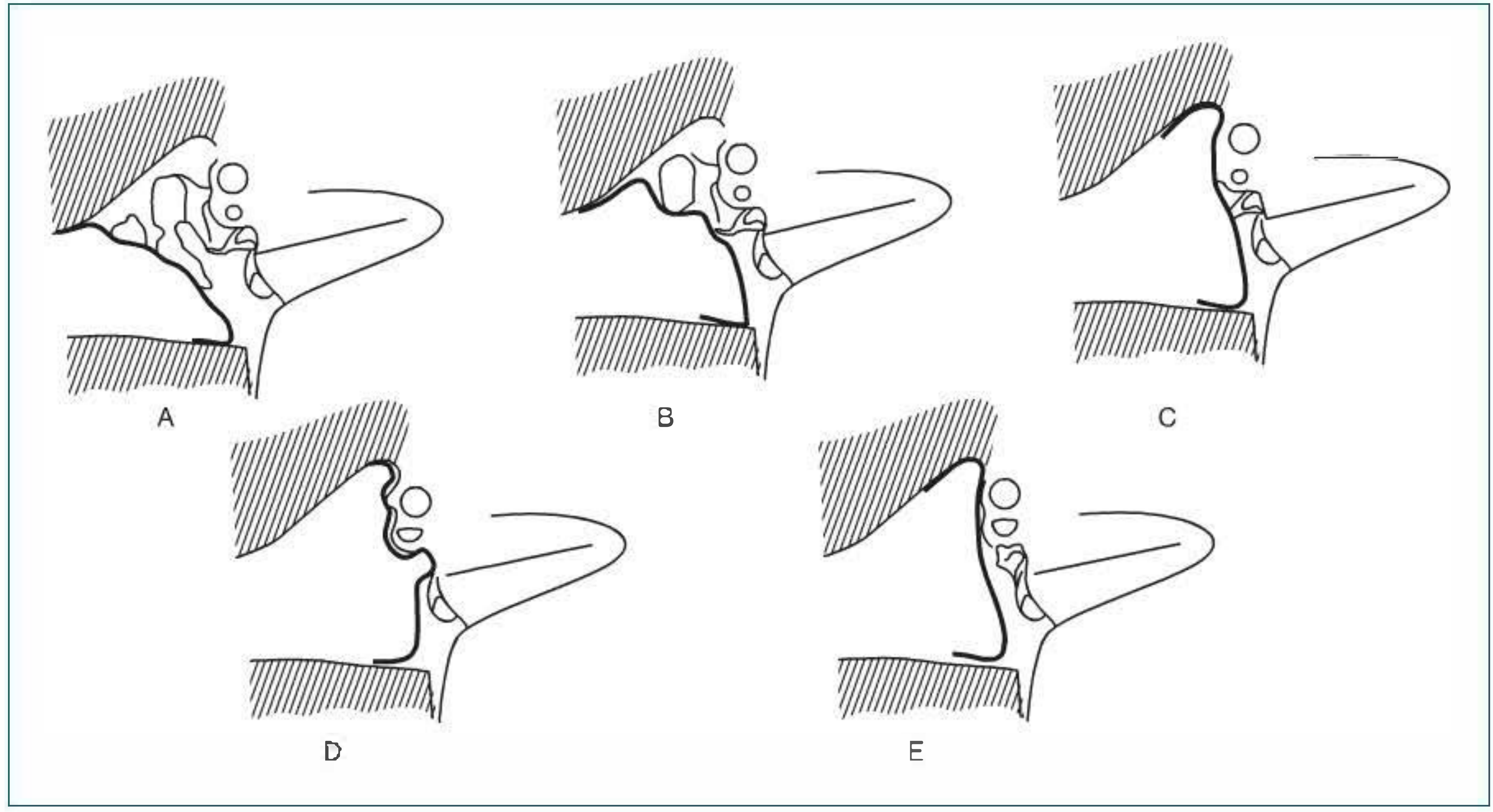
Wullstein's Classification - Type III
In the modern teaching of the Wullstein classification:
| Type | Ossicular Status | Graft Placement | Alternate Names |
|---|---|---|---|
| I | All ossicles intact | Over TM / malleus handle | Myringoplasty |
| II | Malleus eroded | Over incus or remnant malleus | - |
| III | Malleus + incus absent | Directly onto stapes head | Myringostapediopexy, Columella tympanoplasty, Stapes columella tympanoplasty |
| IV | Only stapes footplate present | Between oval and round window | Cavum minor |
| V | Fixed stapes footplate | New window in semicircular canal | Fenestration tympanoplasty |
Shambaugh Surgery of the Ear states explicitly:
"A classical type III or stapes columella tympanoplasty involves placement of a tympanic membrane graft such as temporalis fascia directly onto the stapes head - i.e., the ossicular chain is replaced by the single columella of the stapes."
The Historical Nuance - Wullstein's Own Original Labeling
This is the subtle but important point. In the original Wullstein classification diagram (reproduced in Shambaugh's history chapter), this exact operation - myringostapediopexy with short columella - was labeled "Type I+II", described as:
"Myringostapediopexy producing a shallow middle ear and a columella effect."
In Wullstein's original five-type scheme, what we now call "Type III" was an intermediate category he called "I+II." Over time, the classification was revised and simplified by the otologic community so that this procedure became formally designated Type III.
Summary
- By modern standard teaching (Wullstein classification as it is universally taught today): Yes, myringostapediopexy with a short columella graft = Type III tympanoplasty. The terms are synonymous.
- In Wullstein's original historical paper: This configuration was labeled "Type I+II" - an intermediate hybrid type, not a distinct Type III.
- The "short columella" in this context refers to the intact stapes suprastructure itself acting as the columella (as opposed to a "long columella"/TORP which goes from the footplate to the TM when the stapes arch is also absent).
So for exam and clinical purposes: yes, it is correctly called Type III tympanoplasty.
- Shambaugh: Surgery of the Ear, Chapter on Tympanoplasty, Type III stapes columella section and Figure 28-1
take shambaugh surgery of the ear book as reference and explain in detail the gloms jugular and tympanum chapters especially the surgical management
Finding Sources
Reading File
Reading File
Reading File
Reading File
Reading File
Reading File
Reading File
Reading File
Finding Sources
Reading File
Reading File
Reading File
Reading File
Reading File
Reading File
Reading File
Reading File
Loading Image
Loading Image
Loading Image
Now I have all the material needed for a comprehensive response. Here is the full detailed explanation:
Glomus Jugulare and Glomus Tympanicum - Shambaugh Surgery of the Ear
1. Pathology and Origin
Glomus tumors (GTs) are paragangliomas - benign, highly vascular neoplasms arising from the paraganglion cells of the diffuse neuroendocrine system (DNES). Their cells of origin are the jugulotympanic paraganglions: ovoid, lobulated structures measuring 0.1 to 1.5 mm in diameter, found in association with Jacobson's nerve (tympanic branch of CN IX) and Arnold's nerve (auricular branch of CN X). More than 50% are located in the region of the jugular fossa.
Histologically, chief cells are arranged in clusters called Zellballen, supported by sustentacular cells and small blood vessels. The chief cells contain cytoplasmic granules that store catecholamines. Two ultrastructural subtypes of chief cells - light and dark - are identified.
Biochemistry: Paraganglions lack the enzyme phenylethanolamine-N-methyltransferase, so they predominantly secrete norepinephrine (not epinephrine). Functioning ("secretor") tumors occur in 1-3% of cases. Norepinephrine levels must be elevated 3-5x normal to produce clinical symptoms (headaches, sweating, palpitations, pallor, nausea).
2. Epidemiology and Genetics
- GTs arise more often in Caucasians
- Females are 4-6 times more commonly affected than males
- Peak incidence in the 5th and 6th decades, though any age is possible
- Heredofamilial tendency exists with autosomal dominant transmission
- In familial tumors, associated lesions occur in 25-50% of cases
- 10% of non-familial cases have multiple synchronous tumors
- GTs are characteristically slow-growing and rarely metastasize
- Association exists with pheochromocytoma, thyroid neoplasms, parathyroid adenoma, and MEN syndromes
3. Spread and Invasion
GTs spread along tracts of least resistance: the air cell tracts of the temporal bone, vascular lumina, neurovascular foramina, the Eustachian tube, and direct extension. They invade bone. Cochleovestibular destruction is caused by ischemic necrosis. Spread is multidirectional and simultaneous.
Intracranial extension (ICE) into the posterior cranial fossa occurs directly through dura or along cranial nerve routes - the internal auditory canal is a frequent highway.
Cranial nerve paralysis occurs in:
- 35% of jugulotympanic lesions
- 57% of intravagal paragangliomas
- CN VII through XII and the sympathetic trunk are most commonly involved
4. Classification: Glasscock-Jackson Staging System
This is the classification system used throughout the Shambaugh text (Table 41-1):
Glomus Tympanicum (GTy)
| Stage | Description |
|---|---|
| Type I | Small mass limited to the promontory |
| Type II | Tumor completely filling the middle ear space |
| Type III | Tumor filling middle ear and extending into mastoid process |
| Type IV | Tumor filling middle ear, extending into mastoid or through tympanic membrane to fill EAC; may extend anterior to the ICA |
Glomus Jugulare (GJ)
| Stage | Description |
|---|---|
| Type I | Small tumor involving jugular bulb, middle ear, and mastoid |
| Type II | Tumor extending under internal auditory canal; may have intracranial extension |
| Type III | Tumor extending into petrous apex; may have intracranial extension |
| Type IV | Tumor extending beyond petrous apex into clivus or infratemporal fossa; may have intracranial extension |
5. Clinical Features
The classic presenting symptoms (from Table 41-2):
| Symptom | Glomus Jugulare (n=106) | Glomus Vagale (n=27) |
|---|---|---|
| Pulsatile tinnitus | 84% | 8% |
| Hearing loss | 62% | 4% |
| Aural fullness | 32% | 3% |
| Otalgia | 13% | 3% |
| Hoarseness | 12% | 4% |
| Dysphagia | 8% | - |
- Tumor growth into mesotympanum causes conductive hearing loss; labyrinthine invasion produces a sensorineural component
- TM erosion and bleeding are late symptoms
- Cranial neuropathy indicates extensive disease
- "Idiopathic" cranial neuropathy is an unacceptable diagnosis - it mandates aggressive imaging to exclude a lateral skull base lesion
- Facial paralysis is usually a late sign and an ominous omen for facial nerve outcome
Important warning: Myringotomy or tympanotomy for biopsy is condemned - it causes brisk bleeding that must be packed, risking damage to ear structures. Biopsy of an aberrant ICA can be catastrophic.
6. Diagnosis and Preoperative Workup
The diagnostic process serves as a treatment planning tool. Goals:
- Determine tumor size, type, and extent
- Evaluate histochemical or multicentric lesions
- Identify and assess intracranial extension
- Assess major vasculature involvement
- Assess intracranial collateral circulation
Imaging
CT of temporal bone (with bone windows, axial and coronal):
- Differentiates GTy from GJ: presence of air or bone between the tumor mass and jugular bulb = GTy tumor
- Examine the caroticojugular plate (spine) separating jugular bulb from ICA - eroded early in GJ tumors; a mottled plate is characteristic for GJ tumors
- GTy: well-defined enhancing soft tissue mass over the promontory, hypotympanic floor and jugular fossa usually intact
MRI:
- Characterizes GTs by multiple vascular flow voids ("salt and pepper" appearance) on T2
- Differentiates from schwannomas (homogeneous enhancement, possible cystic areas)
- Best for evaluating ICE and neural/vascular structure involvement
- MRI of head and neck assesses multicentricity
CT angiography / CT venography: Defines vascular supply, ICA involvement, jugular vein patency.
Bilateral carotid angiography: Performed preoperatively at the time of tumor embolization to evaluate ICA involvement and map tumor blood supply (especially important for ICE, which can draw supply from pial vessels, vertebral artery, ICA, AICA, and PICA in addition to ECA sources).
Octreotide scanning: ¹²³I-labeled Tyr3-octreotide. Sensitive and specific for paragangliomas down to 1 cm resolution. Also detects recurrences and other endocrine tumors in MEN syndrome and metastatic disease.
Biochemical Screening
- Every GT patient (except small GTy) gets serum catecholamines and urinary metabolites
- Elevated epinephrine mandates adrenal CT or selective renal vein sampling to exclude pheochromocytoma
- For GTy without family history or multiplicity, biochemical screening can be omitted
7. Treatment Planning
Treatment is palliative or definitive (curative). Key factors:
- Patient age and physiological status
- Tumor type and natural history
- Probability the tumor causes morbidity in the patient's remaining lifespan
Palliation is recommended for patients >65-70 years physiologically, the medically infirm, or select multicentric lesions. Small GTs in elderly patients are unlikely to cause concern in remaining years due to their slow growth. Asymptomatic patients elected for palliation are followed with serial imaging; if growth or symptoms develop, radiation is offered.
Bilateral GJ tumors: The more life-threatening lesion is operated first. Contralateral surgery planned at 6 months if the patient emerges neurologically intact. Extensive bilateral cranial nerve loss can lead to permanent tracheostomy, tracheal diversion, or artificial alimentary support - making further surgery inadvisable.
8. Radiation Therapy
Shambaugh takes a cautious stance on RT:
- RT and stereotactic radiosurgery (SRS) are offered as a minimal, low-morbidity, low-cost conservation strategy
- Virtually any lesion is technically resectable; the primary criticism of surgery is the risk of functional incapacity
- The RT position: "relief of symptoms and failure of tumor to grow during the patient's lifetime is a practical measure of success" (Cummings)
- Shambaugh counters: the assumption that irradiated tumor consists of inert cells is probably inaccurate - RT forces the patient to coexist with a biologically altered tumor
- The distinction between disease "control" and "cure" is more than semantic
- Current data cannot support "disease control" because of tumor rarity, protracted 15-20 year natural history, and evolving RT techniques
9. Surgical Management
Surgical Objectives
"Total tumor removal, with the preservation of structure and function to the greatest extent possible - conservation surgery."
Preoperative Embolization
Performed at the time of diagnostic angiography, 24-48 hours before surgery. Documents the utility of embolization in limiting operative blood loss with predictably low risks.
Anesthetic Goals in Lateral Skull Base Surgery
- Maintenance of hemodynamic stability (especially during tumor manipulation and catecholamine release)
- Prevention of increased intracranial pressure
- Maintenance of cerebral perfusion and oxygenation
- Facilitation of electrophysiologic monitoring
- Replacement of blood loss (autologous blood whenever possible)
- Preoperative identification and treatment of "secretors" for controlled anesthesia induction
- Postoperative airway management
- Invasive monitoring for hemodynamic data
10. Glomus Tympanicum: Surgical Management
Class I GTy - Transcanal Tympanotomy
For a Class I GTy (mesotympanic mass with margins visible 360 degrees + confirmatory imaging):
- Performed via transcanal tympanotomy
- The mass is avulsed from the promontory
- Bleeding controlled by microbipolar coagulation or light packing
- Outpatient procedure with excellent results
Class II-IV GTy - Transmastoid Approach
For Class II-IV GTy (tumor margins not visible on otoscopy, radiologically differentiated from GJ):
- Transmastoid resection is elected
- If intraoperatively the tumor is found to be a GJ tumor (imaging unreliable), the procedure is aborted and definitive lateral skull base surgery is planned for another day
- Procedure: complete mastoidectomy with extended facial recess exposure
- Hypotympanic exposure permits visual assessment of the GTy relative to the jugular bulb, ICA, and temporal bone structures
- Inferior EAC and inferior tympanic ring are drilled substantially for adequate hypotympanic exposure; this bone is reconstructed with bone dust (pâté) collected from the cortical mastoid at the start
- Once tumor is removed, necessary tympanoplastic reconstruction is done
Laser technology: The laser can be defocused to bloodlessly shrink the tumor and identify feeding vessels. Small vessels can be shrunken and cauterized; larger vessels require microbipolar coagulation, light packing, or bone wax packing.
GTy Outcomes (Jackson Series)
- Average follow-up 55 months; average age 53 years; 91% women
- Stage I: 34%, Stage II: 52%, Stage III: 3%, Stage IV: 11%
- Extended facial recess approach used in 73%; transcanal in 16%; canal-wall-down in 11%
- Total tumor removal in 95%
- Long-term tumor control in 92.5%
- Only 2 recurrences (one at 14 years - emphasizing need for long-term follow-up: annually x5 years, then every 5 years thereafter)
- GTy associated with other paragangliomas in only 1 patient (familial history)
11. Glomus Jugulare: Surgical Management
GJ tumor removal requires lateral skull base surgery and is a multidisciplinary team effort.
Facial Nerve Management
Facial nerve neural integrity monitoring (FNNIM) is mandatory. Options in GT surgery:
| Option | Indication | Outcome |
|---|---|---|
| Simple exposure | Small tumors; working between C1 and FN | Minimal morbidity |
| "Short" mobilization | Class I-II; from external genu laterally | Nearly normal postop function; HB Grade 1-2 long-term |
| "Long" mobilization | Class III-IV; from geniculate ganglion distally | Good HB outcomes with stylomastoid fascia cuff technique |
| Selective division + reanastomosis | Rarely necessary today | - |
| Segmental resection + grafting | FN inextricable from tumor (rare) | Required when FN cannot be dissected free |
The stylomastoid foramen segment can be pressed anteriorly while maintaining the stylomastoid fascia cuff, preserving vascular supply to the vertical segment.
FN paralysis preoperatively is an ominous sign - dissection should still be attempted, but resection with end-to-end anastomosis or interpositional grafting may be required.
Internal Carotid Artery Principles
- The ICA is fundamental to every lateral skull base case - the GT always relates to it
- The rate-limiting step in all lateral skull base surgery is dissection of tumor from the ICA
- Proximal and distal control (circumferential access to normal vessel) must be achieved - tympanic, petrous, and intracranial ICA segments all require access complementing easy neck access
- When tumor inextricably involves the ICA, continuity is restored by interpositional vein graft
- ICA sacrifice prediction testing (balloon occlusion) is not commonly done with modern vein grafting capability
GJ Class I and II (Small to Medium)
Tumors confined to the infralabyrinthine chamber with ICA involvement limited to the tympanic segment. A hearing conservation approach that preserves the EAC is used.
Surgical steps:
- Patient supine; anteriorly-based neck/temporal flap incision
- Vital neurovascular neck anatomy isolated and controlled
- Facial nerve extratemporal dissection minimized (preserve vascular supply)
- Internal jugular vein ligated
- Complete mastoidectomy + mastoid tip removal + inferior tympanic bone removal + skeletonization of inferior-anterior EAC
- Access to mesotympanum and complete dissection of tympanic ICA to the Eustachian tube for control
- FN undergoes "short" mobilization
- Proximal control of lateral venous sinus (LVS): achieved by intraluminal or extraluminal packing, or both. A shelf of bone is left overlying the proximal sinus for extraluminal packing between the shelf and the lateral sinus wall
- Caution: Excessive proximal packing into the transverse sinus risks retrograde propagating thrombus with venous congestion, altered consciousness, or aphasia. Occlusion of the vein of Labbé can be fatal
- Tumor dissected from ICA, then mobilized from the infralabyrinthine chamber
- Within the jugular bulb, brisk bleeding from inferior petrosal sinus openings is packed gently with minimal force
GJ Class III and IV (Medium to Large)
When the GT extends into the infratemporal fossa (IFTF) or when control of the petrous ICA is required. A modified IFTF approach (or extension thereof) is used. A complete conductive hearing loss is conceded.
These approaches also provide access to the clivus, nasopharynx, cavernous sinus, and posterior, middle, and anterior cranial fossae.
Surgical steps (in addition to Class I-II steps):
- Same incision as Class I-II but EAC is transected and oversewn
- EAC, TM, and middle ear contents lateral to stapes are resected
- Access to petrous ICA and IFTF requires anterior and inferior dislocation of the mandible by dividing anteromedial ligamentous attachments
- FN undergoes "long" mobilization (from geniculate ganglion distally)
- More recent modification: stylomastoid foramen contents and digastric remain attached to CN VII during translocation (reduces ICA supply risk)
- When anterosuperior extension or distal ICA dissection is extreme: resection of zygoma and TMJ unit with inferior reflection of temporalis muscle
- ET is resected; foramen spinosum contents managed; ICA dissected through pterygoid region to precavernous margin
- Access extended to middle cranial fossa, nasopharynx, foramen rotundum, clivus, posterior cranial fossa, and cavernous sinus
12. Intracranial Extension (ICE)
ICE = transdural spread of tumor into the subarachnoid space. Once considered a criterion for unresectability - now the modern approach correctly treats the tumor and its ICE as a single unit in an unstaged procedure.
ICE management sequence:
- Tumor dissection from ICA/IFTF
- Tumor debulking from temporal bone down to dura
- Removal of ICE
- Defect reconstruction
ICE usually occurs through the posterior fossa dura or along cranial nerve roots. Translabyrinthine and transcochlear adjunctive dissection expands posterior cranial fossa exposure.
Unique obstacles in single-stage ICE resection:
- Wider bony and soft tissue defects
- Local tissue usually rendered unavailable for reconstruction
- CSF pressure enhanced by venous occlusion
- Regional devitalization by RT, ICA exposure, and EAC ligation ischemia
13. Defect Reconstruction
Size of defect determines complexity of repair. General principles:
- Dural defect closed with vascularized tissue
- Tissue bulk (often vascularized) to reinforce and resist CSF pressure
- Lumbar drain for 5-7 days postoperatively for CSF decompression
- Preservation and mobilization of local tissue
Reconstruction options:
| Flap | Use | Notes |
|---|---|---|
| Temporoparietal fascia (superficial temporal fascia) | Small-medium dural defects | Vascularized by EAC; rotated into defect; requires careful zygomatic dissection |
| + free abdominal fat graft | Small defects | Combined with fascia flap |
| SCM fascial flap | Standard closure | Created by cutting along temporal line; mobilized posteriorly and inferiorly; reattached to deep temporal/parotid fascia |
| Rectus abdominis free flap | Large/irradiated defects | Harvested by separate team concurrently; expect 40% atrophy - intentionally oversize |
| Lower trapezius flap | Large defects | Also used for complex defects |
| Serratus muscle | Microvascular free-flap; also for facial reanimation when FN/muscle resected | 3+ slips for nasolabial region; proximal FN anastomosed to motor nerve of serratus |
| Gracilis / radial forearm | Combined defect reconstruction and facial reanimation | - |
14. Rehabilitation of Cranial Nerve Loss
Lateral skull base surgery exposes the patient to potential loss of CN IV through XII and the sympathetic trunk.
- For small lesions: CN preservation >90%
- Single-nerve loss rarely causes clinical problems (most patients compensate)
- Acute aggregate cranial nerve loss is poorly tolerated but generally surgically rehabilitable
- In the elderly, combined CN loss may make swallowing rehabilitation impossible
Key rehabilitation advance: Primary Silastic medialization of the vocal cord has:
- Obviated the need for tracheostomy
- Shortened hospital stay
- Reduced time to oral intake resumption
- "Revolutionized lateral skull base surgery"
15. GJ Skull Base Surgery Outcomes
From a review of 279 skull base procedures in 176 patients (152 GJ, 27 glomus vagale, 3 carotid body tumors):
- Average patient age: 41 years; 2.59:1 female predominance
- Surgical control = complete tumor removal with no recurrence
- For small lesions: CN preservation >90%
- For large tumors: CN loss is a fact of life, but compensation and operative rehabilitation are effective
Summary Table
| Feature | Glomus Tympanicum | Glomus Jugulare |
|---|---|---|
| Origin | Promontory (Jacobson's nerve) | Jugular bulb/fossa |
| Key CT finding | Mass on promontory; intact jugular fossa floor | Jugular fossa enlargement; cortical erosion; mottled caroticojugular plate |
| Distinguishing sign | Air/bone between mass and jugular bulb | Loss of caroticojugular plate integrity |
| Class I surgery | Transcanal tympanotomy | Infralabyrinthine dissection, hearing-sparing lateral skull base |
| Class II-IV surgery | Transmastoid + extended facial recess | Modified IFTF approach; EAC transected; long FN mobilization |
| Hearing outcome | Preserved in most | Conductive loss conceded for Class III-IV |
| Tumor control | 92.5% (Jackson series) | Depends on stage; surgery preferred over RT for cure |
| Biopsy | Condemned | Condemned |
All content sourced from: Shambaugh Surgery of the Ear, Chapter 41, p. 756-769
make ppt with full text you have given above
Reading File
Reading File
Running Command
Running Command
Writing File
Running Command
Running Command
The presentation is ready. Here's your download link:
Glomus Tumors Shambaugh
Presentation · PPTX
What's included - 25 slides total:
| Slide(s) | Content |
|---|---|
| 1 | Title slide (dark navy theme with red accent) |
| 2 | Table of Contents |
| 3 | Pathology & Histological Origin |
| 4 | Epidemiology & Genetics |
| 5 | Routes of Spread & Invasion |
| 6-7 | Glasscock-Jackson Classification (GTy & GJ - separate tables) |
| 8 | Clinical Features + Presenting Symptoms Table |
| 9-10 | Diagnosis, Imaging, Scintigraphy & Biochemical Workup |
| 11 | Treatment Planning Principles |
| 12 | Radiation Therapy vs Surgery |
| 13-15 | GTy Surgical Management (Class I, Class II-IV, Outcomes) |
| 16-18 | GJ Class I-II Surgery (Anesthetic goals, Step-by-step, ICA principles) |
| 19 | GJ Class III-IV Modified IFTF Approach |
| 20 | Intracranial Extension Management |
| 21 | Defect Reconstruction Principles & Flap Options Table |
| 22-23 | Cranial Nerve Rehabilitation & Overall Outcomes |
| 24 | Comparison Summary Table: GTy vs GJ |
| 25 | Key Takeaways (dark closing slide) |
Design: Deep navy + red/teal accent color scheme with alternating table rows, section dividers, two-column layouts for dense content, and consistent footers referencing Shambaugh Chapter 41.
I am unable to open ppt can you make a pdf of whole text please
Reading File
Writing File
Running Command
Running Command
Your PDF is ready:
Glomus Tumors Shambaugh
PDF Document · PDF
What's inside the PDF (A4, fully formatted):
| Section | Content |
|---|---|
| 1 | Pathology & Origin (Zellballen, biochemistry) |
| 2 | Epidemiology & Genetics (demographics, MEN, familial) |
| 3 | Spread & Invasion (routes, ICE, CN involvement) |
| 4 | Glasscock-Jackson Classification tables (GTy + GJ) |
| 5 | Clinical Features + Presenting Symptoms table |
| 6 | Diagnosis & Workup (CT, MRI, angiography, octreotide, biochemical screening) |
| 7 | Treatment Planning (definitive vs palliative, bilateral lesions, embolization) |
| 8 | Radiation Therapy vs Surgery |
| 9 | GTy Surgical Management (Class I, II-IV, outcomes table) |
| 10 | GJ Class I-II (anesthetic goals, FN options table, ICA principles, step-by-step) |
| 11 | GJ Class III-IV (modified IFTF approach, extreme extensions) |
| 12 | Intracranial Extension |
| 13 | Defect Reconstruction (flap options table) |
| 14 | CN Rehabilitation & Outcomes table |
| 15 | Comparison Summary table (GTy vs GJ) |
The PDF uses navy/teal/red colour-coded headings, proper tables, bullet hierarchies, and bold warning callouts throughout.
This is a shared conversation. Sign in to Orris to start your own chat.