
Galezzi fracture
Galeazzi fracture distal radius DRUJ dislocation X-ray
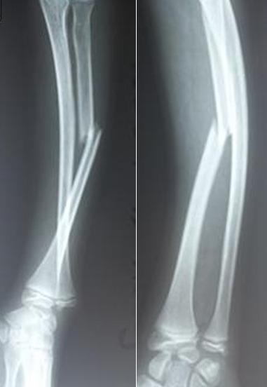
This diagnostic image displays two X-ray views (anteroposterior and lateral) of a pediatric forearm demonstrating a classic Galeazzi fracture-dislocation. The radiograph reveals a complete, displaced fracture of the mid-to-distal third (diaphysis) of the radius bone. Significant lateral and posterior angulation of the proximal radial fragment is visible, along with overriding of the fracture ends. Accompanying this radial injury is a clear disruption of the distal radioulnar joint (DRUJ), characterized by the loss of normal anatomical alignment between the distal radius and the ulnar head at the wrist. The presence of open growth plates (physes) at the distal radius and ulna indicates the skeletal immaturity of the patient. This visual serves as a primary educational example of how a solitary radial shaft fracture necessitates close evaluation of the distal radioulnar joint to identify associated ligamentous instability or luxation, critical for orthopedic trauma management and surgical planning.
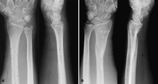
This diagnostic image consists of four x-ray radiographs showing the right (A) and left (B) forearms and wrists in both anteroposterior and lateral projections at a 13-month follow-up. The images demonstrate successful bone union following bilateral Galeazzi fracture-dislocations. The radius shafts in both forearms exhibit consolidated fractures with visible bone remodeling and small, circular radiolucent voids corresponding to screw holes from removed surgical hardware (plate and screw fixation). Specifically, the left radius (B) shows a more complex, formerly comminuted area now bridged by dense bone. The distal radio-ulnar joints (DRUJ) are well-aligned in all views; the distal ulnar heads are appropriately seated within the sigmoid notches of the radii without evidence of dorsal subluxation or dislocation. The carpal bones appear normal in density and configuration. These radiographs illustrate the long-term clinical outcome of anatomical fracture reduction and stabilization of the DRUJ, confirming stable joint congruity and mature bone healing after orthopedic intervention.
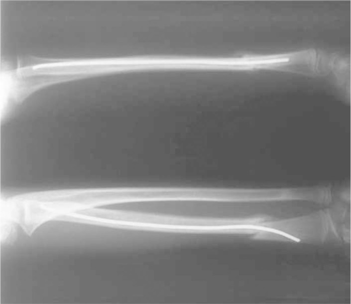
**Modality:** Conventional radiography (X-ray) in two views. **Anatomical Region:** Forearm, specifically the radius and ulna including the distal radioulnar joint (DRUJ) and wrist. **Observed Pathology:** Post-surgical reduction of a Galeazzi fracture-dislocation. There is a mid-to-distal third radial shaft fracture demonstrating interval reduction and stabilization. **Characteristic Features:** - **Internal Fixation:** A radio-opaque flexible intramedullary (IM) nail is visible within the medullary canal of the radius. - **Entry Point:** The IM nail is inserted via the distal radial metaphysis, traversing the fracture site to provide axial alignment. - **Alignment:** The radial shaft shows improved anatomical alignment with visible cortical apposition at the fracture site. - **Associated Findings:** The distal radioulnar joint is visualized to assess for stabilization following the reduction of the radial length. **Diagnostic Context:** This image demonstrates the use of intramedullary nailing as a surgical intervention for pediatric or adolescent Galeazzi-type injuries, focusing on restoring radial length and secondary stability of the distal radioulnar joint.
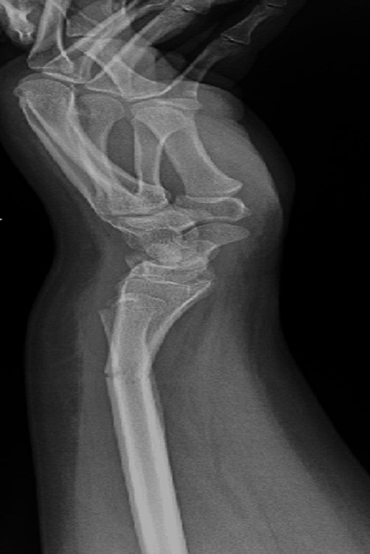
A lateral view X-ray of the wrist and distal forearm demonstrating a Galeazzi-equivalent fracture pattern in a pediatric patient. The radiograph reveals two distinct fractures: a transverse fracture in the distal third of the radial shaft with approximately 21 degrees of dorsal apex angulation, and a concomitant fracture of the distal ulna. The ulnar injury is characterized as a Salter-Harris type II physeal fracture involving the distal ulnar epiphysis, showing approximately 2.5 mm of minimal volar displacement. The anatomical alignment between the radius and ulna is disrupted at the level of the fractures, representing a pediatric variant of the Galeazzi injury where a distal ulnar physeal fracture occurs instead of a distal radioulnar joint (DRUJ) dislocation. Pronounced soft tissue swelling is visible around the wrist and distal forearm. This diagnostic image is intended for educational purposes in orthopedics and trauma radiology, focusing on pediatric fracture classification and the management of forearm shaft injuries involving the growth plate.
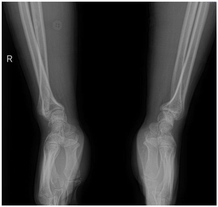
This diagnostic image is a comparative X-ray radiography (lateral view) of the right and left wrists. The right wrist exhibits a clear pathology characterized by a Galeazzi-type injury pattern. Key findings include a fracture of the distal radius with associated shortening and dorsal angulation. Crucially, there is a prominent dorsal dislocation of the ulnar head at the distal radioulnar joint (DRUJ), evidenced by the increased radioulnar distance and displacement relative to the carpal bones. In contrast, the left wrist (shown for comparison) displays normal anatomical alignment of the radius and ulna, with an intact DRUJ and congruent articulation. The image illustrates the clinical significance of assessing the entire forearm unit, as distal radius fractures can frequently be associated with occult or overt DRUJ instability. This visual is typical for orthopedic education focusing on wrist trauma, fracture classification, and joint instability mechanisms.
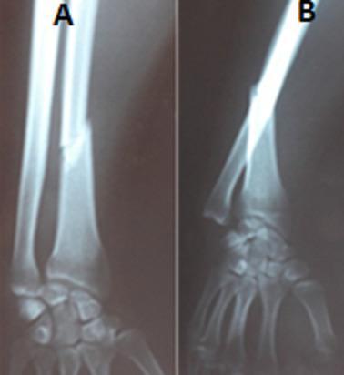
This diagnostic image consists of two radiographic views (X-rays) of the forearm and wrist: an anteroposterior (AP) view (A) and a lateral view (B). The images demonstrate a Galeazzi fracture-dislocation. In view A, a clear transverse-to-oblique fracture of the radial shaft is visible at the junction of the middle and distal thirds, accompanied by shortening and radial angulation. View B highlights the associated disruption of the distal radioulnar joint (DRUJ), showing a prominent dorsal dislocation of the ulnar head relative to the radius and carpus. This combination of a distal radius fracture and DRUJ instability is a hallmark of the Galeazzi injury pattern. The content is clinically categorized as a Mansat stage III fracture-dislocation, indicating significant ligamentous and bony disruption. This material is suitable for orthopedic education regarding forearm trauma, illustrating the mechanical relationship between long bone fractures and adjacent joint stability.
Galeazzi fracture distal radioulnar joint management
Galeazzi Fracture-Dislocation
Definition
- Campbell's Operative Orthopaedics 15th Ed 2026, p. 3633
- Tintinalli's Emergency Medicine, p. (ch. 270)
Mechanism of Injury
- Fall on an outstretched hand with forced pronation
- Direct blow to the forearm
- The radius fracture typically results in dorsal-lateral angulation
Radiographic Signs of DRUJ Injury
| Finding | View |
|---|---|
| Fracture at the base of the ulnar styloid | AP |
| Widened DRUJ space | AP |
| Dorsal dislocation of the ulnar head | Lateral |
| >5 mm shortening of radius relative to ulna | AP (compared to contralateral wrist) |
- Campbell's Operative Orthopaedics 15th Ed 2026, p. 3633-3634
DRUJ Instability - Key Statistic
- DRUJ is unstable in 55% of patients when the radial fracture is less than 7.5 cm from the articular surface
- DRUJ is unstable in only 6% of patients when the fracture is more than 7.5 cm from the articular surface
- Miller's Review of Orthopaedics 9th Ed, p. 906
Treatment
In Adults - Surgical (Standard of Care)
- Pronator quadratus (PQ)
- Brachioradialis
- Thumb abductors and extensors
- ORIF of the radial shaft via the anterior Henry approach (volar) or Thompson approach (dorsal)
- 3.5-mm dynamic compression plate fixation
- After fixation, assess DRUJ under fluoroscopy in neutral, pronation, and supination
- Campbell's Operative Orthopaedics 15th Ed 2026, p. 3634
- Rockwood and Green's Fractures in Adults 10th Ed 2025, p. 1809
Post-fixation DRUJ Assessment Algorithm
| DRUJ Status After Radial Fixation | Action |
|---|---|
| Reduced and stable | Protective splint in supination + early motion |
| Reduced but unstable - large ulnar styloid fragment | ORIF of styloid + immobilize in supination |
| Reduced but unstable - no large fragment | Temporary K-wire transfixation (2 wires, 4 cortices) in supination; remove at ~4-6 weeks |
| Irreducible (soft tissue interposition) | Open reduction of DRUJ |
Pediatric Considerations
Complications
- Nonunion (~5-9% with plate fixation in historical series)
- Malunion - rotational deformity causes pain at DRUJ with end-range pronation/supination
- Infection (0-6%, most commonly Staphylococcus aureus)
- DRUJ instability if not adequately addressed at time of surgery
- Reduced forearm rotation if radial bow is not anatomically restored
Radiographic Examples
Summary Mnemonic
Galezzi vs monteggia
Monteggia fracture ulna radial head dislocation X-ray
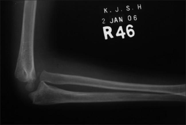
This lateral X-ray radiograph of a pediatric elbow demonstrates a chronic Monteggia fracture-dislocation. The imaging shows a significant anterior dislocation of the radial head, which is no longer aligned with the humeral capitellum. Simultaneously, the proximal ulna exhibits a distinct apex-posterior angulation and cortical irregularity, characteristic of a previous ulnar shaft fracture that has healed in malalignment (malunion). The radiocapitellar line, which normally passes through the center of the capitellum in all views, is clearly disrupted. This diagnostic image is a classic representation used in orthopedic education to illustrate the importance of assessing the entire forearm when an ulnar fracture is present, as well as the long-term sequelae of a missed or untreated Monteggia lesion in a 12-year-old patient. Key educational features include the anatomical relationship between the distal humerus, proximal radius, and ulna, and the structural abnormalities resulting from pediatric forearm trauma.
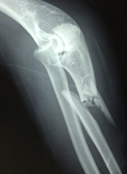
A lateral view X-ray of the elbow and proximal forearm demonstrating a Monteggia fracture-dislocation. The radiograph reveals a comminuted fracture of the proximal ulnar shaft with significant displacement and angulation. Associated with this fracture is an anterior dislocation of the radial head, which no longer articulates correctly with the humeral capitellum. The proximal radius is shifted anteriorly relative to the distal humerus, confirming the diagnostic pattern of a Bado Type I Monteggia injury. The image serves as a clinical example of musculoskeletal trauma, highlighting the importance of assessing the integrity of the radiocapitellar joint in the presence of ulnar fractures. The radiograph clearly illustrates the disruption of the normal anatomical alignment between the humerus, radius, and ulna at the elbow joint.
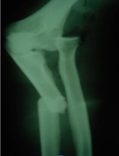
This lateral view X-ray of the elbow and proximal forearm demonstrates a Monteggia fracture-dislocation. Key diagnostic findings include a complete, displaced fracture of the proximal third of the ulnar shaft. Associated with this ulnar injury is an anterior dislocation of the radial head relative to the capitellum of the humerus, consistent with a Bado Type I injury pattern. The alignment of the radiocapitellar line is disrupted, failing to pass through the center of the capitellum. This imaging serves as a classic educational example of the forearm's 'two-bone system' failure, where a displaced fracture of one bone (ulna) necessitates a dislocation or fracture of the second bone (radius or its joints) to accommodate the change in length and mechanical force. Clinical relevance includes the importance of assessing the elbow joint in all isolated-appearing ulnar shaft fractures to prevent missed radial head dislocations.
Galeazzi vs Monteggia - Full Comparison
The Core Concept: Mirror-Image Injuries
| Feature | Galeazzi | Monteggia |
|---|---|---|
| Bone fractured | Radius (distal/middle third) | Ulna (proximal third) |
| Joint dislocated | DRUJ - Distal radioulnar joint (at wrist) | Radial head at the radiocapitellar joint (at elbow) |
| Level of injury | Distal forearm / wrist end | Proximal forearm / elbow end |
| Nickname | "Fracture of necessity" / Piedmont fracture / Reverse Monteggia | "Nightstick fracture dislocation" |
Memory Aid
Galeazzi = Gone at the Grip end (wrist/DRUJ) Monteggia = Missed at the Middle/elbow end (radial head)
Mechanism
| Galeazzi | Monteggia | |
|---|---|---|
| Mechanism | Fall on outstretched hand in forced pronation / direct blow | Fall on outstretched hand with hyperpronation / direct posterior blow to ulna / fall on flexed elbow |
Classification
Galeazzi - by distance from articular surface (Rettig & Raskin)
| Type | Fracture location | DRUJ instability rate |
|---|---|---|
| Type I | <7.5 cm from articular surface | 55% unstable |
| Type II | >7.5 cm from articular surface | 6% unstable |
-
Simple - reduces after anatomic radial fixation
-
Complex - irreducible after radial fixation (ECU or EDM tendon interposition)
-
Rockwood and Green's Fractures in Adults 10th Ed 2025, p. 1766
Monteggia - Bado Classification (4 types)
| Bado Type | Radial Head Direction | Ulnar Fracture | Frequency |
|---|---|---|---|
| I | Anterior dislocation | Apex anterior, any level | ~60% (most common in children) |
| II | Posterior / posterolateral dislocation | Apex posterior | ~15-20% (most common in adults - up to 80%) |
| III | Lateral / anterolateral dislocation | Ulnar metaphysis fracture | Rare - almost exclusively children |
| IV | Anterior dislocation | Fracture of both proximal ulna AND radius at same level | Rare - adults only |
- Rockwood and Green's Fractures in Adults 10th Ed 2025, p. 1765
- Miller's Review of Orthopaedics 9th Ed, p. 906
Clinical Presentation
| Galeazzi | Monteggia | |
|---|---|---|
| Tenderness | Over distal radius and wrist | Along ulnar shaft and elbow |
| Deformity | Dorsal-lateral radial angulation; DRUJ prominence at wrist | Forearm shortening; radial head palpable anterolaterally or posterolaterally |
| Key nerve at risk | None specifically | Posterior interosseous nerve (PIN) - deep branch of radial nerve; causes weakness/paralysis of finger/thumb extension |
Radiographic Diagnosis
Galeazzi - signs of DRUJ injury
- Fracture at the base of the ulnar styloid
- Widened DRUJ on AP view
- Dorsal ulnar head dislocation on lateral view
-
5 mm radial shortening vs. contralateral wrist
Monteggia - signs of radial head dislocation
- Radiocapitellar line (RCL) - a line drawn through the radial shaft should pass through the center of the capitellum on ALL views. If disrupted = radial head dislocation
- Radial head dislocation is easily missed because the obvious ulna fracture overshadows it
Pitfall: The ulna fracture in Monteggia dominates the X-ray and the radial head dislocation is frequently overlooked. Always draw the RCL.
- Rosen's Emergency Medicine, p. ch. 43
Treatment
| Galeazzi | Monteggia | |
|---|---|---|
| Adults | ORIF of radius (Henry anterior approach) + 3.5-mm DCP + assess DRUJ stability | ORIF of ulna + closed reduction of radial head (usually reduces once ulna is fixed) |
| Children | Closed reduction + casting often sufficient | Closed reduction + long arm cast in supination |
| Key principle | Fixing the radius usually reduces the DRUJ | Fixing the ulna anatomically usually reduces the radial head |
| If joint still unstable | K-wire transfixation of DRUJ in supination; ORIF of ulnar styloid if large fragment | Open reduction of radial head via separate approach if it fails to reduce (annular ligament interposition) |
Complications
| Galeazzi | Monteggia | |
|---|---|---|
| Main complications | DRUJ instability, malunion, nonunion, infection | PIN palsy, malunion, nonunion, synostosis, stiffness |
| Nerve injury | Rare | PIN palsy - observe 3 months (usually resolves spontaneously) |
| Missed diagnosis | DRUJ disruption | Radial head dislocation (chronic, irreducible if missed) |
Radiograph Examples
One-Line Summary
| Fracture | Formula |
|---|---|
| Galeazzi | Radius fracture (distal) + DRUJ dislocation (wrist) |
| Monteggia | Ulna fracture (proximal) + Radial head dislocation (elbow) |
Antibiotic coated nailing for tibia
antibiotic coated intramedullary nail tibia fracture infection
PMID: 38143139
PMID: 35613970
antibiotic intramedullary nail SIGN nail open tibial fracture
gentamicin vancomycin coated nail tibia open fracture prevention
antibiotic coated nail infected nonunion tibia outcome
PMID: 37706117
PMID: 36858895
Antibiotic-Coated Nailing for Tibia Fractures
Why It Matters
- Gustilo I-IIIA fractures: infection rate ~3-9% with standard IM nailing
- Gustilo IIIB fractures: infection rate up to 16% (LEAP study)
- Infected nonunion remains one of the most difficult reconstructive problems in orthopaedic surgery
- Rockwood and Green's Fractures in Adults 10th Ed 2025, p. 3186
Indications
| Setting | Indication |
|---|---|
| Open tibial fractures (Gustilo II, IIIA, IIIB) | Primary infection prophylaxis at time of nailing |
| Infected nonunion of tibia | Combined infection control + fracture stabilization |
| Fracture-related infection (FRI) requiring exchange nailing | Definitive single-stage or staged treatment |
| High-risk patients | Diabetes, immunocompromise, contaminated wounds, military/blast injuries |
Coating Methods
1. Custom-Made (Intraoperative) PMMA Coating
- The nail is hand-dipped or painted with polymethyl methacrylate (PMMA) bone cement mixed with antibiotics (most commonly vancomycin, gentamicin, or tobramycin) in the OR
- Cost-effective; can be tailored to culture sensitivities
- Disadvantage: inconsistent coating thickness, risk of debonding, higher reoperation rates vs. commercial nails
- Accounts for the majority (~469/506 patients) in systematic reviews
2. Commercial Coated Nails
- Pre-manufactured with a gentamicin-loaded biodegradable polymer coating (e.g., Expert Tibial Nail with COPAL - Synthes; or similar platforms)
- More reproducible coating; lower reoperation rates than custom-made nails
- Coating degrades and releases antibiotic over days to weeks
3. Other Coating Technologies
- Hydrogel coatings with rifampicin/gentamicin
- Calcium sulfate / calcium phosphate carriers
- Chitosan and PLGA polymer coatings (experimental/emerging)
Antibiotic Choice
| Antibiotic | Coverage | Route |
|---|---|---|
| Gentamicin | Gram-negative, Staph | Commercial nail standard |
| Vancomycin | Gram-positive, MRSA | Custom PMMA nail |
| Tobramycin | Gram-negative | PMMA combination |
| Clindamycin | Gram-positive, anaerobes | Less common |
Clinical Evidence
Meta-Analysis: AC-IMN for Open Tibia Fracture Infection Prevention
| Outcome | Antibiotic-Coated Nail | Standard Nail |
|---|---|---|
| Global infection rate | 8.4% | 22% |
| Deep infection rate | 5.4% | 14% |
| Non-union rate | 3.7% | 14% |
| Deep infection RR | 0.17 (95% CI 0.02-1.31) | - |
| Global infection RR | 0.36 (95% CI 0.10-1.35) | - |
- Trend strongly favors AC-IMN but did not reach statistical significance due to limited number of studies amenable to meta-analysis
- Conclusion: favorable trend; higher-level RCT evidence still needed
Systematic Review: Infected Nonunion Treatment
- Infection eradication rate: 90% (range 68.7-100%)
- Bone consolidation rate: 85.5% (range 57.9-100%)
- No coating-specific side effects reported
- Most studies were retrospective case series - quality remains low
Commercial vs. Custom-Made Nail Comparison
- Overall success rate (union + infection control): 94.4%
- Reoperation rate: 40.7%
- Custom-made (vancomycin-PMMA) nails had 4.71x higher risk of reoperation than commercial gentamicin nails (OR 4.71, p=0.036)
- Suggests commercial nails may be superior in reproducibility and outcomes
Prospective Study: AC-IMN in Open Fractures (Solanki et al., Cureus 2023, PMID 37706117)
- 30 patients (73.3% tibia); predominantly Gustilo IIIA/B fractures
- Infection control: 93.3%
- Bone union: 90% (mean time to union for tibia: 22.13 weeks)
- Excellent bone outcomes (Paley criteria): 76.7%
- Complications: nail cement debonding (10%), proximal nail impingement (10%)
Surgical Technique Considerations
- Thorough irrigation and debridement (I&D) first - AC-IMN does not replace I&D
- Canal reaming is standard - allows nail passage and activates osteogenic response; safe even in open fractures (SPRINT trial)
- Nail is coated (commercially or intraoperatively) and inserted in standard fashion
- In infected nonunion: implant removal + debridement + culture + medullary canal reaming + insertion of antibiotic nail
- Postoperative systemic antibiotics are still given (AC-IMN supplements, not replaces, systemic therapy)
- Gradual weight-bearing is permitted - AC-IMN provides mechanical stability along with local drug delivery
Advantages Over Antibiotic Spacer Alone
| Feature | AC-IMN | Antibiotic Spacer (beads/rod) |
|---|---|---|
| Mechanical stability | Yes (immediate load bearing) | No (requires separate fixation) |
| Staged surgery | Often single-stage | Usually two-stage |
| Local antibiotic delivery | Yes | Yes |
| Bone defect filling | No (if significant defect exists, spacer still needed) | Yes |
Limitations and Current Gaps
- Most evidence is from retrospective case series - randomized data is scarce
- No standardized coating protocol (antibiotic choice, concentration, carrier all vary)
- Risk of antibiotic resistance with local delivery
- Debonding of PMMA-coated nails can cause third-body wear and mechanical issues
- Long-term systemic antibiotic exposure levels not fully characterized
- Best evidence exists for tibia and femur; other bones less studied
Summary
- Strong biological rationale (local delivery bypasses poor vascularity)
- Consistent ~90-94% infection control rates in infected nonunion
- A favorable but not yet statistically proven reduction in FRI in primary open fracture settings
- Commercial nails outperforming custom PMMA nails on reoperation rates
- Evidence base is growing but mostly low-to-moderate quality; RCTs are needed