

Bed sore content like this
NURSING TEACHING PLAN
| SN | Specific Objectives | Contents | Time (min) | Teaching Learning Method | Teaching Aid | Evaluation |
|---|---|---|---|---|---|---|
| Introduction | - Introduction - Self - Topic - Objectives - Pretest | 2 min 3 min 2 min | - | - | - | |
| 1. | At the end of the teaching, participants will be able to: Introduce bed sores | - Introduction of Bed Sores / Pressure Ulcers - Definition: A localized injury to the skin and underlying tissue, usually over a bony prominence, as a result of sustained pressure or pressure in combination with shear | 1 min | Interactive lecture + discussion | - | What is a Bed Sore / Pressure Ulcer? |
| 2. | List risk factors and causes of bed sores | - Risk factors and causes of Bed Sores: 1. Immobility / reduced activity 2. Spinal cord injury 3. Dementia 4. Parkinson disease 5. Congestive heart failure 6. Incontinence 7. Nutritional deficiency (hypoalbuminemia, low protein/calorie intake) 8. Moist skin, friction, shearing forces | 3 min | Interactive lecture + discussion | Flash card | What are the risk factors of Bed Sores? |
| 3. | List signs and symptoms of bed sores | - Signs and Symptoms: 1. Non-blanchable erythema of intact skin (Stage I) 2. Shallow open ulcer, red/pink wound bed (Stage II) 3. Full-thickness tissue loss (Stage III) 4. Full-thickness tissue loss with exposed bone/tendon (Stage IV) 5. Pain, warmth, swelling at site 6. Foul-smelling / purulent discharge in infected ulcers 7. Common sites: sacrum, ischial tuberosity, heels, greater trochanter, lateral malleolus | 2 min | Interactive lecture + discussion | Chart paper | What are the signs and symptoms of Bed Sores? |
| 4. | List diagnostic/assessment criteria for bed sores | - Diagnostic / Assessment Criteria: 1. Braden Scale for risk assessment (mobility, activity, sensory perception, moisture, nutrition, friction/shear) 2. Staging according to NPUAP: Stage I - IV + Unstageable + Deep Tissue Injury 3. Wound assessment: size, depth, exudate, odor, surrounding skin 4. Lab: CBC, albumin, wound culture in infected cases | 1 min | Interactive lecture + discussion | - | What is the diagnostic / assessment criteria for Bed Sores? |
| 5. | List preventive measures for bed sores | - Preventive Measures: 1. Formal risk assessment (Braden Scale) on admission 2. Reposition every 2 hours for high-risk patients; 2-4 times/day for others 3. Position at 30-degree angle to support surface 4. Use pressure-reducing devices: alternating air mattress, gel mattress, water mattress 5. Maintain good nutrition (protein, Vitamin C, zinc) 6. Keep skin clean and dry; manage incontinence 7. Use lifting devices to minimize friction and shear during transfers 8. Patient and caregiver education | 4 min | Interactive lecture + discussion | Chart paper | What are the preventive measures for Bed Sores? |
| 6. | Describe diet for a patient with bed sores | - Diet for Bed Sore Patient: 1. High-protein diet (eggs, milk, dal, fish, chicken) to promote tissue repair 2. Vitamin C-rich foods (citrus fruits, amla, guava) - 84% reduction in ulcer surface area reported 3. Zinc-rich foods (nuts, seeds, whole grains) 4. Adequate calorie intake to prevent cachexia 5. Iron-rich foods to correct anemia 6. Adequate fluids (2-3 litres/day) to maintain skin hydration | 3 min | Interactive lecture + discussion | Chart paper | What is the diet for a Bed Sore patient? |
| 7. | Describe a 1-day meal plan for a bed sore patient | - 1-Day Meal Plan: - Early morning: Warm milk + soaked almonds - Breakfast: Boiled eggs (2) + whole wheat bread + orange juice - Mid-morning: Amla juice / guava - Lunch: Rice + dal + fish/chicken curry + green leafy vegetables + salad - Evening snack: Roasted peanuts + lemon water - Dinner: Chapati + paneer/egg bhurji + vegetable curry + curd - Bedtime: Warm milk | 4 min | Interactive lecture + discussion | Newsprint | What is the 1-day meal plan for a Bed Sore patient? |
| Summary | - Summarization of key points | 2-3 min | Discussion | Discussion | Wh-questions |
Key Points Used for the Plan (Source: Swanson's Family Medicine Review, 9th Ed.)
- Pathophysiology: Pressure + shearing forces + friction + moisture → ischemia of tissues overlying bony prominences → necrosis
- 4 main causative factors: Pressure, shearing forces, friction, moisture
- Staging (NPUAP): Stage I (non-blanchable erythema) → Stage II (partial thickness) → Stage III (full thickness) → Stage IV (bone/tendon exposed)
- Prevention: Reposition every 2 hrs; Braden Scale; pressure-relieving mattresses; nutrition
- Treatment: Normal saline wound cleaning; wet-to-dry dressings; Vitamin C supplementation; debridement; antibiotics for infection (clindamycin + ciprofloxacin for moderate cases)
- Complications: Sepsis, cellulitis, osteomyelitis, peritonitis; mortality 23-36% in hospitalized patients
- Same columns (SN, Specific Objectives, Contents, Time, Teaching Learning Method, Teaching Aid/TLM, Evaluation)
- Same flow (Intro → Definition → Risk Factors → Signs & Symptoms → Diagnosis → Prevention → Diet → Meal Plan → Summary)
- Same teaching methods (Interactive lecture + discussion, Flash cards, Chart paper, Newsprint)
- Same evaluation style (Wh-questions at the end, specific questions per row)
Health teaching on bed sore i need following contents introduction of bed sorr risk factor of bed sore and its causes sign and symptkms diagnostic criteria staging and common sites of developing bed sore preventive measures management and summary
mkdir -p /home/daytona/workspace/bedsore-teaching-plan && ls /home/daytona/workspace/bedsore-teaching-plan/
~/bedsore-teaching-plan/create_plan.js
const {
  Document, Packer, Paragraph, TextRun, Table, TableRow, TableCell,
  AlignmentType, HeadingLevel, BorderStyle, WidthType, VerticalAlign,
  ShadingType, PageOrientation
} = require('docx');
const fs = require('fs');

// ── helpers ────────────────────────────────────────────────────────────────────
const bold = (text, size = 20) =>
  new TextRun({ text, bold: true, size, font: 'Times New Roman' });
const normal = (text, size = 20) =>
  new TextRun({ text, size, font: 'Times New Roman' });
const br = () => new TextRun({ break: 1 });

function para(runs, align = AlignmentType.LEFT, spacing = { before: 40, after: 40 }) {
  return new Paragraph({ children: Array.isArray(runs) ? runs : [runs], alignment: align, spacing });
}

function headerCell(text, shade = '1F4E79') {
  return new TableCell({
    shading: { fill: shade, type: ShadingType.CLEAR, color: shade },
    verticalAlign: VerticalAlign.CENTER,
    margins: { top: 80, bottom: 80, left: 100, right: 100 },
    children: [new Paragraph({
      alignment: AlignmentType.CENTER,
      spacing: { before: 40, after: 40 },
      children: [new TextRun({ text, bold: true, color: 'FFFFFF', size: 20, font: 'Times New Roman' })]
    })]
  });
}

function cell(children, shade = null, align = AlignmentType.LEFT) {
  return new TableCell({
    shading: shade ? { fill: shade, type: ShadingType.CLEAR, color: shade } : undefined,
    verticalAlign: VerticalAlign.TOP,
    margins: { top: 80, bottom: 80, left: 100, right: 100 },
    children: Array.isArray(children) ? children : [children]
  });
}

function dataRow(sn, objective, content, time, method, aid, evaluation, rowShade) {
  return new TableRow({
    children: [
      cell([para([normal(sn)], AlignmentType.CENTER)], rowShade),
      cell([para([normal(objective)])], rowShade),
      cell(content.map(line => para([normal(line)])), rowShade),
      cell([para([normal(time)], AlignmentType.CENTER)], rowShade),
      cell(method.map(line => para([normal(line)])), rowShade),
      cell(aid.map(line => para([normal(line)])), rowShade),
      cell([para([normal(evaluation)])], rowShade),
    ]
  });
}

// ── table rows ─────────────────────────────────────────────────────────────────
const LIGHT = 'EBF3FB';
const WHITE = 'FFFFFF';

const headerRow = new TableRow({
  tableHeader: true,
  children: [
    headerCell('SN'),
    headerCell('Specific Objectives'),
    headerCell('Contents'),
    headerCell('Time\n(min)'),
    headerCell('Teaching\nLearning\nMethod'),
    headerCell('Teaching\nAid'),
    headerCell('Evaluation'),
  ]
});

const rows = [
  // INTRO
  new TableRow({
    children: [
      cell([para([normal('')], AlignmentType.CENTER)], LIGHT),
      cell([para([normal('')])], LIGHT),
      cell([
        para([bold('• Introduction')]),
        para([normal('• Self introduction')]),
        para([normal('• Topic introduction')]),
        para([normal('• Objectives')]),
        para([normal('• Pre-test')]),
      ], LIGHT),
      cell([para([normal('2\n3\n2')], AlignmentType.CENTER)], LIGHT),
      cell([para([normal('—')])], LIGHT),
      cell([para([normal('—')])], LIGHT),
      cell([para([normal('—')])], LIGHT),
    ]
  }),

  // 1 - INTRODUCTION OF BED SORE
  dataRow(
    '1.',
    'At the end of the\nteaching, participants\nwill be able to:\nIntroduce bed sores',
    [
      '• Introduction / Definition:',
      '  A localized injury to the skin',
      '  and/or underlying tissue,',
      '  usually over a bony prominence,',
      '  resulting from sustained pressure',
      '  or pressure combined with shear.',
      '',
      '• Also called: decubitus ulcer,',
      '  pressure sore, pressure injury.',
      '',
      '• Prevalence: 7–9% in acute care;',
      '  11% in long-term care facilities.',
      '  Affects 3 million people annually.',
    ],
    '1',
    ['Interactive', 'lecture +', 'discussion'],
    ['—'],
    'What is a\nBed Sore?',
    WHITE
  ),

  // 2 - RISK FACTORS & CAUSES
  dataRow(
    '2.',
    'List risk factors\nand causes of\nbed sores',
    [
      '• Risk Factors & Causes:',
      '1. Immobility / reduced activity',
      '2. Spinal cord injury',
      '3. Dementia',
      '4. Parkinson disease',
      '5. Congestive heart failure',
      '6. Incontinence',
      '7. Poor nutrition (low protein,',
      '   hypoalbuminemia, low calorie)',
      '8. Anemia',
      '',
      '• Pathogenic factors:',
      '  - Pressure (main factor)',
      '  - Shearing forces',
      '  - Friction',
      '  - Moisture / moist skin',
    ],
    '3',
    ['Interactive', 'lecture +', 'discussion'],
    ['Flash card'],
    'What are the risk\nfactors and causes\nof Bed Sores?',
    LIGHT
  ),

  // 3 - SIGNS & SYMPTOMS
  dataRow(
    '3.',
    'List signs and\nsymptoms of\nbed sores',
    [
      '• Signs & Symptoms:',
      '1. Non-blanchable redness / erythema',
      '   over bony prominence',
      '2. Warmth, swelling, tenderness',
      '3. Shallow open ulcer — red/pink',
      '   wound bed',
      '4. Full-thickness tissue loss;',
      '   subcutaneous fat visible',
      '5. Exposed bone, tendon, or muscle',
      '   (severe cases)',
      '6. Slough or dark eschar on wound',
      '7. Purulent, foul-smelling discharge',
      '   (if infected)',
      '8. Fever, confusion (sepsis sign)',
    ],
    '2',
    ['Interactive', 'lecture +', 'discussion'],
    ['Chart paper'],
    'What are the signs\nand symptoms of\nBed Sores?',
    WHITE
  ),

  // 4 - DIAGNOSTIC CRITERIA + STAGING + COMMON SITES
  dataRow(
    '4.',
    'List diagnostic\ncriteria, staging\nand common sites\nof bed sores',
    [
      '• Assessment Tools:',
      '  - Braden Scale (6 subscales:',
      '    sensory perception, moisture,',
      '    activity, mobility, nutrition,',
      '    friction/shear)',
      '  - Waterlow Score',
      '  - Norton Risk Assessment Scale',
      '',
      '• NPUAP Staging:',
      '  Stage I   – Non-blanchable',
      '              erythema; intact skin',
      '  Stage II  – Partial-thickness skin',
      '              loss; shallow open ulcer',
      '  Stage III – Full-thickness tissue',
      '              loss; fat visible',
      '  Stage IV  – Full-thickness loss;',
      '              exposed bone/tendon',
      '  Unstageable – Covered by slough',
      '              or eschar',
      '  Deep Tissue – Purple/maroon intact',
      '              skin or blood blister',
      '',
      '• Common Sites:',
      '  Sacrum, Ischial tuberosity,',
      '  Greater trochanter, Heel,',
      '  Lateral/medial malleolus, Occiput',
    ],
    '3',
    ['Interactive', 'lecture +', 'discussion'],
    ['Chart paper', '/ Diagram'],
    'What is the\ndiagnostic criteria\nand staging of\nBed Sores?',
    LIGHT
  ),

  // 5 - PREVENTIVE MEASURES
  dataRow(
    '5.',
    'List preventive\nmeasures of\nbed sores',
    [
      '• Preventive Measures:',
      '1. Formal risk assessment on',
      '   admission (Braden Scale)',
      '2. Reposition every 2 hours',
      '   (high-risk); 2–4 times/day',
      '   (moderate risk)',
      '3. Position at 30° angle to surface;',
      '   avoid pressure on bony prominences',
      '4. Pressure-reducing devices:',
      '   alternating air mattress,',
      '   gel/water mattress',
      '5. Use lifting devices (not dragging)',
      '   during transfers to reduce friction',
      '6. Keep skin clean and dry;',
      '   manage incontinence promptly',
      '7. Adequate nutrition: protein,',
      '   Vitamin C, zinc, adequate calories',
      '8. Patient and caregiver education',
      '9. Regular skin inspection',
    ],
    '4',
    ['Interactive', 'lecture +', 'discussion'],
    ['Chart paper'],
    'What are the\npreventive measures\nof Bed Sores?',
    WHITE
  ),

  // 6 - MANAGEMENT
  dataRow(
    '6.',
    'Describe the\nmanagement of\nbed sores',
    [
      '• General / Systemic Management:',
      '  - Treat underlying diseases',
      '  - Nutritional support:',
      '    high protein, Vitamin C',
      '    (84% reduction in ulcer area)',
      '  - Correct anemia',
      '  - Air-fluidized bed therapy',
      '',
      '• Local Wound Care:',
      '  - Clean with normal saline',
      '    (avoid H2O2, povidone-iodine)',
      '  - Wet-to-dry dressings (saline)',
      '  - Hydrocolloid dressings –',
      '    Stage II ulcers',
      '  - Enzymatic debridement for',
      '    necrotic tissue',
      '  - Surgical debridement for',
      '    deep necrotic tissue',
      '',
      '• Antibiotic Therapy:',
      '  Mild: cephalexin / clindamycin',
      '  Moderate: clindamycin +',
      '    ciprofloxacin (2–4 weeks)',
      '  Severe: piperacillin/tazobactam',
      '    or imipenem (up to 12 weeks)',
      '',
      '• Surgical: flap closure for',
      '  Stage III/IV refractory ulcers',
    ],
    '4',
    ['Interactive', 'lecture +', 'discussion'],
    ['Chart paper'],
    'What is the\nmanagement of\nBed Sores?',
    LIGHT
  ),

  // SUMMARY
  new TableRow({
    children: [
      cell([para([normal('')], AlignmentType.CENTER)], WHITE),
      cell([para([bold('Summary')])], WHITE),
      cell([
        para([bold('• Summarization of key points:')]),
        para([normal('- Definition & prevalence')]),
        para([normal('- Risk factors & pathogenic factors')]),
        para([normal('- Signs, symptoms & complications')]),
        para([normal('- NPUAP Staging (I–IV +')]),
        para([normal('  Unstageable + Deep Tissue)')]),
        para([normal('- Common sites')]),
        para([normal('- Prevention & management')]),
      ], WHITE),
      cell([para([normal('3')], AlignmentType.CENTER)], WHITE),
      cell([para([normal('Discussion')])], WHITE),
      cell([para([normal('Discussion')])], WHITE),
      cell([para([normal('Wh-questions')])], WHITE),
    ]
  }),
];

// ── document ───────────────────────────────────────────────────────────────────
const doc = new Document({
  styles: {
    default: {
      document: {
        run: { font: 'Times New Roman', size: 20 }
      }
    }
  },
  sections: [{
    properties: {
      page: {
        size: { orientation: PageOrientation.LANDSCAPE },
        margin: { top: 720, bottom: 720, left: 720, right: 720 }
      }
    },
    children: [
      // Title block
      para([bold('NURSING HEALTH TEACHING PLAN', 28)], AlignmentType.CENTER, { before: 0, after: 100 }),
      para([bold('Topic: Bed Sores (Pressure Ulcers / Pressure Injuries)', 24)], AlignmentType.CENTER, { before: 0, after: 60 }),
      para([
        normal('Subject: Fundamentals of Nursing   |   ', 18),
        normal('Class: GNM / B.Sc. Nursing   |   ', 18),
        normal('Duration: ~20 minutes', 18),
      ], AlignmentType.CENTER, { before: 0, after: 200 }),

      // Main table
      new Table({
        width: { size: 100, type: WidthType.PERCENTAGE },
        columnWidths: [600, 1500, 3200, 600, 1400, 1100, 1800],
        rows: [headerRow, ...rows],
        borders: {
          top:    { style: BorderStyle.SINGLE, size: 6, color: '1F4E79' },
          bottom: { style: BorderStyle.SINGLE, size: 6, color: '1F4E79' },
          left:   { style: BorderStyle.SINGLE, size: 6, color: '1F4E79' },
          right:  { style: BorderStyle.SINGLE, size: 6, color: '1F4E79' },
          insideH:{ style: BorderStyle.SINGLE, size: 4, color: 'AED6F1' },
          insideV:{ style: BorderStyle.SINGLE, size: 4, color: 'AED6F1' },
        }
      }),

      // Footer note
      para([normal(' ')], AlignmentType.LEFT, { before: 200 }),
      para([
        normal('References: Washington Manual of Medical Therapeutics | Bailey & Love\'s Short Practice of Surgery (28th ed.) | Swanson\'s Family Medicine Review', 16)
      ], AlignmentType.LEFT, { before: 0, after: 0 }),
    ]
  }]
});

Packer.toBuffer(doc).then(buffer => {
  fs.writeFileSync('/home/daytona/workspace/bedsore-teaching-plan/BedSore_Health_Teaching_Plan.docx', buffer);
  console.log('SUCCESS: File written.');
});
cd /home/daytona/workspace/bedsore-teaching-plan && node create_plan.js 2>&1
python3 /home/daytona/skills/docx/scripts/office/validate.py /home/daytona/workspace/bedsore-teaching-plan/BedSore_Health_Teaching_Plan.docx 2>&1
python3 -c " import zipfile, os path = '/home/daytona/workspace/bedsore-teaching-plan/BedSore_Health_Teaching_Plan.docx' size = os.path.getsize(path) print(f'File size: {size} bytes') with zipfile.ZipFile(path) as z: names = z.namelist() print('ZIP contents:', names) " 2>&1
NURSING HEALTH TEACHING PLAN
| SN | Specific Objectives | Contents | Time | Teaching Learning Method | Teaching Aid | Evaluation |
|---|---|---|---|---|---|---|
| • Introduction • Self introduction • Topic introduction • Objectives • Pre-test | 2 min 3 min 2 min | — | — | — | ||
| 1. | At the end of the teaching, participants will be able to: Introduce bed sores | • Definition: A localized injury to the skin and/or underlying tissue, usually over a bony prominence, resulting from sustained pressure or pressure combined with shear. • Also called: Decubitus ulcer, Pressure sore, Pressure injury • Prevalence: 7–9% in acute care; 11% in long-term care; affects 3 million people annually | 1 min | Interactive lecture + discussion | — | What is a Bed Sore / Pressure Ulcer? |
| 2. | List risk factors and causes of bed sores | • Risk Factors: 1. Immobility / reduced activity 2. Spinal cord injury 3. Dementia 4. Parkinson disease 5. Congestive heart failure 6. Incontinence 7. Poor nutrition (hypoalbuminemia, low protein/calorie) 8. Anemia • Pathogenic causes: Pressure, Shearing forces, Friction, Moisture | 3 min | Interactive lecture + discussion | Flash card | What are the risk factors and causes of Bed Sores? |
| 3. | List signs and symptoms of bed sores | • Signs & Symptoms: 1. Non-blanchable redness/erythema over bony prominence 2. Warmth, swelling, tenderness at site 3. Shallow open ulcer with red/pink wound bed 4. Full-thickness tissue loss; subcutaneous fat visible 5. Exposed bone, tendon, or muscle (severe) 6. Slough or dark eschar on wound 7. Purulent, foul-smelling discharge (if infected) 8. Fever, confusion (if sepsis develops) | 2 min | Interactive lecture + discussion | Chart paper | What are the signs and symptoms of Bed Sores? |
| 4. | List diagnostic criteria, staging, and common sites of bed sores | • Assessment Tools: - Braden Scale (sensory perception, moisture, activity, mobility, nutrition, friction/shear) - Waterlow Score; Norton Risk Assessment Scale • NPUAP Staging: Stage I - Non-blanchable erythema; intact skin Stage II - Partial-thickness loss; shallow open ulcer Stage III - Full-thickness loss; fat visible, no bone/tendon Stage IV - Full-thickness loss; exposed bone/tendon/muscle Unstageable - Covered by slough or eschar Deep Tissue - Purple/maroon intact skin or blood blister • Common Sites: Sacrum, Ischial tuberosity, Greater trochanter, Heel, Lateral/Medial malleolus, Occiput | 3 min | Interactive lecture + discussion | Chart paper / Diagram | What is the diagnostic criteria and staging of Bed Sores? |
| 5. | List preventive measures of bed sores | • Preventive Measures: 1. Formal risk assessment on admission (Braden Scale) 2. Reposition every 2 hours (high-risk); 2–4 times/day (moderate risk) 3. Position at 30-degree angle to support surface 4. Pressure-reducing devices: alternating air mattress, gel mattress, water mattress 5. Use lifting devices - avoid dragging during transfers 6. Keep skin clean and dry; manage incontinence promptly 7. Adequate nutrition: high protein, Vitamin C, zinc, calories 8. Regular skin inspection at all bony prominences 9. Patient and caregiver education | 4 min | Interactive lecture + discussion | Chart paper | What are the preventive measures of Bed Sores? |
| 6. | Describe management of bed sores | • General/Systemic: - Treat underlying diseases - Nutritional support: Protein + Vitamin C (84% reduction in ulcer area reported) - Correct anemia; air-fluidized bed therapy • Local Wound Care: - Clean with normal saline (avoid H2O2, povidone-iodine) - Wet-to-dry saline dressings - Hydrocolloid dressings for Stage II - Enzymatic debridement for necrotic tissue - Surgical debridement for deep/refractory wounds • Antibiotics: Mild: Cephalexin / Clindamycin Moderate: Clindamycin + Ciprofloxacin (2–4 wks) Severe: Piperacillin/Tazobactam or Imipenem (up to 12 wks) • Surgical: Flap closure for refractory Stage III/IV ulcers | 4 min | Interactive lecture + discussion | Chart paper | What is the management of Bed Sores? |
| Summary | • Key points summarized: - Definition & prevalence - Risk factors & pathogenic causes - Signs, symptoms & complications - NPUAP Staging (I–IV + Unstageable + Deep Tissue) - Common sites - Prevention & management | 3 min | Discussion | Discussion | Wh-questions |
Download your Word Document
BedSore Health Teaching Plan
Word Document · DOCX
- Washington Manual of Medical Therapeutics (NPUAP staging)
- Bailey & Love's Short Practice of Surgery, 28th ed. (common sites, staging table)
- Swanson's Family Medicine Review (risk factors, pathophysiology, prevention, management)
Tell me about peroxide’s inhibitor
No peroxidase inhibitors
Peroxidase Inhibitors
What is Peroxidase?
- Immune defense (myeloperoxidase in neutrophils)
- Plant defense responses
- Thyroid hormone synthesis (thyroid peroxidase)
- Melanin synthesis
Categories of Peroxidase Inhibitors
1. Competitive Inhibitors
- Cyanide (CN⁻) - binds the heme iron at the active site; one of the most potent peroxidase inhibitors
- Azide (N₃⁻) - binds the ferric (Fe³⁺) form of the enzyme; widely used in laboratory settings to inhibit endogenous peroxidase in tissue sections (IHC staining)
- Fluoride (F⁻) - inhibits by binding the heme iron
2. Suicide / Irreversible Inhibitors
- Propylthiouracil (PTU) - irreversibly inhibits thyroid peroxidase (TPO); used clinically to treat hyperthyroidism
- Methimazole (MMI) - also inhibits TPO; the preferred antithyroid drug in most settings
- Carbimazole - prodrug of methimazole
- Phenylhydrazine - reacts with the heme group
3. Substrate Analog Inhibitors
- Hydroxamic acids - inhibit various peroxidases by chelating the active-site iron
- Benzohydroxamic acid - well-studied inhibitor of horseradish peroxidase (HRP) and plant peroxidases
4. Myeloperoxidase (MPO) Inhibitors
- 4-Aminobenzoic acid hydrazide (ABAH) - irreversible, mechanism-based MPO inhibitor; widely used in research
- Tryptamine derivatives
- AZD5904 - a selective, orally bioavailable MPO inhibitor studied in clinical trials for inflammatory diseases
- Verdiperstat (AZD3241) - MPO inhibitor investigated for neurodegenerative diseases (ALS, Parkinson's)
5. Natural / Endogenous Inhibitors
- Ascorbic acid (Vitamin C) - reduces the active oxidized form of peroxidase back to its resting state, effectively inhibiting its cycle
- Glutathione - acts as a reductant, interfering with peroxidase-driven oxidative cycles
- Melatonin - scavenges H₂O₂, indirectly reducing peroxidase activity
6. Laboratory Inhibitors (used in histochemistry)
- Sodium azide (NaN₃) - most commonly added to antibody solutions and used in immunohistochemistry (IHC) to block endogenous peroxidase activity
- 3% H₂O₂ in methanol - used in IHC to quench endogenous peroxidase before staining
Mechanism Summary
| Inhibitor | Type | Target | Use |
|---|---|---|---|
| Cyanide | Competitive / irreversible | Heme iron (Fe³⁺) | Research |
| Azide | Competitive | Heme iron | IHC, research |
| PTU / Methimazole | Suicide (irreversible) | Thyroid peroxidase | Clinical (hyperthyroidism) |
| ABAH | Mechanism-based | Myeloperoxidase | Research |
| Verdiperstat | Irreversible | Myeloperoxidase | Clinical trials |
| Ascorbic acid | Reducing agent | General peroxidases | Physiological |
| NaN₃ | Competitive | General peroxidases | Lab / IHC |
Clinical Relevance
- Antithyroid drugs (PTU, methimazole) work entirely through TPO inhibition - blocking iodination of thyroglobulin
- MPO inhibitors are being explored as treatments for inflammatory conditions including cardiovascular disease, multiple sclerosis, and neurodegeneration
- In wound care, hydrogen peroxide is itself a substrate - using it on wounds also consumes/inhibits local peroxidase activity, which is one reason it can impair healing (another reason it is avoided in bed sore management)
Anterior pituitary hormones
anterior pituitary hormones diagram hypothalamus regulation
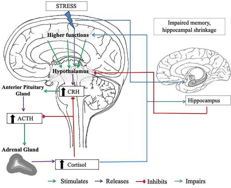
This pathophysiology diagram illustrates the hypothalamic-pituitary-adrenal (HPA) axis and its relationship with the hippocampus under chronic stress conditions. The left side depicts a sagittal section of the brain showing the 'Higher Functions' and the 'Hypothalamus' stimulating the 'Anterior Pituitary Gland' to release corticotropin-releasing hormone (CRH) and adrenocorticotropic hormone (ACTH). These hormones stimulate the 'Adrenal Gland' to secrete cortisol. A negative feedback loop is shown where cortisol inhibits further CRH and ACTH release. The right side of the diagram highlights the clinical impact of chronic cortisol elevation on the 'Hippocampus,' specifically illustrating hippocampal shrinkage and impaired memory. Color-coded arrows define functional relationships: green for stimulation, purple for release, red for inhibition, and blue for impairment. The illustration serves as an educational resource for understanding neuroendocrinology, the physiological stress response, and the long-term neurobiological effects of glucocorticoids on brain morphology and cognitive function.
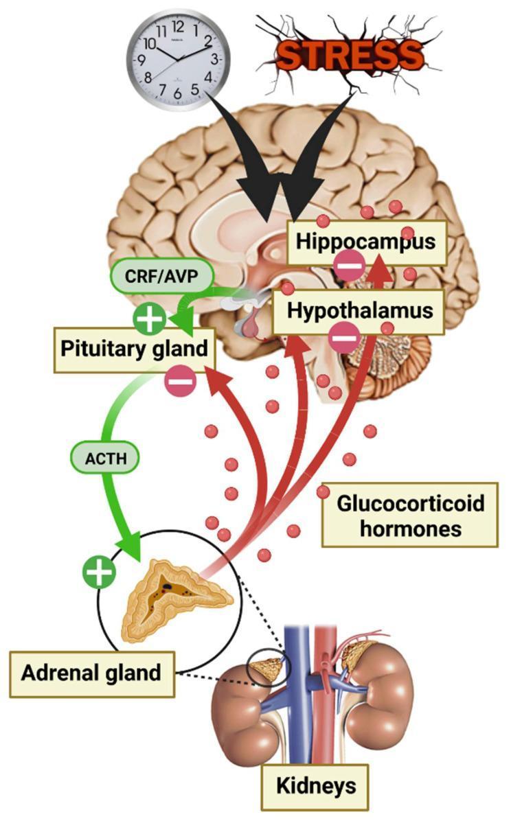
This pathophysiology diagram illustrates the hypothalamic-pituitary-adrenal (HPA) axis regulation in response to stress and circadian rhythms. The top of the image shows external triggers—a clock and a 'STRESS' icon—sending stimulatory signals (black arrows) to the brain, specifically targeting the hippocampus and hypothalamus. The stimulatory pathway proceeds via green arrows: the hypothalamus releases Corticotropin-Releasing Factor (CRF) and Vasopressin (AVP) to stimulate the pituitary gland, which then secretes Adrenocorticotropic Hormone (ACTH). ACTH acts on the adrenal gland (shown in a detailed cross-section above the kidneys) to trigger the release of glucocorticoid hormones, depicted as red spheres. A negative feedback loop is represented by red arrows leading from the glucocorticoids back to the pituitary, hypothalamus, and hippocampus, marked with minus signs (-) to indicate inhibition. The anatomical relationship between the adrenal glands and the kidneys is highlighted at the bottom, showing the adrenal glands' superior position. This diagram serves as an educational tool for understanding neuroendocrine feedback mechanisms and stress physiology.
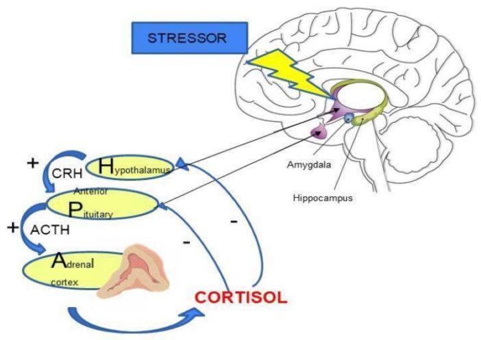
A medical diagram illustrating the regulation of the Hypothalamic-Pituitary-Adrenal (HPA) axis in response to stress. The top-right shows a sagittal cross-section of the human brain, highlighting the amygdala and hippocampus. A 'STRESSOR' box with a yellow lightning bolt icon indicates external stimulus impacting these limbic structures. Below, a flow diagram details the neuroendocrine cascade: positive stimulation (marked with '+') shows the Hypothalamus releasing Corticotropin-Releasing Hormone (CRH) to stimulate the Anterior Pituitary, which in turn releases Adrenocorticotropic Hormone (ACTH) to stimulate the Adrenal Cortex. The Adrenal Cortex releases Cortisol, depicted with blue return arrows labeled with '-' signs, representing the negative feedback loops that inhibit further secretion from both the Hypothalamus and the Anterior Pituitary. This visual resource is used to explain the hormonal response to stress and the homeostatic mechanisms of the endocrine system.
Anterior Pituitary Hormones
Overview Diagram
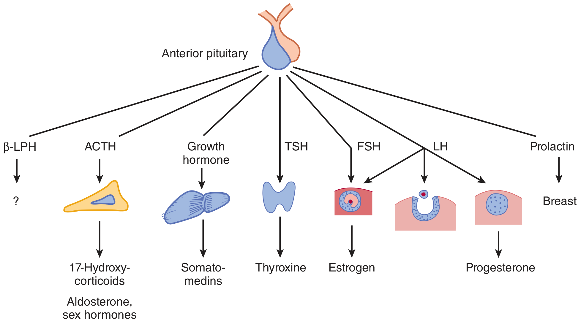
Hypothalamic Control
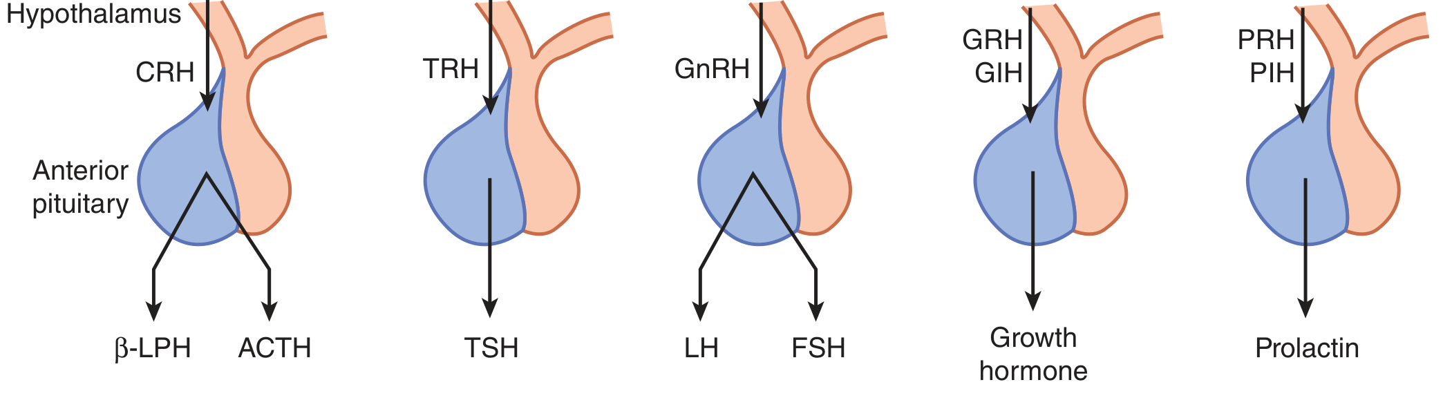
The 6 Major Anterior Pituitary Hormones
1. ACTH - Adrenocorticotropic Hormone (Corticotropin)
| Feature | Detail |
|---|---|
| Cell of origin | Corticotrope |
| Chemical nature | Polypeptide (39 amino acids); cleaved from POMC precursor |
| Hypothalamic stimulator | CRH (Corticotropin-releasing hormone) + AVP (vasopressin) |
| Inhibitor | Glucocorticoids (negative feedback) |
| Target organ | Adrenal cortex |
| Actions | Stimulates secretion of cortisol, aldosterone, and adrenal sex hormones |
| Excess | Cushing's disease (pituitary ACTH-secreting adenoma) |
| Deficiency | Secondary adrenal insufficiency |
Also co-secreted with β-LPH (β-lipotropin) from the same POMC precursor - physiologic role of β-LPH is uncertain.
2. GH - Growth Hormone (Somatotropin)
| Feature | Detail |
|---|---|
| Cell of origin | Somatotrope |
| Chemical nature | Polypeptide (191 amino acids) |
| Hypothalamic stimulator | GHRH (Growth hormone-releasing hormone), Ghrelin |
| Inhibitor | Somatostatin (GIH), IGF-1 (negative feedback) |
| Target organ | Liver, bone, all cells |
| Actions | Stimulates protein synthesis; liver produces IGF-1 (somatomedins) which mediates most growth effects; lipolysis; anti-insulin effect |
| Secretion pattern | Pulsatile; highest pulse during deep sleep (slow-wave) |
| Excess | Gigantism (children), Acromegaly (adults) |
| Deficiency | Short stature (children), altered body composition (adults) |
3. TSH - Thyroid-Stimulating Hormone (Thyrotropin)
| Feature | Detail |
|---|---|
| Cell of origin | Thyrotrope |
| Chemical nature | Glycoprotein - α subunit (shared with LH, FSH, hCG) + unique β subunit; 211 amino acids |
| Hypothalamic stimulator | TRH (Thyrotropin-releasing hormone) |
| Inhibitor | T₃/T₄ (negative feedback), dopamine, somatostatin, glucocorticoids |
| Target organ | Thyroid gland |
| Actions | Stimulates T₃ and T₄ synthesis and secretion; thyroid cell growth |
| Excess | Secondary hyperthyroidism (TSH-secreting adenoma - rare) |
| Deficiency | Secondary hypothyroidism |
4. FSH - Follicle-Stimulating Hormone
| Feature | Detail |
|---|---|
| Cell of origin | Gonadotrope |
| Chemical nature | Glycoprotein - shared α + unique β subunit; 210 amino acids |
| Hypothalamic stimulator | GnRH (pulsatile); activins, estrogen |
| Inhibitor | Sex steroids, inhibin (from gonads) |
| Target organ | Ovary / Testis |
| Actions (female) | Follicle development; estrogen production |
| Actions (male) | Sertoli cell stimulation → spermatogenesis; promotes inhibin release |
| Deficiency | Infertility, hypogonadism |
5. LH - Luteinizing Hormone
| Feature | Detail |
|---|---|
| Cell of origin | Gonadotrope (same cell as FSH) |
| Chemical nature | Glycoprotein - shared α + unique β subunit; 204 amino acids |
| Hypothalamic stimulator | GnRH (pulsatile) |
| Inhibitor | Sex steroids, inhibin |
| Target organ | Ovary / Testis |
| Actions (female) | LH surge triggers ovulation; stimulates corpus luteum → progesterone |
| Actions (male) | Stimulates Leydig cells → testosterone secretion |
| Deficiency | Anovulation (female), low testosterone (male) |
FSH and LH act in sequence: FSH grows the follicle → LH surge causes ovulation → LH maintains corpus luteum. - Ganong's, 26th ed.
6. Prolactin (PRL)
| Feature | Detail |
|---|---|
| Cell of origin | Lactotrope |
| Chemical nature | Polypeptide (198 amino acids) |
| Hypothalamic stimulator | TRH, estrogen, VIP (vasoactive intestinal peptide) |
| Inhibitor | Dopamine (PIH - prolactin inhibiting hormone) - PREDOMINANT control is inhibitory |
| Target organ | Mammary glands |
| Actions | Promotes breast development and milk secretion (lactation); suppresses GnRH → inhibits ovulation (explains lactational amenorrhea) |
| Excess | Hyperprolactinemia → galactorrhea, amenorrhea, infertility; caused by prolactinoma or dopamine antagonist drugs |
| Deficiency | Failure of lactation postpartum |
Unlike all other anterior pituitary hormones, prolactin's predominant hypothalamic control is inhibitory (via dopamine), not stimulatory. - Ganong's, 26th ed.
Summary Table
| Hormone | Cell | Nature | Hypothalamic Control | Target | Key Effect |
|---|---|---|---|---|---|
| ACTH | Corticotrope | Polypeptide | CRH ↑, Glucocorticoids ↓ | Adrenal cortex | Cortisol secretion |
| GH | Somatotrope | Polypeptide | GHRH ↑, Somatostatin ↓ | Liver/bone | Growth, IGF-1 production |
| TSH | Thyrotrope | Glycoprotein | TRH ↑, T₃/T₄ ↓ | Thyroid | T₃/T₄ secretion |
| FSH | Gonadotrope | Glycoprotein | GnRH ↑, Inhibin ↓ | Ovary/Testis | Follicle growth, spermatogenesis |
| LH | Gonadotrope | Glycoprotein | GnRH ↑, Steroids ↓ | Ovary/Testis | Ovulation, testosterone |
| Prolactin | Lactotrope | Polypeptide | Dopamine ↓ (dominant) | Breast | Lactation |
Key Points to Remember
- Shared α-subunit: TSH, LH, FSH, and hCG (placental) all share the same α subunit - their specificity comes from unique β subunits
- Pulsatile secretion: All anterior pituitary hormones are released in pulses - this is why continuous GnRH agonists (like leuprolide) eventually suppress rather than stimulate gonadotropins
- Prolactin is unique: Only hormone predominantly under inhibitory hypothalamic control; dopamine blockade (e.g., metoclopramide, antipsychotics) causes hyperprolactinemia
- Feedback loops: Long-loop (peripheral hormones → hypothalamus/pituitary), short-loop (pituitary hormone → hypothalamus), and ultra-short loop (self-feedback) all regulate secretion
- GH acts indirectly: Most growth-promoting effects of GH are mediated through IGF-1 produced by the liver