
Goal Directed Fluid Therapy - Anaesthesia relevance MD Anaesthetic pg exam answer
Reading File
Now let me search for GDFT content in the library and PubMed simultaneously.
Finding Sources
Finding Sources
Searching PubMed
"goal directed fluid therapy" AND perioperative
Excellent - I have strong hits. Now let me read the key sections from Morgan & Mikhail and Miller's in parallel.
Reading File
Reading File
Reading File
Excellent content. Now let me get the more detailed GDFT sections from Morgan & Mikhail (the main GDFT section at line 4669 and the monitors section) and the Miller's GDT section.
Reading File
Reading File
Finding Sources
Let me now get the Barash dynamic respiratory indices section and the Morgan & Mikhail hemodynamic monitoring for GDFT, plus search for the Frank-Starling and fluid challenge protocol details.
Reading File
Reading File
Finding Sources
I now have rich, comprehensive content. Let me fetch the GDT flowchart image from Miller's and also search for a relevant GDFT diagram for the answer.
Loading Image
Finding Sources
I now have all the material needed for a comprehensive MD Anaesthesia PG exam answer. Let me compose it.
Goal-Directed Fluid Therapy (GDFT) - Anaesthesia Relevance
MD Anaesthesia PG Exam Answer
Definition & Historical Background
Goal-Directed Fluid Therapy (GDFT) is a perioperative fluid management strategy that uses real-time hemodynamic parameters to guide individualized fluid administration, aiming to optimize cardiac output and tissue oxygen delivery while avoiding both hypovolemia and fluid overload.
The concept originated from a 1983 study by Shoemaker et al., which demonstrated lower mortality in critically ill patients when tissue oxygen delivery was optimized through physiological targets related to cardiac output. Modern GDFT evolved from this to become a cornerstone of Enhanced Recovery After Surgery (ERAS) protocols.
- Morgan & Mikhail's Clinical Anesthesiology, 7e, p. 2259
Physiological Rationale
The Problem with Conventional Fluid Therapy
Conventional fluid management uses fixed-rate infusions (often large volumes of 0.9% saline) to treat intraoperative hypotension. This leads to:
- Fluid overload (3-6 kg weight gain)
- Damage to the glycocalyx
- Release of natriuretic peptides
- Anastomotic leakage, pulmonary edema, wound infection
- Postoperative ileus and delayed GI function
- Prolonged hospital stay
Conversely, too little fluid causes mucosal acidosis, hypoperfusion, impaired GI function, and increased anastomotic complications.
The RELIEF Trial (largest multicenter RCT comparing restrictive ≤5 mL/kg/h vs liberal 8 mL/kg/h crystalloid) found acute kidney injury was more frequent with restrictive therapy, highlighting that both extremes are harmful. The optimal target is a positive fluid balance of 1-2 L at end of surgery using isotonic balanced crystalloids.
- Morgan & Mikhail's Clinical Anesthesiology, 7e, p. 2114
Frank-Starling Mechanism - The Core of GDFT
GDFT is built on the Frank-Starling relationship: on the steep portion of the curve, increasing preload significantly increases stroke volume (SV). On the flat portion, additional fluid produces minimal SV gain but causes edema. GDFT keeps the patient at the "knee" of the Starling curve - the optimal preload where SV is maximized without excess.
During positive-pressure ventilation (PPV), there is:
- Inspiratory reduction in RV stroke volume (decreased venous return)
- Subsequent reduction in LV end-diastolic volume during expiration
- LV stroke volume varies cyclically with ventilation
This effect is more pronounced on the steep part of the Starling curve - patients likely to respond to fluid show greater SV variation with each breath.
- Barash's Clinical Anesthesia, 9e, p. 4834
Key Hemodynamic Targets in GDFT
| Parameter | Target / Threshold | Clinical Use |
|---|---|---|
| Stroke Volume (SV) | Maximize (bolus until <10% increase) | Core GDFT endpoint |
| Cardiac Output / Index (CO/CI) | Optimize | Global perfusion |
| Mean Arterial Pressure (MAP) | Maintain ≥65 mmHg | Organ perfusion pressure |
| Stroke Volume Variation (SVV) | <13% = non-responsive | Fluid responsiveness predictor |
| Pulse Pressure Variation (PPV) | >13% = fluid responsive | Fluid responsiveness predictor |
| Flow Time Corrected (FTc) | 350-400 ms | Esophageal Doppler parameter |
| ScvO₂ | >70% | Adequacy of O₂ delivery |
| Lactate | Trending down | Successful resuscitation |
Monitoring Modalities Used in GDFT
1. Esophageal Doppler Monitor (EDM)
- Measures descending aortic blood flow velocity
- Derives stroke volume (SV) and flow time corrected (FTc)
- FTc < 350 ms suggests hypovolemia / vasoconstriction
- FTc > 400 ms + no SV response = euvolemic
- Non-invasive (relatively), provides continuous SV monitoring
- Devices: CardioQ, CardioQ-EDM+
2. Arterial Waveform Analysis / Pulse Contour Analysis
- Analyzes invasive arterial pressure waveform
- Provides SV, CO, SVV, PPV
- Calibrated monitors (require thermodilution or lithium dilution for calibration): PiCCO, LiDCO Plus
- Uncalibrated monitors (trend-based): FloTrac/Vigileo (Edwards), LiDCO Rapid - represent SV trends rather than absolute values
- PPV >13% = fluid responsive; <9% = non-responsive; 9-13% = gray zone (Cannesson et al.)
3. Transesophageal Echocardiography (TEE)
- Gold standard for intraoperative cardiac assessment
- Provides dynamic assessment of ventricular filling, valvular function, cardiac output
- Requires operator expertise; not suitable for continuous automated GDFT
- Best for high-risk cardiac patients
4. Pulmonary Artery Catheter (PAC)
- Historical "gold standard" - measures CO by thermodilution, PCWP, SvO₂
- High invasiveness has led to replacement by less-invasive alternatives
- Still used in complex cardiac cases
5. Pleth Variability Index (PVI)
- Non-invasive photoplethysmographic index
- Derived from pulse oximeter waveform variation
- Predicts fluid responsiveness non-invasively
- Recent meta-analysis (Felippe et al., 2025, PMID 40778974) confirms utility
6. CVP
-
Poor predictor of intravascular volume and fluid responsiveness - should NOT be used as a GDFT target
-
Static measures (CVP, PCWP) are inferior to dynamic measures (SVV, PPV) for predicting fluid responsiveness
-
Miller's Anesthesia, 10e, p. 6393-6394; Barash's Clinical Anesthesia, 9e, p. 4834-4835
Limitations of Dynamic Indices (SVV, PPV) - Critical for PG Exam
These indices are only valid when:
- Patient is intubated and receiving controlled PPV (not spontaneous breathing)
- Tidal volume ≥ 8 mL/kg (validated range)
- Patient is in sinus rhythm (AF/ectopy invalidates results)
- Chest is closed (open chest nullifies the indices)
- Patient is synchronous with ventilator (not fighting the vent)
Situations that invalidate SVV/PPV: Spontaneous ventilation, arrhythmias, right heart failure, pulmonary hypertension, low tidal volume (lung-protective) ventilation, open chest, IAH.
- Barash's Clinical Anesthesia, 9e, p. 4835
The GDFT Protocol - Fluid Bolus Technique
Standard approach (Miller's Anesthesia, 10e):
- Administer 250 mL bolus of colloid or crystalloid rapidly
- Assess SV response - if SV increases by ≥10%, patient is on steep part of Starling curve (fluid responsive)
- Continue boluses until SV increases by <10% - patient is now on the flat part (fluid non-responsive / optimized)
- Maintain on the flat part of the curve with ongoing monitoring
EDM-Based Protocol (Fig. 43.6 - Miller's Anesthesia):
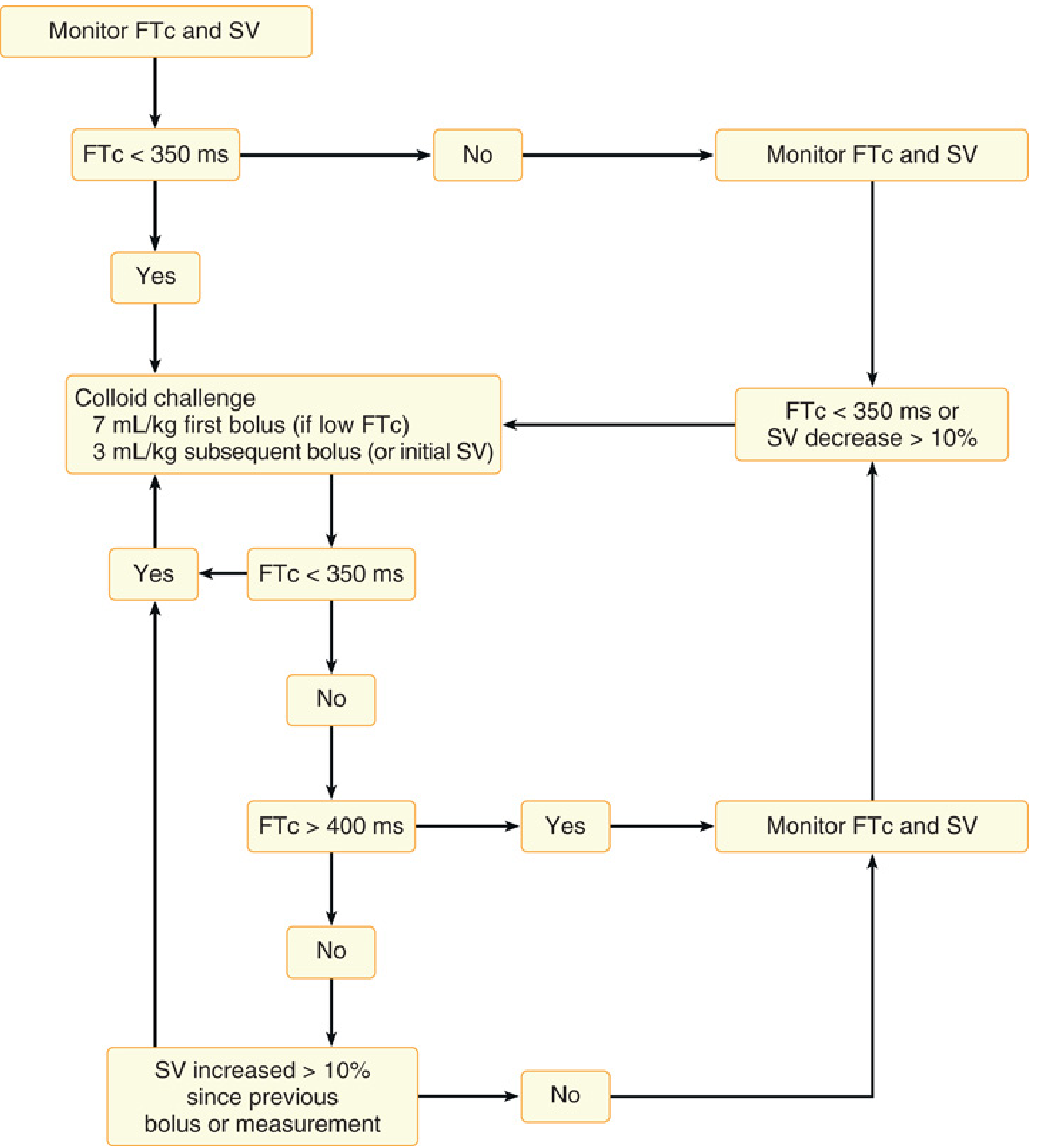
FTc, heart rate-corrected descending aorta flow time; SV, stroke volume - Miller's Anesthesia, 10e
- Miller's Anesthesia, 10e, p. 6394
Fluid Choice in GDFT
| Physiological Loss | Replacement Fluid |
|---|---|
| Insensible (closed abdomen) | Balanced crystalloid - 0.5 mL/kg/h |
| Insensible (open abdomen) | Balanced crystalloid - 1 mL/kg/h |
| Urine output | Balanced crystalloid (ml for ml) |
| Blood loss (intravascular) | Iso-oncotic colloid (estimated volume) |
| Preload deficit | Colloid (guided by monitoring) |
Balanced crystalloids (Lactated Ringer's, PlasmaLyte) preferred over 0.9% saline (which causes hyperchloremic metabolic acidosis).
Iso-oncotic colloids (e.g., 5% albumin, balanced gelatins) preferred for intravascular volume replacement - note that HES (starches) are avoided in sepsis (increased AKI risk) but use in elective surgical setting remains debated.
- Morgan & Mikhail's Clinical Anesthesiology, 7e, Table 48-3, p. 2115
GDFT vs. Restrictive vs. Liberal Fluid Therapy
| Strategy | Characteristics | Issues |
|---|---|---|
| Liberal ("wet") | Fixed high rates (8-10 mL/kg/h + replacement) | Fluid overload, edema, AKI, ileus |
| Restrictive ("dry") | ≤5 mL/kg/h | AKI (RELIEF trial), hypoperfusion |
| GDFT (individualized) | Titrated to hemodynamic response | Optimal for high-risk patients |
Key insight: GDFT is effective in the perioperative phase but appears ineffective in established critical illness (sepsis, ICU) - this distinction is frequently examined.
- Miller's Anesthesia, 10e, p. 6310
Clinical Benefits of GDFT - Evidence Base
Meta-analyses support:
- Reduction in postoperative renal impairment
- Reduction in respiratory failure and wound infection
- Shorter hospital length of stay
- Reduction in overall postoperative morbidity
- The Cochrane systematic review showed reduced hospital/28-day mortality when poorly controlled studies were excluded
Recent meta-analyses (2023-2025):
-
GDFT in thoracic surgery/one-lung ventilation reduces postoperative complications (Li et al., 2023, PMID 37723513)
-
GDFT in oncologic surgeries improves outcomes (Jin et al., 2025, PMID 40305698)
-
GDFT benefits elderly patients intraoperatively (Lasanudin et al., 2024, PMID 40994091)
-
GDFT in kidney transplants reduces delayed graft function (Klonarakis et al., 2024, PMID 38335896)
-
Non-invasive GDFT with PVI is clinically validated (Felippe et al., 2025, PMID 40778974)
-
Miller's Anesthesia, 10e, p. 6394-6395
GDFT and ERAS Protocols
GDFT is a core component of Enhanced Recovery After Surgery (ERAS) protocols:
- Goal: near-zero whole-body water balance (weight gain <2 kg)
- Vasoactive drugs (vasopressors) are used to manage hypotension instead of fluids
- Fluid is reserved for true volume deficit
- Pre- and postoperative oral carbohydrate loading reduces NPO-related deficits
- Weighing pre- and post-operatively documents fluid balance
Note: Recent evidence suggests GDFT may not confer additional benefit when already embedded in a well-structured ERAS pathway in low-to-moderate-risk surgery. The benefit is greatest in high-risk surgical patients and major open surgery.
- Current Surgical Therapy 14e; Sabiston Textbook of Surgery, 21e
Patient Selection - Who Benefits Most from GDFT?
High-risk surgical patients:
- Major open abdominal surgery (colorectal, upper GI, hepatobiliary)
- Emergency surgery
- Patients with significant comorbidities (cardiac, renal, hepatic disease)
- Major vascular surgery
- Thoracic surgery (one-lung ventilation)
- Organ transplantation (kidney, liver)
May not need GDFT:
- Low-risk patients undergoing laparoscopic or minimally invasive procedures
- Outpatient / day-case surgery
- Procedures with minimal physiological stress
Role of Vasopressors in GDFT
Modern GDFT protocols often combine fluid therapy with vasopressors:
- If MAP is low despite optimized SV (fluid non-responsive but hypotensive) → vasopressor (norepinephrine) rather than more fluid
- If SV is low and fluid responsive → fluid bolus
- If SV is adequate but CO is still low → inotrope (dobutamine)
- This tripartite approach (fluids + vasopressors + inotropes) is called Hemodynamic Goal-Directed Therapy
Closed-Loop GDFT - Future Direction
Research has introduced automated closed-loop systems:
-
Algorithms continuously monitor hemodynamic parameters and autonomously titrate fluid delivery and vasopressor infusions
-
Studies by Cannesson and colleagues demonstrated feasibility in intraoperative settings
-
May support clinical decision-making and reduce human error
-
Machine learning integration is being explored
-
Miller's Anesthesia, 10e, p. 6396; Morgan & Mikhail, 7e
Summary Table for Quick Revision
| Aspect | Key Point |
|---|---|
| Origin | Shoemaker (1983) - optimize O₂ delivery in critically ill |
| Core principle | Titrate fluid to Frank-Starling curve using SV response |
| Fluid bolus | 250 mL; repeat if SV increases ≥10% |
| Best monitors | Esophageal Doppler, arterial waveform (FloTrac, PiCCO), TEE |
| Dynamic indices | SVV, PPV - threshold ~13%; only valid in controlled PPV + sinus rhythm |
| Fluid choice | Balanced crystalloid for losses; iso-oncotic colloid for intravascular deficit |
| Avoid | CVP as sole target; 0.9% saline; HES in sepsis |
| Benefit | High-risk major surgery; reduces AKI, infection, LOS |
| ERAS integration | Near-zero balance goal; vasopressors for hypotension, not fluid |
| Limitation | Inconsistent results in some trials; may not add benefit in established ERAS |
Key textbook sources: Morgan & Mikhail's Clinical Anesthesiology 7e (pp. 2114-2115, 2259), Miller's Anesthesia 10e (pp. 6310, 6393-6396), Barash's Clinical Anesthesia 9e (pp. 4834-4835), Sabiston Textbook of Surgery 21e, Current Surgical Therapy 14e.
This is a shared conversation. Sign in to Orris to start your own chat.