


Explain in detail add anything if you want
DIABETES IN PREGNANCY - COMPLETE NOTES
PART 2
1. Gestational Diabetes Mellitus (GDM)
What is it?
Features
- Complications are similar to pre-gestational DM (pre-GDM), EXCEPT:
- No congenital anomalies (GCA) - because GDM develops after the first trimester (organogenesis is already complete)
- No fetal ECHO - as structural heart defects are only a risk in pre-GDM (early hyperglycemia during cardiac development)
- Follow-up and investigations: Same as pre-GDM, with the two exceptions above
2. Diagnosis - DIPSI Criteria
Recommendations
| Test | Timing |
|---|---|
| 1st test | 1st antenatal visit |
| Repeat | 24-28 weeks of pregnancy |
| Minimum time gap between tests | 4 weeks |
| If 1st visit is after 28 weeks | Only 1 test done |
Procedure
- No fasting required (universal screening regardless of meals)
- Give 75 gm glucose in 300 mL water (lime can be added for taste)
- Patient consumes it in 5-10 minutes
- Check 2-hour postprandial (PP) levels using a plasma-calibrated glucometer
If Patient Vomits:
- Within 30 minutes of drinking glucose - repeat test on another day
- After 30 minutes - continue the test (enough absorption has occurred)
2-Hour PP Interpretation
| 2hr PP Value | Interpretation |
|---|---|
| < 140 mg/dL | Normal - Repeat at 24-28 weeks |
| ≥ 140 mg/dL | GDM |
| ≥ 200 mg/dL | Pre-GDM (pre-existing diabetes) |
| 2hr PP Value | Interpretation |
|---|---|
| < 140 mg/dL | Not diabetic |
| ≥ 140 mg/dL | GDM |
| ≥ 200 mg/dL | Start insulin immediately |
3. Management of GDM
Based on 2hr PP Values:
         2hr PP Value
              |
   ┌──────────┴──────────┐
140-199 mg/dL         ≥ 200 mg/dL
   (Initial MNT)      (Immediate Insulin)
       |                     |
  MNT x 2 weeks          8U Insulin
       |                   + MNT
  [Check Metabolic        + Weight counselling
    Goals]               (No role of aspirin)
Metabolic Goals (targets to achieve)
- FBS < 95 mg/dL
- 1hr PP < 140 mg/dL
- 2hr PP < 120 mg/dL
- HbA1c < 6%
- Average capillary glucose < 100 mg/dL
If Goals MET:
- Continue MNT
- Advise 30 min walk daily
- Check 2hr PP levels: 2nd trimester = every 2 weeks, 3rd trimester = weekly, minimum = monthly
If Goals NOT MET:
- Start Metformin → then Insulin + MNT
GOI (Government of India) Treatment Guidelines:
- Diagnosed > 20 weeks → Metformin
- Diagnosed < 20 weeks → Insulin
- 2hr PP > 200 mg/dL (at any time) → Insulin
4. Medical Nutrition Therapy (MNT)
Diet Composition
| Component | Proportion |
|---|---|
| Carbohydrate | 40% |
| Fat | 40% |
| Protein | 20% |
- Distributed over 3 meals + 3 snacks
Caloric Requirements
| BMI | Adjustment |
|---|---|
| All pregnant women | + 350 kcal/day |
| BMI < 18.5 (underweight) | Extra +500 kcal/day |
| BMI > 25 (overweight) | Subtract 500 kcal/day |
5. Oral Hypoglycemic Agents (OHAs)
Metformin
- Recommended by GOI (Government of India)
- Used only in GDM (NOT in pre-GDM)
- Only used > 20 weeks of pregnancy
- 1st line drug
- Advantages:
- Reduces excessive maternal weight gain
- Reduces neonatal hypoglycemia risk
- Reduces chances of Large for Gestational Age (LGA) fetus
- Reduces macrosomia risk
- Preferred in obese patients
- Dose: 500 mg/day → up to 2 g/day
- If dose needed > 2 g/day → Add Insulin
- Side effects:
- Most common (m/c): GI side effects
- Most dangerous: Lactic acidosis
Glyburide
- Dose: 2.5 mg/day → up to 20 mg/day
- Side effect: Increased risk of neonatal hypoglycemia
6. Insulin in Pregnancy
Type Used
- Human pre-mix insulin 30:70
- 30% short-acting + 70% intermediate insulin
- 1 vial = 40 IU
- Storage: 4-8°C
- Syringe reused up to 14 times
Insulin Requirement Trends
- Increases with advancing pregnancy (due to rising insulin resistance)
- Decreases during labor (patient is NPO - nothing by mouth)
Starting Insulin
- Start Human Insulin premix 30:70
- Subcutaneous injection, 30 mins before breakfast, once daily (OD)
- Dose calculated by 2hr PP levels:
| 2hr PP Level | Insulin Dose |
|---|---|
| 120-160 mg/dL | 4 units |
| 160-200 mg/dL | 6 units |
| > 200 mg/dL | 8 units |
Dose Titration Flowchart
- Check levels on Day 3
- If goals NOT met:
- FBS > 95 → Add 2U insulin pre-dinner
- 2hr PP > 120 → Add 2U before breakfast
- Recheck every 3 days
- Titrate until metabolic goals are met
- Once met: Check every 2 weeks (2nd trimester), weekly (3rd trimester)
7. Termination of Pregnancy
| Type | Timing of Delivery |
|---|---|
| Well-controlled on diet (Type A1 GDM) | ≥ 39 weeks |
| GDM on drugs, well-controlled (Type A2) | > 39 weeks |
| GDM on drugs, NOT well-controlled | > 37 weeks |
- Mode of delivery: Vaginal (preferred)
- C-Section indication: Estimated fetal weight ≥ 4.5 kg
8. Intrapartum (During Labor) Management
- Mild GDM on medical management: Skip the morning insulin dose
- Monitor blood sugar every 2 hours with glucometer
- During labor: NPO + IV Normal Saline (NS) @ 100 mL/hr
Insulin in NS Drip (500 mL NS):
| Blood Sugar Level | Insulin Added |
|---|---|
| 90-120 mg/dL | None |
| 120-140 mg/dL | 4 U |
| 140-180 mg/dL | 6 U |
| ≥ 180 mg/dL | 8 U |
- If blood glucose < 70 mg/dL (hypoglycemia) → Start IV 5% Dextrose
9. Postpartum Management
GDM:
- Check blood sugar on Day 3 post-delivery
- At discharge → Follow up at 6 weeks with 75g 2hr OGTT
- Normal → Confirm resolution of GDM → Advise annual 75g OGTT (due to increased risk of developing Type 2 DM)
- Abnormal → Refer to Endocrinologist
Pre-GDM:
- Insulin requirement decreases from Day 2 of delivery
- Shifted back to OHA
- Refer to endocrinologist if needed
PART 3 - COMPLICATIONS & SPECIAL TOPICS
10. Maternal Complications
- Hyperglycemia - primary problem
- Infections (increased risk due to high glucose):
- Asymptomatic bacteriuria
- Candidiasis
- Puerperal sepsis
- Polyhydramnios (excess amniotic fluid) → can cause:
- Preterm labor (PTL)
- Premature rupture of membranes (PROM)
- Cord prolapse
- PPH (postpartum hemorrhage)
- Subinvolution
- Oligohydramnios - due to diabetic vasculopathy or PIH
- Placentomegaly (Big placenta) - swelling of chorionic villi due to hyperglycemia → raises risk of PIH & placenta previa
- Future risks: T2DM, C-section, ketoacidosis
Retinopathy Note
- Pre-gestational diabetes with retinopathy → worsens during pregnancy
- All pre-GDM females must have a baseline fundus examination
Hypoglycemia (Blood sugar < 70 mg/dL)
- 3 teaspoons glucose in 100 mL water, OR
- 6 teaspoons sugar in 100 mL water
11. Fetal Complications - Pedersen's Hypothesis
Maternal Hyperglycemia
        ↓
Fetal Hyperglycemia
        ↓
Stimulates Fetal Pancreas
        ↓
Hyperinsulinemia
    ↙          ↘
↑ Growth    ↓ Lipolysis → Fat deposition around fetal shoulder
    ↓                 ↓
Macrosomia        ↓
         ↘       ↙
        Shoulder Dystocia
Key Fetal Risks:
- Macrosomia → Prolonged labour → Increased C-section rate
- IUGR - only if diabetic vasculopathy or PIH is present
- Increased risk of abortion - in uncontrolled diabetes
- Stillbirth due to:
- Macrosomia → increased O2 demand → hypoxia
- Hyperglycemia → oxidative stress → hypoxia
- Edema of chorionic villi → decreased O2 transport → hypoxia
- (Most common in last 2 weeks of pregnancy)
- Congenital malformations - ONLY in pre-GDM (not GDM), because organogenesis has already occurred by the time GDM develops
- Hormone for fetal growth: Insulin and IGFs
12. Neonatal Complications
1. Neonatal Hypoglycemia (Most Important)
- Mechanism: Maternal hyperglycemia → fetal hyperglycemia → fetal hyperinsulinism
- At birth: glucose supply from mother is cut off, but insulin remains high → Hypoglycemia
2-9: Other Neonatal Complications
| Complication | Mechanism |
|---|---|
| Hypocalcemia | Triggered by hypoglycemia |
| Hypokalemia | Triggered by hypoglycemia |
| Hypomagnesemia | Triggered by hypoglycemia |
| Respiratory Distress Syndrome (RDS) | ↑ Insulin suppresses surfactant production |
| Necrotizing Enterocolitis (NEC) | Direct gut injury |
| Hypoxia → ↑ Erythropoietin → Polycythemia | Chronic intrauterine hypoxia |
| Hyperviscosity | Due to polycythemia |
| Hyperbilirubinemia | RBC breakdown from polycythemia |
| HOCM (Hypertrophic Obstructive Cardiomyopathy) | Insulin-driven cardiac growth |
Important: Anemia is NOT a complication of diabetic pregnancy.
Late Complications in the Child:
- T2DM: 1-3% risk
- Obesity
- Metabolic syndrome (Syndrome X)
13. Lung Maturity Tests
| Test | Details |
|---|---|
| L/S Ratio (Most common test) | Mature ≥ 2:1, Immature < 2:1 |
| Phosphatidyl Glycerol (Best test, done ≥ 35 weeks) | Present = Mature, Absent = Immature |
| Lamellar Body Count | < 15,000/mL = Not mature; > 50,000/mL = 100% mature; 30,000-40,000/mL = Positive |
| Bedside/Shake/Bubble test | Obsolete |
| Nile Blue Sulphate test | Obsolete |
Nile Blue Sulphate Test (Principle)
- AF fetal skin cells mature at the same time as lungs
- Orange cells = Mature skin cells (stained with dye)
- Blue cells = Immature skin cells
-
50% orange cells = Lungs mature
14. Shoulder Dystocia
Management - Mnemonic: HELPERR
| Step | Action | Notes |
|---|---|---|
| H | Call for Help | Immediate |
| E | Liberal Episiotomy | Creates more space |
| L | Legs (McRobert's Manoeuvre) | 1st & most effective - flex legs against abdomen; straightens sacrum, increases functional space (does not increase pelvic diameter) |
| P | Suprapubic Pressure + McRobert's | Sustained thrusts to dislodge shoulder |
| E | Enter manoeuvre | Manual rotation: Wood's Corkscrew (both shoulders) or Rubin's (one shoulder) |
| R | Remove posterior arm | Jacquemier manoeuvre |
| R | Roll onto all 4s | Gaskin's manoeuvre |
| Zavanelli | Last resort | Push head back into uterus → Emergency C-section |
Other Notes:
- Dead baby: Cleidotomy (fracture clavicle of baby)
- Symphysiotomy: Dividing pubic symphysis of mother - now Obsolete
- CONTRAINDICATED: Fundal pressure (worsens impaction)
- Each manoeuvre performed for a maximum of 30 seconds before moving to the next
Complications of Shoulder Dystocia:
- Erb's Palsy (C5, C6 injury) - most common - arm is internally rotated, adducted, and pronated ("waiter's tip" position)
- Injury to C5, C6 nerve roots
- Injury from manoeuvres
Summary Table - Quick Recall
| Topic | Key Point |
|---|---|
| DIPSI | 75g glucose, 2hr PP, no fasting |
| GDM diagnosis cut-off | ≥ 140 mg/dL |
| Pre-GDM cut-off | ≥ 200 mg/dL |
| MNT duration before reassessment | 2 weeks |
| Immediate insulin indication | 2hr PP ≥ 200 mg/dL |
| GOI: < 20 weeks DM → | Insulin |
| GOI: > 20 weeks DM → | Metformin |
| Metformin max dose | 2 g/day |
| Metformin most dangerous SE | Lactic acidosis |
| Insulin type in pregnancy | Human premix 30:70 |
| Delivery timing (well-controlled diet) | ≥ 39 weeks |
| C-section threshold | Baby weight ≥ 4.5 kg |
| Pedersen's hypothesis key product | Fetal hyperinsulinemia |
| Most common neonatal complication | Hypoglycemia |
| RDS mechanism | Insulin suppresses surfactant |
| Most common lung maturity test | L/S ratio |
| Best lung maturity test | Phosphatidyl glycerol |
| Most effective shoulder dystocia manoeuvre | McRobert's |
| Contraindicated in shoulder dystocia | Fundal pressure |
| Most common fetal complication in shoulder dystocia | Erb's palsy (C5, C6) |
MNT ?
Medical Nutrition Therapy (MNT) in Gestational Diabetes
What is MNT?
Diet Composition
| Macronutrient | Proportion |
|---|---|
| Carbohydrate | 40% |
| Fat | 40% |
| Protein | 20% |
Notice: Carbohydrates are reduced (normally 55-60% in a regular diet) and fat is kept relatively high - this is intentional to blunt postprandial glucose spikes.
Meal Distribution
- 3 main meals + 3 snacks throughout the day
- This is important because:
- Spreading carbs over 6 smaller portions prevents large glucose spikes
- Prevents prolonged fasting (which can cause ketosis/ketoacidemia, harmful in pregnancy)
- Keeps insulin demand steady rather than in large surges
Caloric Requirements
| Situation | Calories |
|---|---|
| All pregnant females (baseline) | +350 kcal/day added to normal requirement |
| BMI < 18.5 (underweight) | Additional +500 kcal/day |
| BMI > 25 (overweight/obese) | Subtract 500 kcal/day |
The reason overweight women subtract calories is to limit excessive gestational weight gain, which worsens insulin resistance and increases macrosomia risk - but never starve a pregnant diabetic woman as ketones cross the placenta and harm the fetus.
What is the Duration of MNT Trial?
- MNT is given a trial of 2 weeks
- After 2 weeks, check if metabolic goals are met:
| Goal | Target |
|---|---|
| FBS | < 95 mg/dL |
| 1hr PP | < 140 mg/dL |
| 2hr PP | < 120 mg/dL |
| HbA1c | < 6% |
| Average capillary glucose | < 100 mg/dL |
- Goals met → Continue MNT + 30 min daily walk
- Goals not met → Add Metformin (or Insulin if indicated)
Additional Lifestyle Advice with MNT
- 30 minutes of moderate walking daily is advised alongside diet
- This improves insulin sensitivity
- Combined with MNT, many GDM patients can be managed without medications
Why Carbs are 40% (Not Lower)?
- Very low carb diets in pregnancy risk ketoacidosis even at normal blood sugars (starvation ketosis)
- Fetal brain runs on glucose - complete carb restriction is dangerous
- 40% is the "sweet spot" - low enough to reduce glucose load, safe enough to prevent ketosis
Key Takeaway
MNT is not just a "diet" - it is a structured, calculated therapeutic intervention. It is always the first step in GDM management and continues even after medications are started. It is NEVER stopped.
2hr PP Value | ┌──────────┴──────────┐ 140-199 mg/dL ≥ 200 mg/dL (Initial MNT) (Immediate Insulin) | | MNT x 2 weeks 8U Insulin | + MNT [Check Metabolic + Weight counselling Goals] (No role of aspirin)
GDM Management Based on 2hr PP Value - Full Explanation
The Two Pathways
              2hr PP Value (DIPSI Test)
                        |
         ┌──────────────┴──────────────┐
         │                             │
   140–199 mg/dL                  ≥ 200 mg/dL
   (Mild GDM)                    (Severe GDM)
         │                             │
    START MNT                  IMMEDIATE Insulin
    for 2 weeks                  (8 Units) + MNT
         │                      + Weight Counselling
   Check Metabolic              (No role of Aspirin)
      Goals...
PATH 1 - 140 to 199 mg/dL
Why MNT first?
- Blood sugar is elevated but not severely so
- Diet control alone has a good chance of bringing it to target
- Avoids unnecessary medication in pregnancy
What happens after 2 weeks of MNT?
         After 2 weeks of MNT
                  |
     ┌────────────┴────────────┐
     │                         │
Goals MET                 Goals NOT MET
     │                         │
Continue MNT            Step 1: Add Metformin
+ 30 min walk daily     (500mg → up to 2g/day)
+ Monitor 2hr PP                │
                        If still not controlled
                                │
                        Step 2: Add Insulin
                                │
                          Continue MNT
                        throughout all steps
Metabolic Goals to check:
| Parameter | Target |
|---|---|
| FBS | < 95 mg/dL |
| 1hr PP | < 140 mg/dL |
| 2hr PP | < 120 mg/dL |
| HbA1c | < 6% |
| Average capillary glucose | < 100 mg/dL |
PATH 2 - ≥ 200 mg/dL
Why Immediate Insulin? Why not MNT first?
- Blood sugar this high cannot be controlled by diet alone
- At ≥ 200 mg/dL, the fetus is already at serious risk:
- Macrosomia already beginning
- Fetal hyperinsulinism already triggered
- Every day of delay = fetal harm → so insulin starts same day
Starting Dose = 8 Units
- Human premix insulin 30:70
- Subcutaneous, 30 mins before breakfast
Why 8 Units specifically?
- Based on the insulin dose table from 2hr PP:
| 2hr PP | Dose |
|---|---|
| 120-160 mg/dL | 4 units |
| 160-200 mg/dL | 6 units |
| > 200 mg/dL | 8 units |
- Since ≥ 200 falls in the highest bracket → 8U is the starting dose
What else is given alongside?
| Component | Reason |
|---|---|
| MNT | Diet control is always maintained - never stopped even on insulin |
| Weight counselling | Obesity worsens insulin resistance; excessive weight gain increases macrosomia risk |
| No Aspirin | Aspirin has no role in GDM management (it is used in PIH/preeclampsia prevention, not GDM) |
Why "No Role of Aspirin" is Specifically Mentioned
- Prevention of preeclampsia (PIH)
- Antiphospholipid syndrome
Complete Picture Together
DIPSI Test (75g glucose, 2hr PP)
              │
    ┌─────────┼──────────┐
< 140      140-199     ≥ 200
  │           │           │
Normal      MNT         8U Insulin
Repeat at   x 2wks      + MNT
24-28 wks      │         + Weight counselling
            Goals?       (No aspirin)
           ↙    ↘
        Met    Not met
          │       │
       Continue  Metformin
        MNT      → Insulin
One-Line Summary
140-199 = give diet 2 weeks to fix it first. ≥ 200 = start insulin immediately, no waiting - diet alone will never be enough at this level.
5F is shape of abd
5F's of Abdominal Distension (Shape/Swelling)
The Classic Mnemonic
5 F's
| F | Cause | Details |
|---|---|---|
| 1. Fat | Obesity | Generalized fat deposition; uniformly distended abdomen; no fluid thrill, no shifting dullness |
| 2. Fluid | Ascites | Free fluid in peritoneal cavity; fluid thrill +ve, shifting dullness +ve |
| 3. Flatus | Gas / Intestinal obstruction | Tympanitic (drum-like) on percussion; bowel sounds high-pitched in obstruction |
| 4. Feces | Constipation / Fecal impaction | Doughy feel, indentable mass along colon |
| 5. Fetus | Pregnancy | Uterine enlargement; fetal parts palpable, uterine souffle heard |
Extended Version - "9F" or "Additional F's" (Some textbooks add)
| Extra F | Cause |
|---|---|
| Fibroid | Large uterine fibroid mimicking pregnancy |
| Full bladder | Urinary retention - suprapubic swelling, dull on percussion |
| Fatal growth | Tumor / malignancy (ovarian cyst, hepatomegaly, splenomegaly) |
How to Differentiate Clinically
Fat
- Uniform distension
- Umbilicus sunken (pulled inward)
- No percussion change with position
- No fluid thrill
Fluid (Ascites)
- Umbilicus everted (pushed outward)
- Shifting dullness - dull flanks, resonant center (supine); shifts when patient turns
- Fluid thrill - tap one flank, feel impulse on other
- Puddle sign - earliest sign of ascites
Flatus
- Tympanitic (resonant) all over
- Abdomen uniformly distended
- Moves around with position changes
Feces
- Indentable, "putty-like" consistency
- Follows the line of the colon (ascending → transverse → descending)
- Usually in left iliac fossa (sigmoid)
Fetus
- Uterus felt as central pelvic-abdominal mass
- Grows upward from pelvis
- Fundal height corresponds to weeks of gestation:
12 weeks → Pubic symphysis
20 weeks → Umbilicus
36 weeks → Xiphisternum
40 weeks → Slightly below xiphisternum (lightening)
Exam Tip
If a question says "female, abdominal distension, first thing to rule out" → always think pregnancy first before ascites or any other cause.
If question says "shifting dullness + fluid thrill" → Ascites (Fluid)
If question says "tympanitic abdomen + high-pitched bowel sounds" → Flatus / obstruction
Cause of visible pulasation in abd
Causes of Visible Pulsation in the Abdomen
Two Types of Pulsation
1. Expansile Pulsation (True Pulsation)
- Pulsates outward in all directions
- Indicates the structure itself is pulsating
- Classic example: Abdominal Aortic Aneurysm (AAA)
2. Transmitted Pulsation (False Pulsation)
- Pulsation transmitted from aorta to an overlying structure
- The structure itself is not pulsating - just receiving the beat
- Example: A solid mass (like pancreatic cyst) sitting over the aorta
Causes - Organized
A. Vascular Causes (Most Important)
| Cause | Details |
|---|---|
| Abdominal Aortic Aneurysm (AAA) | Most common cause; expansile + pulsatile mass in epigastric/central abdomen; palpable in thin patients |
| Normal aortic pulsation | Visible in thin/cachectic patients - no pathology |
| Tortuous aorta | In elderly, hypertensive patients |
B. Transmitted Pulsation (from Aorta)
| Cause | Region |
|---|---|
| Pancreatic cyst / pseudocyst | Epigastric - sits over aorta |
| Retroperitoneal mass | Any mass overlying the aorta |
| Enlarged lymph nodes (para-aortic) | Central abdomen |
| Hepatomegaly | Right hypochondrium - transmitted cardiac pulsation |
C. Hepatic Pulsation (Special Case)
| Cause | Mechanism |
|---|---|
| Tricuspid regurgitation (TR) | Systolic pulsation of liver; retrograde flow from RV → RA → IVC → Hepatic veins → Liver |
| Right heart failure | Congestive hepatomegaly - may show presystolic pulsation |
| Constrictive pericarditis | Pulsatile liver |
Exam point: Pulsatile liver = Tricuspid Regurgitation until proven otherwise
D. Epigastric Pulsation
| Cause | Details |
|---|---|
| Normal | Visible in thin people, especially after meals |
| Right ventricular hypertrophy/enlargement | Epigastric pulsation due to RV sitting against diaphragm |
| AAA | As above |
| Hyperdynamic circulation | Fever, anemia, thyrotoxicosis - increased cardiac output makes pulsation visible |
E. Other Causes
| Cause | Details |
|---|---|
| Pregnancy | Uterine pulsation visible in late pregnancy |
| Splenic artery aneurysm | Left hypochondrium - rare |
| Renal artery aneurysm | Flank - rare |
| Hyperdynamic states | Anemia, thyrotoxicosis, fever - aorta pulsates more forcefully |
Clinical Differentiation
Expansile vs Transmitted
Place both hands on either side of mass:

Expansile (AAA):
  ← hands pushed apart on both sides →
  TRUE pulsation

Transmitted (overlying mass):
  Both hands move UP together (not apart)
  FALSE / transmitted pulsation
Epigastric Pulsation - Quick Rule
Epigastric pulsation
        │
   ┌────┴────┐
Increases    Decreases
on lying     on sitting
flat         forward
   │              │
Normal /        RV
Aortic       enlargement
If pulsation increases on lying flat and decreases on leaning forward → likely aortic If pulsation better felt in epigastrium + increases on inspiration → likely RV hypertrophy
Most Important for Exams
| Scenario | Answer |
|---|---|
| Expansile pulsatile central abdominal mass | AAA |
| Pulsatile liver | Tricuspid Regurgitation |
| Epigastric pulsation + signs of RVH | Right Ventricular Hypertrophy |
| Pulsatile mass over epigastrium, transmitted | Pancreatic cyst / retroperitoneal mass |
| Visible pulsation in thin person, no mass | Normal transmitted aortic pulsation |


Expalin everything in detail and anything if you want
OVARIAN CANCER - COMPLETE NOTES
1. OVARIAN CYSTS
Functional Ovarian Cysts
- Most common (m/c) type of ovarian cyst
- Occur due to hormonal disturbances
- Mostly temporary - resolve spontaneously on their own
- No specific treatment required in most cases
Types of Functional Cysts
| Type | Key Feature |
|---|---|
| Follicular cyst (m/c) | Size of follicle ≥ 3 cm; occurs when follicle fails to rupture at ovulation |
| Corpus Luteal cyst | m/c cyst to rupture (can cause acute abdomen) |
| Theca Lutein cyst | Due to ↑ hCG levels - seen in molar pregnancy, twin pregnancy, infertility treatment (Clomiphene, HMG) |
Why Theca Lutein cyst in molar pregnancy? Molar pregnancy produces massive amounts of hCG → overstimulates theca cells → multiple bilateral cysts. They are the LARGEST functional cysts.
2. FEATURES OF OVARIAN CANCERS
- Present late (hence screening is NOT routinely done in the general population)
- Symptoms are vague: Nausea, vomiting, loss of appetite, bloating - often dismissed as IBS
- Adnexal mass found on examination
- IOC (Investigation of Choice): TVS (Transvaginal Sonography)
This is why ovarian cancer has a poor prognosis - it is the "silent killer." Most patients are Stage III or IV at diagnosis.
3. BENIGN vs MALIGNANT ADNEXAL MASS
| Feature | Benign | Malignant |
|---|---|---|
| Age | Reproductive age (m/c) | Extremes of age - Pre-pubertal OR Postmenopausal |
| Pain | Present (due to inflammation) | Absent (grows silently) |
| History | Longer duration | Short history + rapid progression + weight loss |
| Side | Unilateral | Bilateral |
| Consistency | Cystic | Solid |
| Tenderness | Present | Absent |
| USG features | Anechoic, unilocular, no solid component | Bilateral, >10 cm, solid components, thick septa, papillary outgrowths, vascular, ascites, enlarged lymph nodes, matted bowel loops |
USG Features of Malignancy (Red Flags):
- Thick septa (>3 mm)
- Papillary excrescences (projections from inner wall)
- Solid component within the cyst
- Mixed echogenicity
- Vascularity on Doppler
- Ascites
- Enlarged para-aortic lymph nodes
- Matted bowel loops
4. MANAGEMENT OF OVARIAN CYST
              Patient with Ovarian Cyst
                         │
          ┌──────────────┴──────────────┐
          │                             │
   Reproductive age group         Extremes of age
          │                             │
       Cyst size              ┌─────────┴──────────┐
          │                   │                    │
   ┌──────┼──────┐      Post-menopausal        Pre-pubertal
   │      │      │            │                    │
3-5cm  5-7cm  >7cm     Check CA-125          Check AFP,
   │      │      │       ≥35 IU?              hCG, LDH
   │      │      │            │                    │
Wait & Follow Surgery    Surgery after        Surgery after
watch  up   (high risk   further              investigations
       USG  torsion/     investigations
            rupture)
Key Notes on CA-125:
- Limited value in reproductive age because CA-125 rises in many benign conditions:
- Fibroid, PID, Genital TB, endometriosis, even menstruation
- In reproductive age: CA-125 >200 IU = significant
- In postmenopausal women: any rise in CA-125 ≥35 IU is significant (fewer false positives)
Risk of Malignant Transformation:
- Postmenopausal: 30%
- Premenopausal: 7%
5. OVARIAN PATHOLOGIES IN PREGNANCY
| Scenario | Most Common |
|---|---|
| m/c benign ovarian tumor in pregnancy | Dermoid cyst (mature cystic teratoma) |
| m/c cyst to undergo torsion | Dermoid cyst |
| m/c time for torsion | End of 1st trimester / Puerperium |
| m/c ovarian cancer in pregnancy | Dysgerminoma |
Management of Ovarian Cyst in Pregnancy:
         Ovarian cyst in Pregnancy
                    │
       ┌────────────┴────────────┐
       │                         │
  Asymptomatic               Symptomatic
       │                  (Rupture/Torsion)
 ┌─────┴──────┐                  │
 │            │           Remove cyst
1st T       2nd T         immediately
 │            │        (regardless of GA)
Wait &     Malignant
watch      features on
 │         USG OR >10cm
m/c =           │
Corpus        Surgery
Luteum
(resolves
spontaneously)

NOTE: Removal in 1st T is C/I
→ ↓ Progesterone → Abortion
Why wait in 1st trimester? The corpus luteum produces progesterone which maintains the pregnancy until the placenta takes over (~10-12 weeks). Removing an ovarian cyst in the 1st trimester risks removing the corpus luteum → progesterone drops → abortion.
6. RISK FACTORS FOR OVARIAN CANCER
Incessant Ovulation Theory
Risk Factor Table
| Category | Proven Risk Factors | Controversial |
|---|---|---|
| Excessive estrogen | Early menarche, Late menopause, Obesity, Endometriosis | PCOS, HRT |
| Excessive ovulation | Nulliparity | Infertility + ovulation-inducing drugs |
| Genetic syndromes | Lynch syndrome, BRCA1, BRCA2 | - |
| Carcinogens | Asbestos | Talc, Smoking (→ mucinous adenocarcinoma) |
Protective Factors
| Category | Examples |
|---|---|
| Related to estrogen | Physical exercise |
| Reducing ovulation | Multiparity, OCP, Anovulation, Breastfeeding |
| Surgery | Hysterectomy, Tubal ligation, Salpingectomy |
Why does OCP protect? It suppresses ovulation → fewer total ovulations in lifetime → less surface trauma to ovarian epithelium.
Why does breastfeeding protect? Prolactin suppresses GnRH → anovulation during breastfeeding period.
7. GENETIC SYNDROMES
Lynch Syndrome (HNPCC)
- Gene: MLH1 / MSH2 (mismatch repair genes)
- Most common associated cancer: Colorectal cancer > Endometrial cancer (60-70% risk)
- Risk of ovarian cancer: 20%
BRCA1
- Highest risk of ovarian cancer: 40%
- Also increases endometrial cancer risk
- Prevention: TAH + BSO (Total Abdominal Hysterectomy + Bilateral Salpingo-Oophorectomy) after family completion - ideally at 35-40 years of age
BRCA2
- Risk of ovarian cancer: 15%
- More associated with breast cancer than BRCA1
Hereditary vs Sporadic Ovarian Cancer
| Feature | Hereditary | Sporadic |
|---|---|---|
| Age group | Younger (~50 years) | Older (60-70 years) |
| Proportion | 5-10% of all ovarian cancers | 90-95% |
| Screening | Annual TVS + CA-125 (from 35-40 yrs) | Reassurance only |
8. WHO CLASSIFICATION OF OVARIAN TUMORS
              OVARIAN TUMORS
                    │
    ┌───────────────┼───────────────┐
    │               │               │
Epithelial      Germ Cell       Sex Cord Stromal    Metastatic
  (90%)         (5-15%)            (3-5%)
  m/c            2nd m/c           3rd m/c
Most Common Overall:
- m/c ovarian tumor (all types): Serous cystadenoma
- m/c ovarian tumor in reproductive age: Dermoid cyst
- m/c ovarian cancer: Serous cystadenocarcinoma
9. EPITHELIAL OVARIAN TUMORS
Types:
- Serous tumors (m/c)
- Mucinous tumors
- Brenner's tumor
- Endometrioid tumor
- Clear cell tumor
General Features of Epithelial Tumors:
- m/c age: 60 years
- m/c bilateral
- Worst prognosis due to non-specific symptoms (nausea, vomiting, IBS, weight loss)
- HPE hallmark: Psammoma bodies (calcified concentric rings)
- Mutations:
- Low grade: KRAS, PTEN mutations
- High grade: p53 mutation
10. SEROUS vs MUCINOUS TUMORS
| Feature | Serous | Mucinous |
|---|---|---|
| % Benign | 60% (serous cystadenoma) | 80% (mucinous cystadenoma) |
| Occurrence | Mostly bilateral | Mostly unilateral |
| Associated mutation | BRCA1, BRCA2, p53 | KRAS mutation |
| Smoking | NOT a risk factor | Risk factor |
| Malignant counterpart | Serous cystadenocarcinoma | Mucinous cystadenocarcinoma |
| Tumor marker | CA-125 | CEA, CA 19-9 |
| HPE | Psammoma bodies | None specific |
| Gross | Unilocular, clear/serous fluid | Honeycomb appearance - multiloculated, mucinous material |
| Microscopy | Resembles Fallopian tube lining | Resembles endocervix lining |
| Associated complication | - | Pseudomyxoma Peritonei |
11. PSEUDOMYXOMA PERITONEI
- Peritoneum filled with mucinous material ("jelly belly")
- Causes:
- Appendix cancer: most common cause
- Mucinous ovarian tumor (metastatic)
- Mucocele of appendix
- Bad prognosis due to high recurrence rate
- Treated surgically with cytoreduction + HIPEC (heated intraperitoneal chemotherapy)
12. BRENNER'S TUMOR
- Mnemonic: "We BUST"
- HPE: Walthard cell nest + Coffee bean nuclei (nuclear grooving)
- Always benign
- Always solid (not cystic)
- Always unilateral (100%)
- Lined by transitional epithelium (resembles bladder lining)
Coffee bean nuclei (central grooving) is seen in two tumors:
- Brenner's tumor
- Granulosa cell tumor
13. ENDOMETRIOID vs CLEAR CELL TUMOR
| Feature | Endometrioid Tumor | Clear Cell Tumor (Malignant) |
|---|---|---|
| HPE | Resembles endometrial glands | Hobnail cells (cells with nuclei bulging into lumen like hobnails) |
| Associations | a. Endometriosis b. Endometrial cancer | a. Endometriosis b. In-utero DES (diethylstilbestrol) exposure |
Key Notes:
- m/c ovarian tumor associated with endometriosis: Clear cell > Endometrioid
- m/c ovarian tumor associated with endometrial cancer: Endometrioid > Granulosa cell
- Solid ovarian tumors: Brenner's & Fibroma
14. GERM CELL TUMORS (GCT)
Types:
- Teratoma:
- Benign: Mature cystic teratoma (Dermoid cyst)
- Malignant: Immature teratoma
- Monodermal: Struma ovarii (all thyroid tissue)
- Dysgerminoma
- Yolk sac tumor (Endodermal sinus tumor)
- Embryonal carcinoma
- Choriocarcinoma
- Mixed type
General GCT Features:
- m/c in young girls (10-30 years)
- Usually unilateral
- Better prognosis if diagnosed early (compared to epithelial tumors)
15. TUMOR MARKERS - GCT
| GCT | Markers SEEN | Markers NEVER SEEN |
|---|---|---|
| Dysgerminoma | LDH, hCG, PLAP | AFP (never) |
| Yolk sac tumor | AFP, LDH, α1-antitrypsin | hCG (never) |
| Embryonal carcinoma | AFP, hCG | LDH (never) |
| Choriocarcinoma | hCG | AFP, LDH |
| Teratoma | No tumor marker | - |
Exam trick: AFP is NEVER seen in dysgerminoma. hCG is NEVER seen in yolk sac tumor. These negatives are frequently tested.
16. DERMOID CYST (Mature Cystic Teratoma)
Features:
- m/c ovarian tumor in reproductive age
- m/c ovarian tumor in pregnancy
- m/c ovarian cyst to undergo torsion
- Usually unilateral (bilateral in only 10% cases)
- Contains all 3 germ cell layers (ectoderm is most common component: hair, skin, teeth, sebaceous glands)
- Mostly benign
Structure:
- Rokitansky protuberance - a raised prominence (plug) inside the cyst from which hair, teeth, and other structures arise
- This is the most common site of malignant transformation
Malignancy Risk:
- Risk: 0.2-2% (very low)
- m/c type of malignancy: Squamous cell carcinoma
- m/c site: Rokitansky protuberance
USG Findings:
- Tip of iceberg sign - echogenic component obscures deeper structures (like an iceberg, only tip visible)
- Dot and dash appearance - hair shafts and sebaceous material create characteristic pattern
Contents:
- Sebaceous material (yellow, oily)
- Hair (white/yellow)
- Teeth (hyperechoic on USG)
- Bone
Management:
- Cystectomy (preferred) - removes cyst, preserves ovary; especially important in young patients (risk of torsion is high so needs removal)
- Oophorectomy - if family is completed
17. DYSGERMINOMA
Features:
- 2nd most common GCT
- Usually unilateral (bilateral in 15-20%)
- Highest risk of bilaterality among GCTs
- Best prognosis among all GCTs
- Only radiosensitive ovarian tumor (very important exam point)
- m/c ovarian cancer in dysgenetic gonads (Turner syndrome, Swyer syndrome)
- m/c ovarian cancer in pregnancy
Gross Appearance:
- Fleshy, solid, lobulated, tan/pink color
HPE (Histopathology):
- Nests of clear cells separated by fibrous septa
- Lymphocyte infiltration in the septa (characteristic finding)
- Resembles testicular seminoma (they are counterparts of each other)
Tumor Markers:
- LDH, hCG, PLAP
- AFP is NEVER elevated (key exam point)
18. YOLK SAC TUMOR (Endodermal Sinus Tumor)
Features:
- Most malignant GCT
- Worst prognosis - rapidly progressive
- Presents with acute abdomen (due to rapid growth)
- 100% unilateral - never bilateral
Tumor Marker:
- AFP (Alpha-fetoprotein) - most important marker
- Also: LDH, α1-antitrypsin
- hCG is NEVER elevated
HPE:
- Schiller-Duval body (Glomeruloid body)
- Appearance: central capillary surrounded by tumor cells, contained within cystic spaces
- Resembles a primitive kidney glomerulus under microscope
19. OCCURRENCE SUMMARY - GCTs
| Category | Most Common |
|---|---|
| m/c GCT overall | Dermoid cyst > Dysgerminoma |
| m/c germ cell cancer | Immature teratoma |
| m/c tumor in dysgenetic gonads | Gonadoblastoma |
| m/c cancer in dysgenetic gonads | Dysgerminoma |
| Best prognosis GCT | Dysgerminoma |
| Worst prognosis GCT | Yolk sac tumor |
MASTER COMPARISON TABLE - Quick Recall
| Tumor | Age | Side | Marker | HPE | Special Feature |
|---|---|---|---|---|---|
| Serous cystadenoma | 60 yrs | Bilateral | CA-125 | Psammoma bodies | m/c ovarian tumor overall |
| Mucinous cystadenoma | - | Unilateral | CEA, CA19-9 | Endocervix-like | Pseudomyxoma peritonei |
| Brenner's | - | Unilateral | None | Coffee bean nuclei | Solid, benign, transitional epithelium |
| Clear cell | - | - | - | Hobnail cells | DES exposure, endometriosis |
| Dermoid cyst | Reproductive | Unilateral (90%) | None | All 3 layers | m/c to undergo torsion |
| Dysgerminoma | 10-30 yrs | Unilateral (80%) | LDH, hCG, PLAP | Nests + lymphocytes | Only radiosensitive; best GCT prognosis |
| Yolk sac tumor | 10-30 yrs | Always unilateral | AFP | Schiller-Duval body | Worst prognosis GCT |
| Choriocarcinoma | - | - | hCG | - | hCG only, AFP never |
Can you make proper flow chart for mx of ovarian cyst
A clean, professional medical flowchart titled "Management of Ovarian Cyst" on white background. The flowchart uses boxes, arrows, and color coding. Start with a top box: "OVARIAN CYST DETECTED (IOC: TVS)" Then splits into TWO main branches: LEFT BRANCH (blue header): "REPRODUCTIVE AGE GROUP" - Splits by cyst size into three sub-branches: - "3–5 cm" → green box "Wait & Watch" - "5–7 cm" → yellow box "Follow up with Serial USG" - "> 7 cm" → orange box "Surgery (High risk of torsion or rupture)" RIGHT BRANCH (purple header): "EXTREMES OF AGE" - Splits into two sub-branches: - "POST-MENOPAUSAL" → check "CA-125 ≥ 35 IU?" → red box "Surgery after further investigations" - "PRE-PUBERTAL" → check "Any raised: AFP, hCG, LDH?" → red box "Surgery after investigations" At bottom, two note boxes: NOTE 1 (grey): "% Malignant transformation: Post-menopausal = 30% | Pre-menopausal = 7%" NOTE 2 (grey): "CA-125 limited in reproductive age (rises in Fibroid, PID, TB). >200 IU = Significant" Use clear arrows connecting all boxes, color-coded branches, rounded rectangle boxes, sans-serif font, medical diagram style
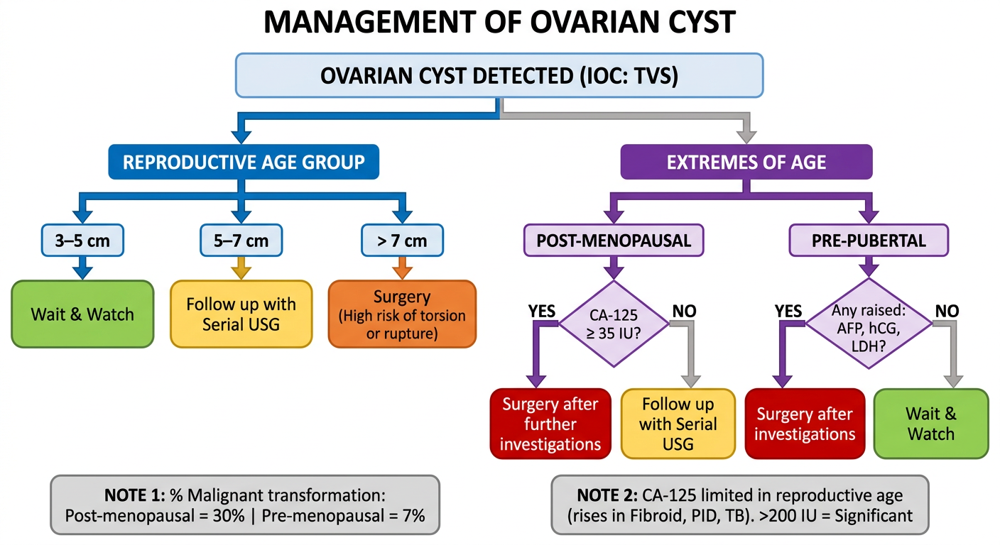
Management of Ovarian Cyst - Full Flowchart
                    OVARIAN CYST DETECTED
                        (IOC: TVS)
                             │
            ┌────────────────┴────────────────┐
            │                                 │
   REPRODUCTIVE AGE                    EXTREMES OF AGE
            │                                 │
        Cyst Size                   ┌─────────┴──────────┐
            │                       │                    │
   ┌────────┼────────┐        POST-MENOPAUSAL        PRE-PUBERTAL
   │        │        │               │                    │
 3-5cm   5-7cm    >7cm          CA-125 ≥35 IU?     AFP/hCG/LDH
   │        │        │               │              raised?
   ▼        ▼        ▼               ▼                    │
Wait &   Follow   Surgery        Surgery              Surgery
Watch    up USG   (torsion/      after further        after
                  rupture        investigations       investigations
                  risk)
Key Decision Points Explained
Reproductive Age - Why Size Matters?
| Size | Reason for Decision |
|---|---|
| 3-5 cm | Most likely functional cyst → will resolve on its own in 4-8 weeks; no intervention needed |
| 5-7 cm | Too big to ignore but not urgent; monitor with serial USG every 4-6 weeks to see if growing or shrinking |
| >7 cm | High risk of torsion (twisting cuts off blood supply) or rupture → surgery before complication |
Post-Menopausal - Why CA-125?
- After menopause, ovaries should be quiet - any cyst is suspicious
- CA-125 ≥ 35 IU in this age group is significant (few false positives post-menopause)
- If raised → operate
Pre-Pubertal - Why AFP/hCG/LDH?
- Ovarian cyst in a child is most likely a Germ Cell Tumor
- GCTs produce AFP (Yolk sac tumor), hCG (Choriocarcinoma), LDH (Dysgerminoma)
- Any raised marker → operate immediately
Important Notes
- Malignant transformation risk: Post-menopausal = 30% | Pre-menopausal = 7%
- CA-125 in reproductive age is unreliable (rises in Fibroid, PID, TB, endometriosis) - only >200 IU is significant in this group
- In pregnancy + 1st trimester → wait and watch (removing cyst risks progesterone loss → abortion)
- If symptomatic at any age (torsion/rupture) → immediate surgery regardless of age or size
Ovarian cyst in Pregnancy │ ┌────────────┴────────────┐ │ │ Asymptomatic Symptomatic │ (Rupture/Torsion) ┌─────┴──────┐ │ │ │ Remove cyst 1st T 2nd T immediately │ │ (regardless of GA) Wait & Malignant watch features on │ USG OR >10cm m/c = │ Corpus Surgery Luteum (resolves spontaneously) NOTE: Removal in 1st T is C/I → ↓ Progesterone → Abortion Make it clear & proper
A clean professional medical flowchart on white background titled "Management of Ovarian Cyst in Pregnancy" with clear boxes, arrows, and color coding. TOP BOX (dark blue, wide): "OVARIAN CYST IN PREGNANCY" Splits into TWO branches with a horizontal divider line: LEFT BRANCH - green header box: "ASYMPTOMATIC" Arrow down splits into TWO sub-branches: Sub-branch LEFT (light blue box): "1st TRIMESTER" Arrow down to yellow box: "WAIT & WATCH" Arrow down to green note box: "m/c = Corpus Luteum cyst (resolves spontaneously)" Arrow down to RED warning box: "⚠ REMOVAL IS CONTRAINDICATED — Removing cyst may remove corpus luteum → ↓ Progesterone → ABORTION" Sub-branch RIGHT (light orange box): "2nd TRIMESTER" Arrow down to decision diamond (yellow): "Malignant features on USG? OR Size > 10 cm?" YES arrow → red box: "SURGERY" NO arrow → green box: "Continue monitoring with USG" RIGHT BRANCH - red header box: "SYMPTOMATIC (Rupture / Torsion)" Arrow down to bold red box: "REMOVE CYST IMMEDIATELY — Regardless of gestational age" At the very bottom, a grey note box spanning full width: "KEY NOTES: 1st trimester removal C/I — corpus luteum maintains progesterone until placenta takes over at 10-12 weeks | In 2nd trimester surgery is safest if needed | Symptomatic = always operate regardless of trimester" Use rounded rectangle boxes, clean sans-serif font, color coded branches, professional medical diagram style, plenty of white space, clear readable text
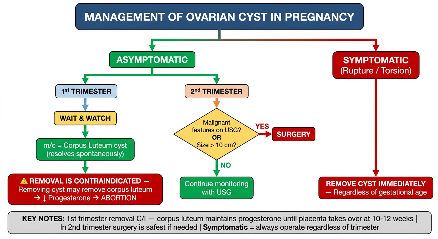
Complete Explanation of the Flowchart
Step 1 - First Question: Is she Symptomatic?
Symptomatic (torsion/rupture)  →  Operate IMMEDIATELY
                                   (no matter what trimester)

Asymptomatic                   →  Go to Step 2
Step 2 - Asymptomatic: Which Trimester?
1st Trimester - WAIT & WATCH
| Point | Explanation |
|---|---|
| m/c cyst found | Corpus luteum cyst (normal) |
| Why it resolves | Corpus luteum naturally involutes after 10-12 weeks when placenta takes over progesterone production |
| Why NOT to remove | Corpus luteum = only source of progesterone in early pregnancy. Remove it → progesterone drops → uterus contracts → abortion |
| Exception | Only operate if symptomatic (torsion/rupture) |
Key Rule: Removal of ovarian cyst in 1st trimester is absolutely contraindicated unless there is an emergency.
2nd Trimester - Best time to operate if needed
| Criteria | Action |
|---|---|
| Malignant features on USG (thick septa, solid component, papillary projections, ascites) | Surgery |
| Size > 10 cm | Surgery (risk of torsion, rupture, obstruction during labour) |
| None of the above | Continue monitoring with serial USG |
Why 2nd trimester is the safest time for surgery in pregnancy?
- Organogenesis is complete (less teratogenic risk from anaesthesia)
- Placenta has taken over progesterone (no abortion risk)
- Uterus not yet too large to obstruct the surgical field
- Risk of preterm labour from surgery is lowest
Symptomatic Branch - Always Operate
| Symptom | Cause | Action |
|---|---|---|
| Acute pain, vomiting, peritonism | Torsion | Emergency laparotomy/laparoscopy |
| Sudden pain + haemodynamic instability | Rupture | Emergency surgery |
| No matter the trimester | - | Remove cyst regardless of gestational age |
Summary in One Line per Step
- Symptomatic (any trimester) → Operate NOW
- 1st trimester + asymptomatic → Wait, watch, do NOT touch
- 2nd trimester + suspicious/large → Operate
- 2nd trimester + normal → Monitor
What is the meaning of CA125 more than 35 in post menopausal