
corpora quadrigemina
Reading File
Searching sources...
Finding Sources
Finding Sources
Finding Sources
Reading File
Finding Sources
Finding Sources
Reading File
Reading File
Finding Sources
Loading Image
Here is a comprehensive overview of the corpora quadrigemina, drawn from authoritative neuroscience and neuroanatomy textbooks.
Corpora Quadrigemina
Definition and Location
The corpora quadrigemina (Latin: "four bodies") are four rounded eminences on the dorsal surface (tectum) of the midbrain (mesencephalon). They are arranged as two pairs and together form the roof of the midbrain, sitting above the cerebral aqueduct of Sylvius:
- Superior pair - the superior colliculi (optic tectum)
- Inferior pair - the inferior colliculi
The cross-section below (upper midbrain, at level of the superior colliculus) shows their relationship to surrounding structures:
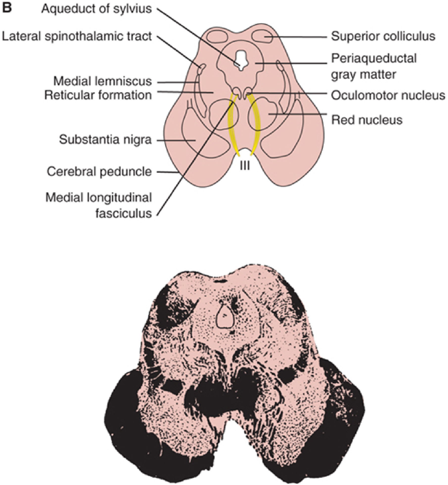
Figure: Upper midbrain cross-section. The superior colliculus sits dorsal to the aqueduct of Sylvius, with the oculomotor nucleus (CN III), red nucleus, substantia nigra, and cerebral peduncles visible ventral to it. - Localization in Clinical Neurology, 8e
The Two Pairs
1. Superior Colliculi (Visual Centers)
- Receive direct input from the retina via the optic tract (extrageniculate pathway through the brachium of the superior colliculus)
- Also called the optic tectum
- Each contains a retinotopic map of the contralateral visual field
- Key functions:
- Controls rapid (saccadic) eye movements - electrical stimulation commands a saccade to the corresponding point in space
- Directs reflex orientation toward visual stimuli
- Mediates the pupillary light reflex (fibers project to the pretectal nuclei)
- Receives descending input from the frontal eye fields via cortical projections
A minority of optic tract fibers bypass the lateral geniculate nucleus (LGN) and travel directly to the superior colliculus and pretectal area, forming the extrageniculate visual pathway that governs reflexive (non-conscious) visual responses. - Neuroanatomy through Clinical Cases, 3rd Ed.
2. Inferior Colliculi (Auditory Relay)
- Receive auditory information ascending from the cochlear nuclei and superior olivary complex via the lateral lemniscus
- Key functions:
- Major relay station for auditory information en route to the medial geniculate nucleus (thalamus) and then auditory cortex
- Important in sound localization (together with the superior olivary complex)
- Also interact with the superior colliculi - important for auditory-visual integration and orienting responses
- Superior cerebellar peduncle fibers (brachium conjunctivum) decussate at the level of the inferior colliculus before ascending to the thalamus
Surrounding Structures (Midbrain Context)
| Structure | Location | Key Function |
|---|---|---|
| Tectum (dorsal) | Contains corpora quadrigemina | Visual & auditory reflexes |
| Tegmentum (middle) | Ascending/descending tracts, reticular nuclei | Red nucleus, CN nuclei, periaqueductal gray |
| Cerebral peduncles (ventral) | Corticospinal (middle 3/5), frontopontine (medial 1/5), temporopontine (lateral 1/5) | Motor descending pathways |
| Substantia nigra | Between peduncles & tegmentum | Dopaminergic, movement control |
| CN III nucleus | Beneath superior colliculus | Oculomotor (eye movement, pupil) |
| CN IV nucleus | At level of inferior colliculus | Trochlear (superior oblique) |
- Localization in Clinical Neurology, 8e
Key Clinical Correlation: Parinaud's Syndrome
Lesions compressing the dorsal midbrain/pretectal area (i.e., the region of the corpora quadrigemina) produce Parinaud's syndrome, characterized by four components:
- Impairment of vertical gaze, especially upgaze - due to compression of the dorsal vertical gaze center
- Light-near dissociation - large, irregular pupils that do not react to light but may react to near-far accommodation; this results from disruption of optic fibers to the Edinger-Westphal nuclei via the posterior commissure, while convergence fibers are relatively spared
- Eyelid abnormalities - bilateral lid retraction (Collier's sign) or ptosis
- Impaired convergence and convergence-retraction nystagmus on attempted upgaze
Common causes:
-
Pineal region tumors (compressing dorsally)
-
Hydrocephalus - dilated suprapineal recess pushes down onto the collicular plate
-
Multiple sclerosis or vascular disease of the midbrain/pretectal area
-
In children with hydrocephalus: the "setting-sun sign" (eyes deviated downward and inward)
-
Neuroanatomy through Clinical Cases, 3rd Ed.; The Wills Eye Manual
Vascular Supply
The corpora quadrigemina receive blood from:
-
Medial branches of the posterior cerebral artery (supply lateral cerebral peduncle, lateral tegmentum, corpora quadrigemina, and pineal gland)
-
Also contributions from the superior cerebellar artery and posterior choroidal branches
-
Adams and Victor's Principles of Neurology, 12th Ed.
Summary
| Superior Colliculi | Inferior Colliculi | |
|---|---|---|
| Sensory input | Visual (retina, optic tract) | Auditory (cochlear nuclei, lateral lemniscus) |
| Primary function | Saccadic eye movements, visual orienting, pupillary reflex | Auditory relay to thalamus (MGB), sound localization |
| CN nucleus at same level | CN III (oculomotor) | CN IV (trochlear) |
| Notable crossing | - | Brachium conjunctivum (superior cerebellar peduncle) decussates here |
| Clinical lesion | Parinaud's syndrome (compression of dorsal midbrain) | Hearing pathway disruption |
in easy acc. to veterinary anatomy
Searching sources...
Finding Sources
The library doesn't contain veterinary anatomy textbooks - it's a human medical library. Let me search the web for veterinary-specific information on the corpora quadrigemina.
Searching the Web
corpora quadrigemina veterinary anatomy animals easy explanation
Searching the Web
corpora quadrigemina rostral caudal colliculi domestic animals veterinary neuroscience
Reading Web Page
https://veteriankey.com/the-brain
Here is the corpora quadrigemina explained simply, in veterinary anatomy terms:
Corpora Quadrigemina - Veterinary Anatomy (Easy Version)
What is it?
"Corpora quadrigemina" literally means "four bodies" (Latin). It is the name given to four rounded bumps on the top (dorsal) surface of the midbrain (mesencephalon) - the middle part of the brainstem.
Think of it like two pairs of small hills sitting on the roof of the midbrain.
Veterinary Terminology - Important Difference!
In veterinary anatomy, the names are different from human anatomy:
| Human Term | Veterinary Term | Position |
|---|---|---|
| Superior colliculi | Rostral colliculi | Front pair (toward nose) |
| Inferior colliculi | Caudal colliculi | Back pair (toward tail) |
This is because in four-legged animals, "superior/inferior" (up/down) doesn't make as much sense as "rostral/caudal" (front/back).
Simple Diagram (Dorsal View of Brainstem)
        ← ROSTRAL (toward nose)

         [Pineal body]
    [Rostral colliculus] [Rostral colliculus]   → VISION
    [Caudal  colliculus] [Caudal  colliculus]   → HEARING

         CAUDAL (toward tail) →
The paired rostral and caudal colliculi mark the dorsal surface of the midbrain in all domestic animals (dog, cat, horse, cow). - Veterian Key
What Do They Do? (Simple)
Rostral Colliculi (Front Pair) = "Eye Reflex Centers"
- Receive input from the eyes (optic tract)
- Control reflex eye movements - like when an animal quickly looks toward something that moved
- Help direct the head/eyes toward a visual stimulus
- Connected to the thalamus via the brachium of the rostral colliculus
Caudal Colliculi (Back Pair) = "Ear Relay Stations"
- Receive input from the ears (auditory pathway via lateral lemniscus)
- Act as a relay station for sound information going up to the thalamus (medial geniculate body) and then to the auditory cortex
- Important for sound localization (which direction did that sound come from?)
- Connected to the thalamus via the brachium of the caudal colliculus
- The commissure of the caudal colliculi connects the two sides
Neighboring Structures (Easy)
- Just above (dorsal): the cerebral aqueduct (connects 3rd and 4th ventricles)
- Just in front: pineal body (epiphysis cerebri) - sits between the thalami and the corpora quadrigemina
- Just below: CN IV (trochlear nerve) exits at the level of the caudal colliculus and decussates in the rostral medullary velum - this is the only cranial nerve to exit from the dorsal surface of the brainstem
- Ventral to them: cerebral peduncles (crura cerebri), substantia nigra, red nucleus
Easy Memory Trick
| Rostral Colliculi | Caudal Colliculi | |
|---|---|---|
| Sense | Sight | Sound |
| Input from | Optic tract (eyes) | Lateral lemniscus (ears) |
| Relay to | Lateral geniculate body | Medial geniculate body |
| CN at same level | CN III (oculomotor) - rostral | CN IV (trochlear) - caudal |
Simple rule: Rostral = refleX for eYes. Caudal = auditorY relay.
Clinical Relevance in Vet Practice
- A lesion compressing the dorsal midbrain/tectum (e.g., a brain tumor, hydrocephalus) in animals can cause:
- Loss of menace response or abnormal pupillary light reflexes (rostral colliculus/pretectal area involvement)
- Vertical gaze deficits - similar to Parinaud's syndrome in humans
- Hearing pathway disruption if caudal colliculi are affected
- The corpora quadrigemina are visible as four distinct bumps when you examine the dorsal brainstem of a dissected animal brain (dog, cat, etc.)
In short: the corpora quadrigemina = the midbrain's sensory reflex hub - the front pair watches the eyes, the back pair listens to the ears. In vet anatomy, just remember rostral colliculi (vision) + caudal colliculi (hearing).
This is a shared conversation. Sign in to Orris to start your own chat.