
can you tell me all the important things need to be studied for upper limb anatomy for exams in detail
upper limb anatomy important topics for medical exams MBBS
Upper Limb Anatomy - Complete Exam Guide
1. BONES OF THE UPPER LIMB
Pectoral Girdle
- Clavicle - only bony connection between upper limb and axial skeleton; S-shaped; medial 2/3 convex anteriorly, lateral 1/3 concave anteriorly
- Scapula - triangular flat bone; key landmarks: acromion, coracoid process, glenoid cavity, spine of scapula, suprascapular notch (suprascapular nerve passes through it; suprascapular artery passes over it - "Army over the bridge")
Arm
- Humerus - head (articular), greater & lesser tubercles, bicipital groove (contains long head of biceps), surgical neck (vulnerable to fractures - axillary nerve injury), anatomical neck, deltoid tuberosity, medial & lateral epicondyles, lateral supraepicondylar ridge, capitulum (articulates with radius), trochlea (articulates with ulna), olecranon fossa, coronoid fossa, radial fossa
Forearm
- Radius - head (articulates with capitulum), radial tuberosity (insertion of biceps), styloid process, dorsal tubercle of Lister
- Ulna - olecranon process, coronoid process, trochlear notch, radial notch, head, styloid process
Hand (27 bones)
- 8 carpal bones (proximal row: Scaphoid, Lunate, Triquetrum, Pisiform; distal row: Trapezium, Trapezoid, Capitate, Hamate)
- Mnemonic: "Some Lovers Try Positions That They Can't Handle"
- 5 metacarpals
- 14 phalanges (thumb has 2; fingers have 3 each)
Exam tip: Scaphoid fracture - tenderness in anatomical snuffbox; avascular necrosis risk because blood supply enters distally. Colles' fracture - distal radius, dinner fork deformity.
2. BRACHIAL PLEXUS (Most Important Topic)

Structure: Roots → Trunks → Divisions → Cords → Branches

Trunks
| Trunk | Roots |
|---|---|
| Superior | C5 + C6 |
| Middle | C7 alone |
| Inferior | C8 + T1 |
Cords (named by relation to axillary artery 2nd part)
| Cord | From |
|---|---|
| Lateral | Anterior divisions of superior + middle trunks |
| Medial | Anterior division of inferior trunk |
| Posterior | Posterior divisions of all three trunks |
Key Branches (Exam Favorites)
- From roots: Dorsal scapular nerve (C5 - rhomboids), Long thoracic nerve (C5,6,7 - serratus anterior)
- From superior trunk: Suprascapular nerve (C5,6 - supraspinatus + infraspinatus), Nerve to subclavius (C5,6)
- From lateral cord: Lateral pectoral nerve, Musculocutaneous nerve, Lateral root of median nerve
- From medial cord: Medial pectoral nerve, Medial cutaneous nerve of arm, Medial cutaneous nerve of forearm, Ulnar nerve, Medial root of median nerve
- From posterior cord: Upper subscapular nerve, Thoracodorsal nerve, Lower subscapular nerve, Axillary nerve, Radial nerve
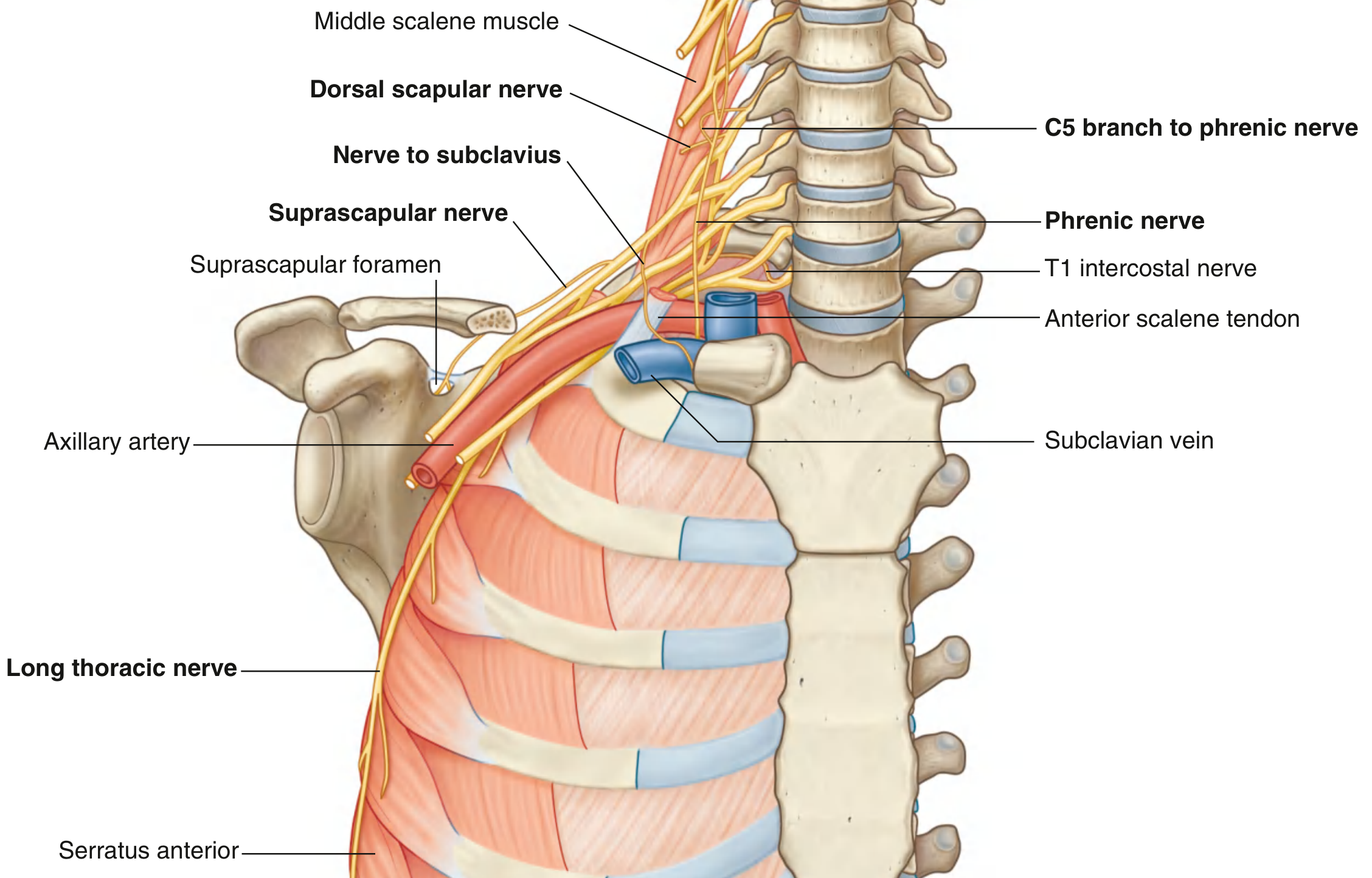
Brachial Plexus Injuries (High-Yield Clinical)
| Injury | Roots | Cause | Deformity |
|---|---|---|---|
| Erb's palsy (upper) | C5, C6 | Birth (shoulder dystocia), fall on shoulder | "Waiter's tip" - arm adducted, medially rotated, wrist flexed |
| Klumpke's palsy (lower) | C8, T1 | Pulling upward arm, cervical rib | "Claw hand" + Horner's syndrome (if T1 root torn) |
3. AXILLA
- Axillary artery (3 parts, divided by pectoralis minor)
- Axillary vein
- Brachial plexus (cords)
- Axillary lymph nodes (5 groups: anterior/pectoral, posterior/subscapular, lateral, central, apical)
- Long thoracic nerve (on serratus anterior)
- Intercostobrachial nerve
- 1st part (1 branch): Superior thoracic artery
- 2nd part (2 branches): Thoraco-acromial + Lateral thoracic artery
- 3rd part (3 branches): Subscapular, Anterior circumflex humeral, Posterior circumflex humeral
4. MUSCLES - BY COMPARTMENT
Shoulder (Rotator Cuff) - "SITS"
| Muscle | Origin | Insertion | Action | Nerve |
|---|---|---|---|---|
| Supraspinatus | Supraspinous fossa | Greater tubercle | Initiates abduction (0-15°) | Suprascapular (C5,6) |
| Infraspinatus | Infraspinous fossa | Greater tubercle | Lateral rotation | Suprascapular (C5,6) |
| Teres minor | Lateral border scapula | Greater tubercle | Lateral rotation | Axillary (C5,6) |
| Subscapularis | Subscapular fossa | Lesser tubercle | Medial rotation | Upper + Lower subscapular (C5-7) |
Arm - Anterior Compartment (Flexors)
| Muscle | Action | Nerve |
|---|---|---|
| Biceps brachii | Flexion + supination (main supinator) | Musculocutaneous (C5,6) |
| Brachialis | Main flexor of elbow | Musculocutaneous (C5,6) |
| Coracobrachialis | Flexion + adduction of arm | Musculocutaneous (C5,6,7) |
Arm - Posterior Compartment
| Muscle | Action | Nerve |
|---|---|---|
| Triceps brachii (3 heads) | Extension of elbow | Radial nerve (C6,7,8) |
| Anconeus | Assists extension | Radial nerve |
Forearm - Anterior Compartment (Flexors - Median & Ulnar nerves)
- Pronator teres, Flexor carpi radialis, Palmaris longus, Flexor carpi ulnaris, Flexor digitorum superficialis
- Flexor digitorum profundus (FDP) - lateral half (index + middle fingers) = median nerve; medial half (ring + little fingers) = ulnar nerve
- Flexor pollicis longus - anterior interosseous nerve (branch of median)
- Pronator quadratus - anterior interosseous nerve
Forearm - Posterior Compartment (Extensors - all radial nerve)
Exam tip: All forearm extensors = radial nerve. Radial nerve injury at spiral groove causes "wrist drop."
5. MAJOR NERVES AND INJURIES
Median Nerve (C6-T1)
- Course: Lateral cord (C6,7) + medial cord (C8,T1) → down medial arm → cubital fossa (medial to brachial artery) → between heads of pronator teres → anterior interosseous nerve branch → carpal tunnel → palm
- In forearm: Innervates all flexors EXCEPT flexor carpi ulnaris and medial half of FDP
- In hand: Thenar muscles (LOAF = Lumbricals 1&2, Opponens pollicis, Abductor pollicis brevis, Flexor pollicis brevis), palmar and digital sensation of lateral 3.5 fingers
- Lesion at wrist (carpal tunnel): Ape hand (loss of opposition), sensory loss lateral 3.5 fingers, no wrist drop
- Lesion at elbow: "Pointing index" with loss of hand grasp + loss of pronation
- Clinical test: "OK sign" (inability = AIN injury)
Ulnar Nerve (C8-T1)
- Course: Medial cord → behind medial epicondyle (very vulnerable!) → between heads of FCU → Guyon's canal (lateral to pisiform) → deep and superficial branches in hand
- In forearm: FCU + medial half FDP
- In hand: Hypothenar muscles, all interossei, medial 2 lumbricals, adductor pollicis + half flexor pollicis brevis; sensation of medial 1.5 fingers
- Lesion: Claw hand (ring + little fingers mostly), froment's sign (uses FPL to compensate), loss of finger abduction/adduction (interossei)
- "Claw worse with wrist lesion" - paradox of ulnar nerve - more claw with distal lesion because intrinsics are non-functional but FDP still pulling
Radial Nerve (C5-T1)
- Course: Posterior cord → spiral groove of humerus (behind lateral intermuscular septum) → lateral epicondyle → divides into superficial (sensory) and deep (posterior interosseous nerve, PIN)
- Muscles: All posterior arm + forearm extensors
- Lesion at axilla: Saturday night palsy - wrist drop + finger drop + loss of triceps (triceps reflex lost), sensory loss dorsal forearm/hand
- Lesion at spiral groove: Wrist drop (triceps spared - supplied above lesion)
- PIN injury (at radial tunnel): Finger drop without wrist drop (ECRL spared because innervated above PIN)
Musculocutaneous Nerve (C5-C7)
- From lateral cord; pierces coracobrachialis; supplies all 3 anterior arm muscles; continues as lateral cutaneous nerve of forearm
Axillary Nerve (C5-C6)
- From posterior cord; exits through quadrangular space with posterior circumflex humeral artery
- Supplies: Deltoid + teres minor; skin over deltoid (regimental badge area)
- Injury: Surgical neck of humerus fracture or shoulder dislocation → loss of shoulder abduction
6. JOINTS
Sternoclavicular Joint
- Only true joint between upper limb and axial skeleton
- Saddle joint (functionally), fibrocartilage articular disc present
- Ligaments: anterior + posterior sternoclavicular, costoclavicular, interclavicular
Acromioclavicular Joint
- Plane joint; fibrocartilage articular disc (may be absent)
- Key stabilizer: coracoclavicular ligament (conoid + trapezoid ligaments) - prevents superior displacement of clavicle
- Injury: AC separation (falls on shoulder tip)
Glenohumeral (Shoulder) Joint
- Ball-and-socket synovial joint
- Most mobile but least stable joint of the body
- Stability provided by: Rotator cuff muscles (primary dynamic stabilizers), glenoid labrum (deepens socket), glenohumeral ligaments, long head of biceps
- Weak point: Inferior wall - dislocation almost always anterior and inferior
- Bursae: Subacromial/subdeltoid bursa (most important - supraspinatus tendon lies below it)
- Movements: Flexion, extension, abduction, adduction, medial/lateral rotation, circumduction
- Abduction: 0-15° = supraspinatus; 15-90° = deltoid; 90-180° = trapezius + serratus anterior (scapular rotation)
Elbow Joint
- Hinge joint (flexion/extension); includes proximal radioulnar joint (pronation/supination)
- Medial collateral ligament (anterior bundle most important) resists valgus stress
- Lateral collateral ligament (radial collateral + lateral ulnar collateral) resists varus
- Carrying angle: Normal 5-15° valgus; increased = cubitus valgus (delayed ulnar nerve palsy)
- Pulled elbow (nursemaid's elbow) - radial head subluxation in children
- Fat pad sign on X-ray = joint effusion (intracapsular fracture)
Wrist (Radiocarpal) Joint
- Condyloid joint; between radius + articular disc proximally, and scaphoid + lunate distally
- Movements: Flexion, extension, abduction (radial deviation), adduction (ulnar deviation)
- Carpal tunnel syndrome: Compression of median nerve - pain/tingling lateral 3.5 fingers, worse at night, thenar wasting; Tinel's + Phalen's test positive
7. CARPAL TUNNEL
- Boundaries: Floor = carpal bones (arch); Roof = flexor retinaculum
- Lateral attachment: Scaphoid tubercle + trapezium tubercle
- Medial attachment: Pisiform + hook of hamate
- Contents: 9 tendons + 1 nerve
- 4 tendons FDS + 4 tendons FDP + 1 tendon FPL + Median nerve
- Ulnar nerve and artery pass ANTERIOR to the retinaculum (in Guyon's canal) - NOT through carpal tunnel
8. HAND MUSCLES AND SPACES
Thenar Eminence (Median nerve - C8, T1)
- Abductor pollicis brevis (most superficial)
- Flexor pollicis brevis (superficial head median, deep head ulnar)
- Opponens pollicis
- (Adductor pollicis = ulnar nerve)
Hypothenar Eminence (Ulnar nerve)
- Abductor digiti minimi
- Flexor digiti minimi
- Opponens digiti minimi
Lumbricals
- 4 lumbricals; arise from FDP tendons
- Action: Flex MCP joints, extend IP joints ("intrinsic plus" position)
- Lateral 2 (index + middle) = median nerve; Medial 2 (ring + little) = ulnar nerve
Interossei
- 4 dorsal (abduct fingers = "DAB"); 3 palmar (adduct = "PAD")
- All = ulnar nerve
- Reference finger for abduction/adduction = middle finger
Palmar Arches
- Superficial palmar arch = mainly ulnar artery; lies superficial to long flexor tendons; gives common palmar digital arteries
- Deep palmar arch = mainly radial artery; lies on metacarpal bases; gives palmar metacarpal arteries; 1 cm proximal to superficial arch
9. BLOOD SUPPLY
Axillary Artery (continuation of subclavian at lateral border of 1st rib)
- 3 parts (by pectoralis minor)
- Becomes brachial artery at lower border of teres major
Brachial Artery
- Main artery of arm; medial to humerus → cubital fossa (medial to biceps tendon, lateral to median nerve)
- Gives: Profunda brachii (deep brachial artery) → accompanies radial nerve in spiral groove
- Bifurcates at neck of radius into radial + ulnar arteries
Radial Artery
- Travels under brachioradialis → anatomical snuffbox → dorsum of hand → between heads of 1st dorsal interosseous → deep palmar arch
Ulnar Artery
- Larger branch; gives anterior + posterior interosseous arteries via common interosseous → Guyon's canal → superficial palmar arch
Clinical: Allen's Test
- Compress both radial and ulnar arteries; release one; assess filling of hand. Tests adequacy of palmar arch anastomosis.
10. VEINS
Superficial Veins (Exam Favorite for IV access)
- Cephalic vein: Begins from dorsal venous arch (lateral) → lateral forearm → lateral to biceps → deltopectoral groove → pierces clavipectoral fascia → drains into axillary vein
- Basilic vein: Begins from dorsal venous arch (medial) → medial forearm → pierces deep fascia at mid-arm → becomes axillary vein at lower border of teres major
- Median cubital vein: In cubital fossa - connects cephalic and basilic; most common IV access site; brachial artery is deep to bicipital aponeurosis here
11. ANATOMICAL SPACES (Clinical High-Yield)
Quadrangular Space
- Boundaries: Teres minor (superior), Teres major (inferior), Long head of triceps (medial), Surgical neck of humerus (lateral)
- Contents: Axillary nerve + posterior circumflex humeral artery
Triangular Space
- Boundaries: Teres minor (superior), Teres major (inferior), Long head of triceps (lateral)
- Contents: Circumflex scapular artery
Triangular Interval (Lower triangular space)
- Boundaries: Teres major (superior), Long head of triceps (medial), Lateral head of triceps/humerus (lateral)
- Contents: Radial nerve + Profunda brachii artery
Cubital Fossa
- Triangle: Brachioradialis (lateral), Pronator teres (medial), imaginary line between epicondyles (base)
- Contents (lateral to medial): Radial nerve, Brachial artery, Median nerve, Biceps tendon, Brachialis (floor)
- Mnemonic: "Really Nice Beverage Makes Me Happy" (Radial N, Biceps tendon (via Median n), Brachial artery, Median nerve)
12. SURFACE ANATOMY AND CLINICAL LANDMARKS
| Structure | Location |
|---|---|
| Radial pulse | Lateral to FCR tendon at wrist (also in anatomical snuffbox) |
| Ulnar pulse | Lateral to pisiform / FCU tendon |
| Median nerve at wrist | Deep to palmaris longus tendon, lateral to FCR |
| Anatomical snuffbox | Thumb extended/abducted; floor = scaphoid + trapezium |
| Guyon's canal | Between pisiform (medial) and hook of hamate (lateral) |
13. KEY CLINICAL CORRELATIONS FOR EXAMS
| Condition | Nerve/Structure | Key Features |
|---|---|---|
| Carpal tunnel syndrome | Median nerve | Night pain, thenar wasting, Tinel's + Phalen's |
| Saturday night palsy | Radial nerve (spiral groove) | Wrist drop, triceps spared |
| Cubital tunnel syndrome | Ulnar nerve (medial epicondyle) | Ring/little finger clawing, Froment's sign |
| Winged scapula | Long thoracic nerve (serratus anterior) | Serratus anterior palsy, scapula wings on pushing |
| Erb's palsy | C5, C6 | Waiter's tip; shoulder dislocation / birth injury |
| Klumpke's palsy | C8, T1 | Claw hand + Horner's if T1 root involved |
| Anterior shoulder dislocation | Axillary nerve | Deltoid weakness, loss of shoulder contour |
| "Fallen shoulder" (winging) | Accessory nerve (trapezius) | Different from serratus winging |
| Pulled elbow | Radial head subluxation | Annular ligament slips over head in children |
| Dupuytren's contracture | Palmar fascia (not nerve) | Ring + little finger flexion deformity |
| De Quervain's tenosynovitis | APL + EPB in 1st dorsal compartment | Radial wrist pain; Finkelstein's test |
| Trigger finger | Flexor tendon sheath | Catching/locking finger; stenosing tenosynovitis |
14. LYMPHATICS
- Superficial lymphatics follow superficial veins
- Deep lymphatics follow deep arteries
- All lymph from upper limb drains ultimately to apical axillary nodes → subclavian lymph trunk
- Lateral group of axillary nodes: receive most limb lymph
- Apical group: receives all other axillary nodes
Summary: Highest-Yield Topics for Exams
- Brachial plexus - roots, trunks, divisions, cords, branches
- Nerve injuries - median (ape hand/CTS), ulnar (claw/Froment's), radial (wrist drop), axillary
- Rotator cuff - SITS muscles, supraspinatus most commonly injured
- Carpal tunnel - contents, boundaries, CTS
- Axilla - walls, contents, lymph nodes, axillary artery branches
- Hand muscles - thenar (median), hypothenar (ulnar), lumbricals, interossei
- Superficial veins - cephalic and basilic courses
- Anatomical snuffbox - contents, scaphoid fracture
- Cubital fossa - contents from lateral to medial
- Glenohumeral joint - stability, dislocation pattern
few gram negative rods present and this conclusion. Ceftriaxone,cefotaxime and ceftizoxime result indicates cross_resistance/susceptibility with each other. -Bacterial isolates with an intermediate colistin result have colistin MIC of <= 2 µg/mL and those with resistant result have colistin MIC of >= 4 µg/mL. -Clinical and PK-PD data demonstrate colistin has limited clinical efficacy, even if an intermediate result is obtained. Alternative agents are strongly preferred. Colistin or polymyxin B should be used in combination with one or more active antimicrobial agents. Consultation with an infectious disease specialist is recommended. -Colistin or polymyxin B should be given with a loading dose, and with maximal renally adjusted doses. -Colistin or polymyxin-B given systematically are unlikely to be effective for pneumonia Reference: CLSI. Performance Standards for Antimicrobial Susceptibility Testing. 30th ed. CLSI supplement M100. Clinical and Laboratory Standards Institute; 2020. Ceftazidime/avibactam does not have activity against Metallo-Beta-Lactamase producers. To guide therapy, testing for the presence of Metallo-Beta-Lactamase(MBLs) is advised (Please contact the laboratory within 2 days of this report to requisition a test on this isolate). For MBL producers, combining ceftazidime-avibactam with aztreonam is preferred. Ampicillin resistant Enterococci are resistant to amoxicillin, piperacillin, and imipenem.
1. Ceftriaxone / Cefotaxime / Ceftizoxime - Cross-Resistance
- Resistance in all three usually signals ESBL (Extended-Spectrum Beta-Lactamase) or AmpC beta-lactamase production
- ESBL producers should be treated with carbapenems regardless of in-vitro susceptibility shown to cephalosporins
2. Colistin Comments
| Comment | Interpretation |
|---|---|
| Intermediate MIC ≤ 2 µg/mL, Resistant MIC ≥ 4 µg/mL | Breakpoint definitions per CLSI 2020 |
| Limited clinical efficacy even if intermediate | PK-PD data shows colistin is a last-resort drug with narrow therapeutic window |
| Use in combination | Monotherapy leads to rapid resistance emergence; combine with meropenem, rifampicin, fosfomycin, etc. |
| Loading dose + maximal renally adjusted dose | Colistin is a prodrug (colistimethate sodium); slow onset requires a loading dose; nephrotoxicity risk requires renal adjustment |
| Unlikely effective for pneumonia | Colistin achieves poor lung penetration when given IV/systemically; inhaled colistin may be used as an adjunct |
3. Ceftazidime/Avibactam + MBL Warning
- Ceftazidime-avibactam (CAZ-AVI) covers KPC, OXA-48, and some serine beta-lactamases - but it has no activity against Metallo-Beta-Lactamases (MBLs) such as NDM, VIM, IMP
- If the isolate is suspected or confirmed to be an MBL producer, CAZ-AVI alone will fail
- The lab is offering to run MBL confirmatory testing if requested within 2 days
- CAZ-AVI + Aztreonam combination is the preferred regimen
- Aztreonam is not hydrolyzed by MBLs, but it is usually destroyed by co-existing ESBL/KPC
- Avibactam (in CAZ-AVI) inhibits the co-existing ESBL/KPC, protecting aztreonam
- This combination effectively overcomes dual resistance mechanisms
4. Ampicillin-Resistant Enterococci - Extended Resistance
| Drug | Reason |
|---|---|
| Amoxicillin | Same mechanism, same beta-lactam ring vulnerability |
| Piperacillin | Also an aminopenicillin derivative with the same target (PBP5) |
| Imipenem | Enterococcal PBP5 has intrinsically low affinity for carbapenems; ampicillin resistance predicts carbapenem resistance in E. faecium especially |
- Ampicillin-resistant Enterococcus faecium (ARE) is very common
- Treatment options shift to vancomycin (if susceptible), or linezolid/daptomycin for VRE (Vancomycin-Resistant Enterococci)
Summary Table
| Comment | Key Takeaway |
|---|---|
| CAZ/CTX/CZX cross-resistance | Test one, predict all three; resistance = likely ESBL |
| Colistin intermediate/resistant | Last resort; always use in combination + loading dose; poor for pneumonia |
| CAZ-AVI + MBL warning | Request MBL testing; if positive, use CAZ-AVI + aztreonam combo |
| Ampicillin-R Enterococcus | Also resistant to amoxicillin, piperacillin, imipenem by inference |
also few pus cells, gram positive negative rods and gram gram positive cooci few present along with this MRI report Clinical indication: Right gluteal cellulitis now patient spiking fever. Need MRI pelvis to look for collection. Procedure details: MRI pelvis with contrast examination. Comparison: None. Findings: At least two right-sided perianal sinus tracts are identified arising from the right lateral aspect of the anal canal at approximately the 9 o'clock and 6 o'clock positions. The tracts extend laterally and inferiorly through the right ischioanal fossa into the right gluteal soft tissues with multiple secondary ramifications. They demonstrate T2/STIR hyperintensity with peripheral post-contrast enhancement, consistent with active inflammatory sinus tracts. The superior tract (9 o'clock) extends approximately 67 mm from the anal verge, while the inferior tract (6 o'clock) extends approximately 50 mm from the anal verge. An irregular peripherally enhancing fluid- and gas-containing collection is present along the superior (9 o'clock) sinus tract within the right perianal region at the level of the pubic symphysis, measuring approximately 38 x 21 mm, consistent with an abscess. An additional communicating peripherally enhancing abscess is noted within the right ischioanal fossa, measuring approximately 30 x 29 mm. There is extensive edema and avid post-contrast enhancement involving the subcutaneous tissues and superficial and deep fascial planes of the right perianal region, right ischioanal fossa, and right gluteal soft tissues, consistent with extensive cellulitis. Multiple punctate foci of hypointense signals suggesting soft-tissue gas are seen tracking along the sinus tracts and throughout the adjacent soft tissues, with anterior extension into the right hemiscrotum. There is associated diffuse fascial thickening and enhancement. Multiple small peripherally enhancing intramuscular collections are also identified within the right gluteus maximus muscle, consistent with microabscesses, with associated diffuse intramuscular edema and enhancement in keeping with infective myositis. The constellation of extensive fascial involvement, associated abscesses, and soft-tissue gas is concerning for necrotizing soft-tissue infection (necrotizing fasciitis). No supralevator extension is identified. There is no transsphincteric extension to the contralateral side. The internal and external anal sphincter complexes are preserved. No evidence of pelvic osteomyelitis is seen on the current examination. The visualized urinary bladder, prostate gland, and seminal vesicles are unremarkable. The visualized pelvic osseous structures demonstrate normal marrow signal without evidence of osteomyelitis. No significant pelvic lymphadenopathy is identified. Conclusion: 1. Complex right-sided perianal soft tissue infection with at least two enhancing perianal sinus tracts arising at the 9 o'clock and 6 o'clock positions, extending through the right ischioanal fossa into the right gluteal soft tissues with multiple secondary ramifications. Associated right perianal and right ischioanal abscesses. 2. Extensive cellulitis involving the right perianal, ischioanal, gluteal, and right hemiscrotal soft tissues. 3. Infective myositis involving the right gluteal musculature with multiple intramuscular microabscesses. 4. Extensive soft-tissue gas tracking along the sinus tracts, fascial planes, and into the right hemiscrotum, with diffuse fascial thickening and enhancement, concerning for necrotizing soft-tissue infection (necrotizing fasciitis). Clinical and surgical consultation is recommended.
OVERALL CLINICAL PICTURE
- MRI findings - necrotizing fasciitis, perianal fistulae/abscesses, myositis, scrotal extension, soft-tissue gas
- Microbiology - polymicrobial infection (Gram-negative rods + Gram-positive cocci + Gram-positive rods + pus cells)
- Clinical - spiking fever = systemic sepsis in progress
MRI FINDINGS - CLINICAL INTERPRETATION
What the radiologist is telling you, plain language:
| Finding | Clinical Meaning |
|---|---|
| Two perianal sinus tracts (9 o'clock, 6 o'clock) | Source of infection - likely cryptoglandular anal fistulae as the entry point |
| Tracts extending 50-67 mm into right ischioanal fossa and gluteal tissue | Infection has tracked far beyond the anal region |
| Perianal abscess 38x21 mm + ischioanal abscess 30x29 mm | Two discrete pus collections requiring surgical drainage |
| Soft-tissue gas in fascial planes + right hemiscrotum | Gas-forming organisms = hallmark of necrotizing fasciitis |
| Diffuse fascial thickening + enhancement | Fasciitis - infection running along fascial planes |
| Multiple intramuscular microabscesses in gluteus maximus | Infective myositis - muscle involvement, not just fascia |
| Scrotal extension | Classic Fournier's gangrene territory |
| No supralevator extension | Good - infection has not gone above the levator ani (would mean intraabdominal spread) |
| Sphincter complexes preserved | Surgically important - sphincters may be salvageable |
| No osteomyelitis | Bone not yet involved |
Severity Markers on this MRI:
- Gas in soft tissues = highest-risk sign; gas-forming bacteria (anaerobes, E. coli, Klebsiella) producing CO2/hydrogen
- Fascial plane tracking = explains why fever is so high and systemic
- Scrotal involvement = confirms Fournier's classification
- Myositis with microabscesses = deeper infection than simple cellulitis
MICROBIOLOGY CORRELATION
Pus/wound culture showing:
| Organism Type | Likely Organisms in Fournier's | Role |
|---|---|---|
| Gram-negative rods (few) | E. coli, Klebsiella, Pseudomonas, Proteus, Bacteroides | Endogenous gut flora from anorectal source |
| Gram-positive cocci (few) | Staphylococcus, Streptococcus, Enterococcus | Skin flora + gut flora |
| Gram-positive rods (few) | Clostridium spp. (anaerobes), Corynebacterium | Anaerobic gas-forming organisms |
| Pus cells | Polymorphonuclear cells | Active bacterial infection confirmed |
This is classic polymicrobial synergistic infection:
- Aerobic bacteria consume oxygen → create anaerobic environment
- Anaerobes then proliferate → produce gas (soft-tissue gas seen on MRI)
- Synergy between aerobes and anaerobes = more destructive than either alone
- Streptococcus pyogenes alone can also cause monomicrobial necrotizing fasciitis (Type II) - needs to be ruled out
ANTIBIOTIC STRATEGY
This is a Type I (polymicrobial) necrotizing fasciitis - requires broad-spectrum coverage:
Recommended Empirical Regimen (pending full culture sensitivity):
| Coverage Needed | Drug Options |
|---|---|
| Gram-negative rods (including Pseudomonas) | Piperacillin-tazobactam OR Meropenem (if ESBL suspected) |
| Gram-positive cocci (including MRSA) | Vancomycin OR Linezolid |
| Anaerobes | Metronidazole (if using carbapenem, may be redundant but often added) |
Standard Protocol:
Meropenem 1g IV q8h (if ESBL/resistant GNR on prior culture)
+ Vancomycin IV (weight-based, target trough 15-20 or AUC-guided)
+ Metronidazole 500mg IV q8h (additional anaerobic cover)
Piperacillin-Tazobactam 4.5g IV q6h (extended infusion over 4h)
+ Vancomycin IV
+ Metronidazole 500mg IV q8h
Relating to the previous report comments:
- If colistin needed for pan-resistant GNR - use with loading dose + combination (as per comments)
- If ESBL present - use carbapenem, not cephalosporins
- If Enterococcus is ampicillin-resistant - use vancomycin; if VRE - use linezolid or daptomycin
- Antifungal (Candida coverage) should be added if immunocompromised or diabetic patient
IMMEDIATE MANAGEMENT PRIORITIES
This patient needs to go to the operating theatre TODAY - not tomorrow.
Step-by-step urgent management:
- Emergency wide surgical debridement of all necrotic tissue
- Drainage of both abscesses (perianal 38x21mm + ischioanal 30x29mm)
- Fecal diversion (defunctioning colostomy) - strongly recommended given:
- Perianal fistula tracts as source
- Scrotal/perineal involvement
- Prevents ongoing contamination of wound
- Scrotal involvement - scrotal skin usually preserved (good vascular supply from tunica vaginalis); testes usually salvageable
- Re-look debridement in 24-48 hours (often multiple trips to theatre required)
- Wound VAC (negative pressure wound therapy) after initial debridement
- IV fluids - aggressive resuscitation (sepsis protocol / surviving sepsis campaign)
- Monitor lactate - elevated lactate = worse prognosis
- ICU admission - this patient will likely need ITU/HDU
- Blood cultures x2 before antibiotics
- Check: FBC, CRP, ESR, LFT, RFT, coagulation (DIC screen), blood gas, glucose
- Start immediately after blood cultures - do NOT delay for sensitivities
- Broaden based on culture results as above
- Hyperbaric oxygen therapy (HBO) - if available, evidence suggests benefit in necrotizing fasciitis; inhibits anaerobic bacterial growth; improves tissue oxygenation
- IVIG (IV Immunoglobulin) - consider if streptococcal toxic shock suspected
- Glycemic control - diabetes is the #1 risk factor for Fournier's; tight glucose control
- Nutritional support early (high-calorie, high-protein for wound healing)
PROGNOSTIC SCORING
Fournier's Gangrene Severity Index (FGSI):
- FGSI > 9 = mortality ~75%
- FGSI < 9 = mortality ~9%
Upsala University Hospital Score (LRINEC Score - for necrotizing fasciitis generally):
- Score ≥ 6 = high risk; ≥ 8 = very high risk
SUMMARY - PRIORITY ACTION LIST
| Priority | Action | Timeframe |
|---|---|---|
| 1 | Call surgery + colorectal + urology + ID team NOW | Immediate |
| 2 | ICU/HDU admission | Within 1 hour |
| 3 | Blood cultures + send wound swab for culture | Immediately |
| 4 | Start IV broad-spectrum antibiotics | Within 1 hour |
| 5 | Emergency surgical debridement + abscess drainage | Within hours, same day |
| 6 | Consider fecal diversion intraoperatively | At surgery |
| 7 | Re-look debridement | 24-48 hours post-op |
| 8 | Adjust antibiotics based on sensitivities | As results available |
| 9 | Consider HBO therapy | If available, post-stabilization |
| 10 | Wound VAC + nutritional support + glycemic control | Post-debridement |
The soft-tissue gas on MRI combined with spiking fever and polymicrobial pus is the definition of a surgical emergency. The mortality of Fournier's gangrene rises significantly with every hour of delay to the operating theatre. Antibiotics alone will not control this infection.
Hey can you give a guide on how to study the lower limb anatomy from KLm along with netters atlas. I have studied upperlimb already too can you give a road map and all the important topics need to be covered and compare the difficulty of upper and lkwer limb too
KLM anatomy lower limb important topics lumbosacral plexus hip knee ankle
Lower Limb Anatomy - Complete Study Roadmap
Using KLM (Krishnamurthy/Langman's/Moore) + Netter's Atlas
PART A: UPPER LIMB vs. LOWER LIMB - DIFFICULTY COMPARISON
| Parameter | Upper Limb | Lower Limb |
|---|---|---|
| Plexus complexity | Brachial plexus - very complex (5 roots, 3 trunks, 6 divisions, 3 cords, 5 terminal nerves) | Lumbosacral plexus - simpler conceptually but spread over 2 separate plexuses |
| Number of muscles | More muscles, especially hand intrinsics | Fewer intrinsics but thigh/leg muscles are larger and more clinically tested |
| Joint complexity | Shoulder (most mobile) is complex | Knee joint is THE most complex joint - more tested in exams than shoulder |
| Clinical nerve injuries | 5+ distinct nerve injury patterns (median/ulnar/radial/axillary/musculocutaneous) | Fewer patterns but sciatic/common fibular/femoral are very high-yield |
| Anatomical spaces | Axilla, cubital fossa, carpal tunnel - all different from lower limb | Femoral triangle, adductor canal, popliteal fossa, tarsal tunnel - same concept, easier |
| Veins | Cephalic + basilic tricky to track | Great saphenous vein - longer course but simpler + very high clinical yield |
| Overall difficulty | ⭐⭐⭐⭐⭐ (harder - hand anatomy, brachial plexus) | ⭐⭐⭐⭐ (hard - but knee joint, lumbosacral plexus are manageable) |
Key insight for you:
| Upper Limb | Lower Limb Equivalent |
|---|---|
| Brachial plexus | Lumbosacral plexus |
| Axilla | Femoral triangle + gluteal region |
| Cubital fossa | Popliteal fossa |
| Carpal tunnel | Tarsal tunnel |
| Median/Ulnar/Radial nerve injuries | Sciatic/Common fibular/Femoral nerve injuries |
| Shoulder joint | Hip joint |
| Elbow joint | Knee joint (much more complex than elbow) |
| Cephalic/Basilic veins | Great/Small saphenous veins |
| Rotator cuff | Gluteal muscles (no direct equivalent but similar concept) |
| Anatomical snuffbox | Anatomical snuffbox of foot (less tested) |
PART B: HOW TO USE KLM + NETTER'S TOGETHER
The Right Workflow (Do this for every region):
Step 1: Read KLM TEXT for the region (understand concepts/relations)
Step 2: Open Netter's PLATE for that region (visualize it)
Step 3: Go back to KLM and re-read - now it clicks visually
Step 4: Draw/label the diagram from memory (most effective retention step)
Step 5: Read clinical boxes in KLM (these become exam questions)
Netter's Plates to prioritize for Lower Limb:
| Region | Netter's Plate Numbers (8th Ed) |
|---|---|
| Bones of lower limb | 481-489 |
| Lumbosacral plexus | 540-541 |
| Gluteal region / sciatic nerve | 505-508 |
| Femoral triangle | 532-534 |
| Thigh muscles | 520-531 |
| Knee joint | 493-499 |
| Leg muscles | 543-550 |
| Popliteal fossa | 557-558 |
| Ankle + tarsal tunnel | 565-568 |
| Foot - dorsum + sole | 569-578 |
| Great saphenous vein | 538-539 |
| Dermatomes + cutaneous nerves | 542, 580 |
PART C: COMPLETE STUDY ROADMAP - REGION BY REGION
Recommended Study Order (follow this sequence):
Bones → Lumbosacral Plexus → Gluteal Region → Thigh →
Femoral Triangle → Knee Joint → Leg → Popliteal Fossa →
Ankle/Tarsal Tunnel → Foot → Veins/Lymphatics → Dermatomes
WEEK-BY-WEEK PLAN (assuming 3-4 weeks for lower limb)
WEEK 1 - Foundation
DAY 1-2: BONES OF THE LOWER LIMB
- Ilium: iliac crest, ASIS, AIIS, PSIS, PIIS, greater sciatic notch, iliac fossa
- Ischium: ischial tuberosity (sit on this), ischial spine (separates greater from lesser sciatic notch), lesser sciatic notch
- Pubis: pubic tubercle, pubic crest, pubic symphysis, pectineal line
- Acetabulum: lunate surface + acetabular fossa + acetabular notch (becomes acetabular foramen)
- Head (2/3 sphere; fovea capitis = ligamentum teres attachment)
- Neck (angle of inclination ~126°; angle of anteversion ~12-15°)
- Greater and lesser trochanters + intertrochanteric line (anterior) + crest (posterior)
- Shaft: linea aspera (medial + lateral lips), pectineal line, gluteal tuberosity
- Distal: medial + lateral condyles, epicondyles, intercondylar notch, patellar surface, adductor tubercle (medial epicondyle - for adductor magnus)
- Tibia: medial + lateral condyles, tibial plateau, tibial tuberosity, Gerdy's tubercle (IT band), soleal line, medial malleolus
- Fibula: head, neck (common fibular nerve wraps here!), lateral malleolus
- Mnemonic: "The Cunning Nag Can't Trot" (Talus, Calcaneus, Navicular, Cuboid, Cuneiforms)
- Calcaneus = largest tarsal bone; sustentaculum tali (shelf supporting talus)
Netter's plates 481-492 - spend time on these; bone landmarks are the foundation of everything else.
DAY 3-4: LUMBOSACRAL PLEXUS
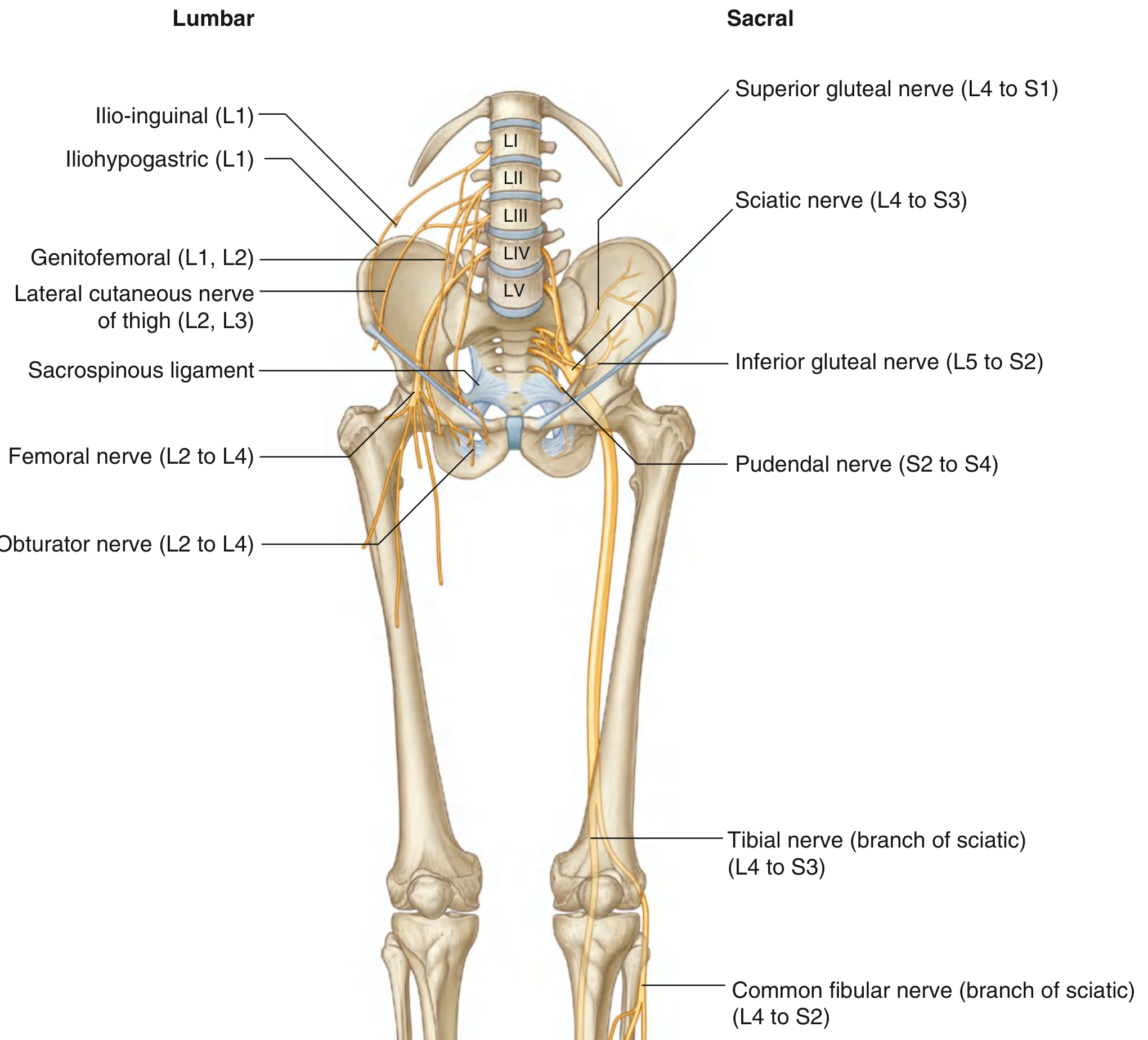
Lumbar Plexus (L1-L4) - inside psoas major muscle
| Nerve | Roots | Distribution |
|---|---|---|
| Iliohypogastric | L1 | Skin of lower abdomen + gluteal region |
| Ilioinguinal | L1 | Medial thigh, scrotal/labial skin |
| Genitofemoral | L1, L2 | Genital branch (cremaster) + femoral branch (upper thigh skin) |
| Lateral cutaneous nerve of thigh | L2, L3 | Lateral thigh skin (meralgia paraesthetica when compressed) |
| Femoral nerve | L2, L3, L4 | Anterior thigh muscles + skin, medial leg + foot (saphenous) |
| Obturator nerve | L2, L3, L4 | Medial thigh (adductors) + medial thigh skin |
| Lumbosacral trunk | L4 + L5 | Contributes to sacral plexus |
Sacral Plexus (L4-S4) - on anterior surface of piriformis
| Nerve | Roots | Distribution |
|---|---|---|
| Sciatic nerve | L4-S3 | Posterior thigh, entire leg + foot (largest nerve in body) |
| Superior gluteal nerve | L4-S1 | Gluteus medius, minimus, TFL |
| Inferior gluteal nerve | L5-S2 | Gluteus maximus |
| Posterior cutaneous nerve of thigh | S1-S3 | Posterior thigh skin |
| Pudendal nerve | S2-S4 | Perineum, external anal sphincter, genitalia |
| Nerve to obturator internus | L5-S2 | Obturator internus + gemellus superior |
| Nerve to quadratus femoris | L4-S1 | Quadratus femoris + gemellus inferior |
Key comparison with upper limb: There is NO single plexus diagram as neat as the brachial plexus. Instead, think of it as lumbar plexus (front/medial of limb) + sacral plexus (back/lateral of limb). The sciatic nerve = the "brachial plexus equivalent" in terms of importance.
WEEK 2 - Gluteal Region + Thigh
DAY 5-6: GLUTEAL REGION
- Superior: iliac crest
- Inferior: gluteal fold (not the crease - the fold is formed by gluteus maximus lower border)
- Lateral: greater trochanter
- Above piriformis: Superior gluteal nerve + artery + vein (only these 3)
- Below piriformis: Inferior gluteal nerve/vessels, Sciatic nerve, Posterior cutaneous nerve of thigh, Pudendal nerve/vessels, Nerve to obturator internus, Nerve to quadratus femoris
- Tendon of obturator internus
- Pudendal nerve + internal pudendal vessels (re-entering after looping through greater foramen)
- Nerve to obturator internus
| Muscle | Nerve | Action |
|---|---|---|
| Piriformis | S1, S2 (direct branches) | Lateral rotation + abduction of thigh |
| Obturator internus + gemelli | Nerve to OI (L5-S2) | Lateral rotation |
| Quadratus femoris | Nerve to QF (L4-S1) | Lateral rotation |
| Gluteus minimus | Superior gluteal (L4-S1) | Abduction + medial rotation |
| Gluteus medius | Superior gluteal (L4-S1) | Primary abductor - stabilizes pelvis in walking |
| Gluteus maximus | Inferior gluteal (L5-S2) | Powerful extension - climbing stairs, rising from chair |
Trendelenburg test: When gluteus medius is weak (superior gluteal nerve injury), the pelvis drops to the opposite side when standing on the affected leg. This is the "antalgic gait" you must know.
Safe zone for gluteal injection: Upper outer quadrant of the gluteal region - avoids sciatic nerve which runs through the lower half.
DAY 7-9: THIGH - THREE COMPARTMENTS
Anterior Compartment (Femoral nerve L2-L4)
| Muscle | Action |
|---|---|
| Iliopsoas (iliacus + psoas major) | Hip flexion (most powerful) |
| Sartorius | Hip flexion + abduction + lateral rotation; knee flexion + medial rotation ("tailor's muscle") |
| Quadriceps femoris (rectus femoris + vastus lateralis + medialis + intermedius) | Knee extension (most powerful in body) |
| Pectineus | Hip flexion + adduction |
- Femoral nerve also gives: medial cutaneous nerve of thigh + intermediate cutaneous nerve of thigh; saphenous nerve (medial side of knee, leg, foot)
Medial Compartment (Obturator nerve L2-L4)
| Muscle | Note |
|---|---|
| Adductor longus | Most anterior; forms medial border of femoral triangle |
| Adductor brevis | Deep to adductor longus |
| Adductor magnus | Largest - two parts: adductor part (obturator nerve) + hamstring part (sciatic nerve); distal attachment at adductor tubercle; has adductor hiatus (femoral vessels pass through) |
| Gracilis | Medial side of knee; part of pes anserinus |
| Obturator externus | Lateral rotator of hip |
Posterior Compartment - Hamstrings (Sciatic nerve)
| Muscle | Origin | Note |
|---|---|---|
| Biceps femoris (long head) | Ischial tuberosity | Tibial part of sciatic |
| Biceps femoris (short head) | Lateral lip of linea aspera | Common fibular part (only hamstring innervated by common fibular) |
| Semitendinosus | Ischial tuberosity | Long thin tendon; part of pes anserinus |
| Semimembranosus | Ischial tuberosity | Broad flat tendon (mem = membrane-like) |
All hamstrings = extend hip + flex knee, EXCEPT they all cross two joints. Biceps femoris also laterally rotates leg; semi-muscles medially rotate.
Pes anserinus ("goose's foot") - combined insertions of Sartorius + Gracilis + Semitendinosus on medial tibia. Common site of pes anserine bursitis.
WEEK 2 (continued): KEY ANATOMICAL SPACES
DAY 10: FEMORAL TRIANGLE + ADDUCTOR CANAL
- Base: Inguinal ligament
- Lateral border: Medial margin of sartorius
- Medial border: Medial margin of adductor longus
- Floor: Iliopsoas (lateral) + pectineus (medial)
- Roof: Fascia lata + cribriform fascia (contains saphenous opening)
- Nerve (femoral) - lateral to sheath
- Artery (femoral)
- Vein (femoral)
- Empty space (lymphatics/femoral canal)
- Lymphatics
Femoral sheath surrounds artery + vein + femoral canal (lymphatics) but NOT the femoral nerve (lateral to sheath).
Femoral canal = most medial compartment; femoral ring is its superior opening = weak point = site of femoral hernia. Femoral hernias are more common in females (wider pelvis).
- Subsartorial canal; continuation of femoral triangle
- Bounded by: Vastus medialis (lateral), adductor longus + magnus (floor/medial), sartorius (roof/anterior)
- Contents: Femoral artery + vein, saphenous nerve, nerve to vastus medialis
- Ends at adductor hiatus in adductor magnus → femoral vessels become popliteal vessels
WEEK 3 - The Knee + Leg
DAY 11-13: KNEE JOINT (Most Important Joint in Lower Limb)
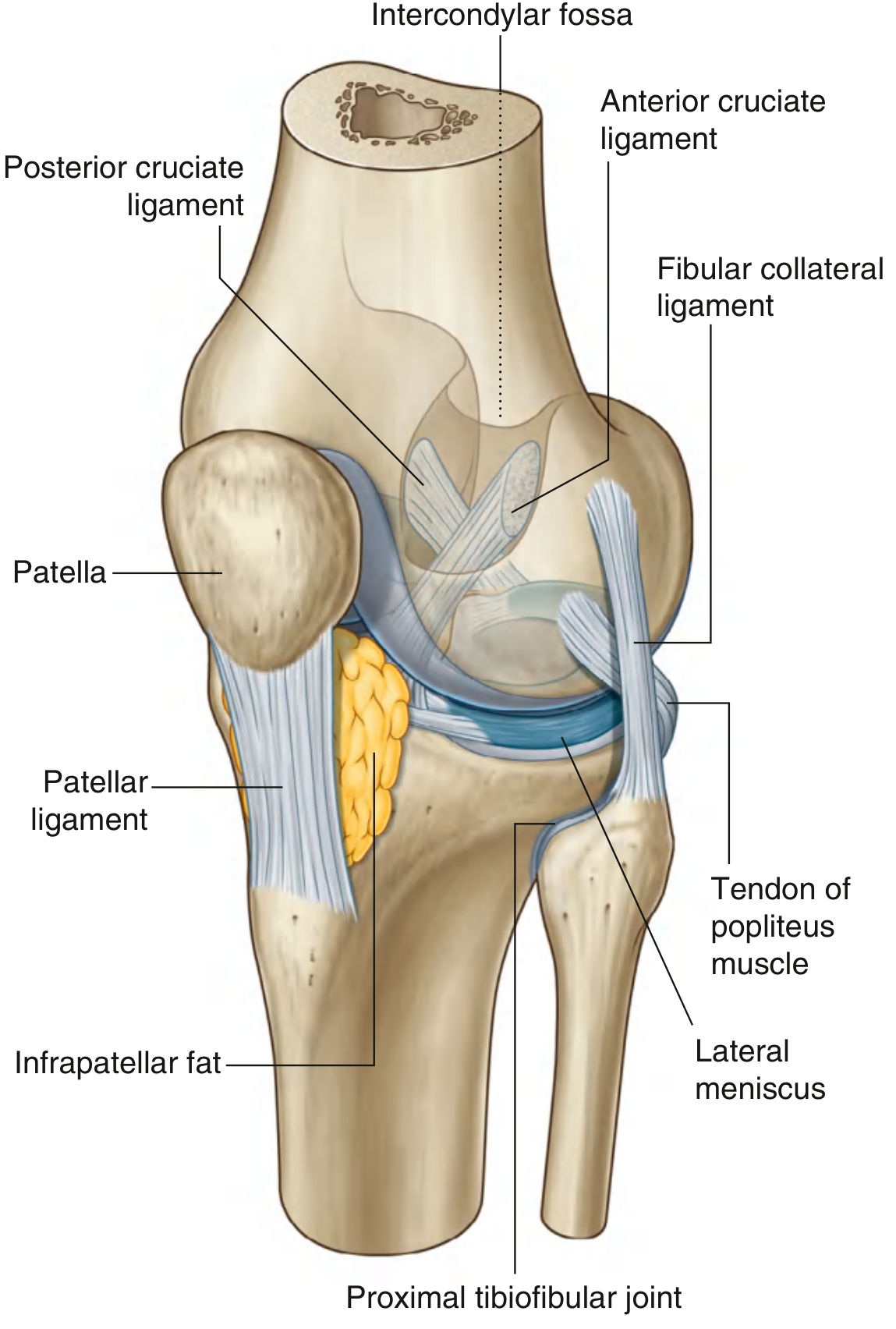
- Femoral condyles + tibial condyles (weight-bearing)
- Patella + patellar surface of femur (patellofemoral joint)
| Medial Meniscus | Lateral Meniscus | |
|---|---|---|
| Shape | C-shaped, less curved | Nearly circular (O-shaped) |
| Size | Larger | Smaller |
| Mobility | Less mobile (attached to capsule + tibial collateral ligament) | More mobile (not attached to fibular collateral ligament) |
| Injury frequency | More commonly injured | Less commonly injured |
| Reason | Less mobile = more vulnerable to tear | More mobile = can escape injury |
| ACL | PCL | |
|---|---|---|
| Attachment (femur) | Posterior part of medial surface of lateral condyle | Anterior part of lateral surface of medial condyle |
| Attachment (tibia) | Anterior intercondylar area | Posterior intercondylar area |
| Function | Prevents anterior displacement of tibia on femur; limits hyperextension | Prevents posterior displacement of tibia; most important knee stabilizer |
| Test | Anterior drawer test / Lachman test | Posterior drawer test |
| Common injury | Valgus force + rotation (sports) | Dashboard injury (car accidents) |
- Tibial (medial) collateral ligament = attached to medial meniscus (so MCL tear often = medial meniscus tear too)
- Fibular (lateral) collateral ligament = cord-like; NOT attached to lateral meniscus
Unhappy triad (O'Donoghue's triad): ACL + MCL + medial meniscus tear from valgus twisting force.
- In full extension, tibia rotates laterally (screw-home mechanism) - locks the knee
- Popliteus muscle unlocks the knee by medially rotating tibia (internally rotating tibia = unlocks)
- Prepatellar bursa - "housemaid's knee" (repeated kneeling)
- Infrapatellar bursa (deep) - "clergyman's knee" (kneeling more upright)
- Suprapatellar bursa - communicates with knee joint cavity
- Semimembranosus bursa - can communicate with joint, called Baker's cyst when distended
DAY 14: POPLITEAL FOSSA
- Superomedial: Semimembranosus + semitendinosus
- Superolateral: Biceps femoris
- Inferomedial: Medial head of gastrocnemius
- Inferolateral: Lateral head of gastrocnemius + plantaris
- Floor: Popliteal surface of femur, posterior joint capsule, popliteus (with its fascia)
- Roof: Popliteal fascia (continuation of deep fascia)
- Popliteal lymph nodes
- Common fibular nerve (lateral side)
- Tibial nerve (medial, more central)
- Popliteal vein (deep to nerve)
- Popliteal artery (deepest - directly against joint capsule)
Mnemonic for depth (superficial → deep): "Very Nervous People Act Deeply" (V=vein...no, actually: Nerve - Vein - Artery from superficial to deep)
Popliteal artery aneurysm = most common peripheral aneurysm; presents with knee mass + ischemic symptoms
DAY 15-17: LEG - THREE COMPARTMENTS
Anterior Compartment (Deep fibular nerve - L4, L5)
| Muscle | Action | Note |
|---|---|---|
| Tibialis anterior | Dorsiflexion + inversion | Main dorsiflexor |
| Extensor hallucis longus | Extends big toe + dorsiflexion | |
| Extensor digitorum longus | Extends toes 2-5 + dorsiflexion | |
| Fibularis (peroneus) tertius | Dorsiflexion + eversion | Often absent |
Deep fibular nerve injury (at neck of fibula or in anterior compartment syndrome): foot drop + inability to dorsiflex/extend toes; sensory loss only on first web space
Lateral Compartment (Superficial fibular nerve - L5, S1)
| Muscle | Action |
|---|---|
| Fibularis (peroneus) longus | Eversion + plantarflexion; plantar crosses foot obliquely to support transverse arch |
| Fibularis (peroneus) brevis | Eversion + plantarflexion |
Common fibular nerve injury at neck of fibula (most vulnerable nerve in lower limb - same concept as radial nerve at spiral groove): foot drop + eversion loss. Sensory loss = dorsum of foot + lateral leg.
Posterior Compartment
- Gastrocnemius (two heads) + Soleus + Plantaris = Triceps surae
- All insert via calcaneal (Achilles) tendon onto calcaneus
- Action: Plantarflexion (S1 reflex = ankle jerk)
- Gastrocnemius also flexes knee (crosses two joints)
| Muscle | Action | Note |
|---|---|---|
| Popliteus | Unlocks knee | |
| Flexor hallucis longus | Flexes big toe | "Sprinter's muscle"; supports medial arch; tendon passes below sustentaculum tali |
| Flexor digitorum longus | Flexes toes 2-5 | Tendon divides in foot |
| Tibialis posterior | Inversion + plantarflexion | Supports medial longitudinal arch; tendinopathy = flat foot |
Tibial nerve injury (rare; in tarsal tunnel or popliteal fossa): Loss of plantarflexion, toe flexion, intrinsic foot muscles; sensory loss = sole of foot.
Anterior compartment syndrome: Raised pressure in anterior compartment after fracture/exercise → deep fibular nerve + tibial anterior artery compression → foot drop + severe pain. Emergency fasciotomy needed.
WEEK 4 - Ankle, Foot, Vessels, Veins
DAY 18-19: ANKLE JOINT + TARSAL TUNNEL
- Mortise and tenon joint (hinge type)
- Mortise = formed by medial malleolus + tibia (superior) + lateral malleolus
- Tenon = trochlea of talus
- Main movements: Dorsiflexion + plantarflexion
- Lateral: 3 ligaments - ATFL (anterior talofibular - most commonly sprained), CFL (calcaneofibular), PTFL (posterior talofibular - least often injured)
- Medial (deltoid ligament): Fan-shaped; very strong; rarely torn; avulsion fracture of medial malleolus more common
Inversion sprain = most common ankle injury; tears ATFL first, then CFL. "Lateral ankle sprain."
- Bounded by: medial malleolus (medially), calcaneus (laterally), flexor retinaculum (roof)
- Contents (from anterior to posterior - mnemonic: "Tom, Dick ANd Harry"):
- Tibialis posterior tendon
- Digitorum (FDL tendon)
- Artery (posterior tibial artery)
- Nerve (tibial nerve - divides into medial + lateral plantar nerves here)
- Hallucis (FHL tendon)
Tarsal tunnel syndrome: Tibial nerve compression → pain/tingling in sole of foot.
DAY 20-21: FOOT
| Arch | Key Support Structures |
|---|---|
| Medial longitudinal arch (highest, most important) | Calcaneus-talus-navicular-medial cuneiform-1st metatarsal; supported by plantar calcaneonavicular (spring) ligament, tibialis posterior tendon, FHL |
| Lateral longitudinal arch | Calcaneus-cuboid-5th metatarsal; low arch |
| Transverse arch | 3 cuneiforms + cuboid at midfoot; peroneus longus tendon |
Pes planus (flat foot) = loss of medial arch, often due to tibialis posterior tendinopathy Pes cavus (high arch) = associated with Charcot-Marie-Tooth disease
| Layer | Muscles |
|---|---|
| 1st (superficial) | Abductor hallucis, Flexor digitorum brevis, Abductor digiti minimi |
| 2nd | Quadratus plantae, Lumbricals (4) |
| 3rd | Flexor hallucis brevis, Adductor hallucis, Flexor digiti minimi brevis |
| 4th (deepest) | Dorsal interossei (4), Plantar interossei (3) |
Same concept as hand: Lumbricals + interossei; medial plantar nerve (= median nerve equivalent) innervates medial 3.5 toes; lateral plantar nerve (= ulnar nerve equivalent) innervates lateral 1.5 toes + all interossei
DAY 22: BLOOD SUPPLY
- Branches: Superficial epigastric, superficial circumflex iliac, superficial external pudendal, deep external pudendal
- Profunda femoris (deep femoral artery) = largest branch; origin ~4 cm below inguinal ligament; gives medial + lateral circumflex femoral arteries + 3-4 perforating arteries
- Medial circumflex femoral artery = main blood supply to head of femur (important in neck of femur fractures!)
- Femoral artery → adductor hiatus → becomes popliteal artery
- Branches: Superior/inferior genicular arteries (5 total) + middle genicular
- Divides at lower border of popliteus into:
- Anterior tibial artery → anterior compartment → dorsum of foot → dorsalis pedis artery (palpable between EHL and EDL tendons)
- Posterior tibial artery → posterior compartment → tarsal tunnel → divides into medial + lateral plantar arteries
- Fibular (peroneal) artery = branch of posterior tibial → lateral compartment
Dorsalis pedis pulse - felt on dorsum of foot between 1st + 2nd metatarsals Posterior tibial pulse - felt behind medial malleolus
DAY 23: VEINS + LYMPHATICS
- Begins at medial end of dorsal venous arch → anterior to medial malleolus → medial side of leg → behind medial condyles of tibia + femur → anteromedial thigh → saphenous opening in fascia lata → drains into femoral vein ~4 cm below inguinal ligament
- Accompanied by saphenous nerve (terminal branch of femoral nerve) throughout
- Contains multiple valves
- Used in CABG (coronary artery bypass grafting)
- Varicose veins when valves incompetent
- Begins at lateral dorsal venous arch → posterior to lateral malleolus → up posterior leg → pierces deep fascia in popliteal fossa → drains into popliteal vein
- Accompanied by sural nerve
- Superficial lymphatics follow GSV → superficial inguinal lymph nodes
- Deep lymphatics follow femoral vessels → deep inguinal nodes → external iliac nodes
- Popliteal nodes drain foot and posterior leg
DAY 24-25: DERMATOMES + CUTANEOUS NERVES (Very Exam-Heavy)
| Dermatome | Autonomous Zone |
|---|---|
| L1 | Inguinal region + upper medial thigh |
| L2 | Lateral thigh (LCNT) |
| L3 | Lower medial thigh + medial knee |
| L4 | Medial great toe (big toe) - tests L4 |
| L5 | Medial side of 2nd toe + dorsum of foot |
| S1 | Little toe (5th toe) + lateral foot - tests S1 |
| S2 | Posterior thigh |
| S3 | Gluteal fold skin |
| Reflex | Tests |
|---|---|
| Knee jerk (patellar) | L3, L4 (femoral nerve) |
| Ankle jerk (Achilles) | S1, S2 (tibial nerve) |
PART D: TOP HIGH-YIELD TOPICS FOR EXAMS
| Rank | Topic | Why Important |
|---|---|---|
| 1 | Lumbosacral plexus | Nerve roots + terminal nerves + lesion patterns |
| 2 | Knee joint - cruciate ligaments, menisci | Most common sports injury + clinical tests |
| 3 | Sciatic nerve - course + injury | Most common nerve injury question |
| 4 | Femoral triangle - contents, femoral hernia | Very standard short answer |
| 5 | Common fibular nerve at neck of fibula | Foot drop - most common lower limb nerve injury |
| 6 | Hip joint - type, stability, dislocation | Femoral neck fractures, blood supply |
| 7 | Gluteal region - greater/lesser sciatic foramina, piriformis relations | IM injection site, sciatica |
| 8 | Great saphenous vein - course + tributaries | CABG, varicose veins, venous cutdown |
| 9 | Tarsal tunnel - contents (Tom Dick ANd Harry) | Tarsal tunnel syndrome |
| 10 | Adductor canal | Femoral artery access, adductor hiatus |
| 11 | Calcaneal (Achilles) tendon | Rupture, anatomy, blood supply |
| 12 | Ankle ligaments + inversion sprain | Most common ankle injury |
| 13 | Popliteal fossa - contents, depth order | Baker's cyst, popliteal aneurysm |
| 14 | Arches of foot | Flat foot, clinical relevance |
| 15 | Dermatomes + reflexes | Clinical neurology cases |
PART E: DIRECT UPPER vs. LOWER LIMB COMPARISON TABLE
| Feature | Upper Limb | Lower Limb |
|---|---|---|
| Plexus mnemonic | Robert Taylor Drinks Cold Beer (RTDCB) | No standard mnemonic needed - just lumbar + sacral |
| Most complex joint | Shoulder (mobility) | Knee (ligament complexity) |
| Most important nerve | Median nerve | Sciatic nerve |
| Most common nerve injury | Radial nerve (spiral groove) | Common fibular nerve (neck of fibula) |
| "Gateway" space | Axilla | Femoral triangle |
| Transition space | Cubital fossa | Popliteal fossa |
| Carpal tunnel equiv. | Carpal tunnel (median nerve) | Tarsal tunnel (tibial nerve) |
| Most important vein | Cephalic (IV access) | Great saphenous (CABG + varicose veins) |
| Long tendon of biceps | Long head biceps (intertubercular groove) | Long head of rectus femoris (less tested) |
| "Funny bone" | Ulnar nerve at medial epicondyle | Common fibular at neck of fibula |
| Triangle/fossa mnemonic | NAVY (cubital fossa: N-A-V from lateral-medial) | NAVEL (femoral triangle) |
| Sesamoid bone | Pisiform | Patella |
| Primary abductor | Deltoid (arm) | Gluteus medius (thigh) |
| Key clinical test | Froment's sign, Tinel's | Trendelenburg test, Lachman's |
| Difficulty rating | ⭐⭐⭐⭐⭐ | ⭐⭐⭐⭐ |
PART F: QUICK-REVISION MNEMONICS SUMMARY
| Topic | Mnemonic |
|---|---|
| Tarsal bones | "The Cunning Nag Can't Trot" |
| Tarsal tunnel contents | "Tom Dick ANd Harry" (TP, FDL, Artery, Nerve, FHL) |
| Femoral triangle contents | NAVEL (Nerve, Artery, Vein, Empty, Lymphatics) |
| Structures passing above piriformis | Only superior gluteal nerve/vessels (3 only) |
| Rotator cuff = SITS | Supraspinatus, Infraspinatus, Teres minor, Subscapularis |
| Gluteal abductors (Trendelenburg) | Gluteus medius = "medial = medal = champion abductor" |
| Short head biceps femoris = common fibular | Only hamstring NOT from tibial nerve |
| Medial meniscus = more injured | Medial meniscus is MARRIED to the capsule = less free = more torn |
| ACL prevents anterior tibia slip | ACL = Anterior = prevents Anterior displacement |
| Popliteal fossa depth | Nerve - Vein - Artery (superficial to deep) |
| Pes anserinus | SGS = Sartorius, Gracilis, Semitendinosus ("Say Grace before Supper") |
I am about to start to study the anatomy of the lower limb. I've already covered the anatomy of the upper limb. I have the roadmap, I have the plan, I have the books. I'm about to study with KLM and would use Netter's Atlas for visual representation. Secondly, I just wanted to know that I have all the resources, all the plans, but I'm con- But I am confused where to start. Although I would get rid of this confusion, but I need to prepare myself mentally for what I am about to study, so that when I start to study, everything makes sense.
Mental Orientation for Lower Limb Anatomy
Before You Open a Single Page of KLM
First - Understand What the Lower Limb IS Built For
- Why is the hip socket deeper than the shoulder? Stability.
- Why does the knee have cruciate ligaments when the elbow doesn't? Stability under weight-bearing.
- Why are the foot muscles smaller and the leg muscles massive? Locomotion needs power, not fine control.
- Why does the sciatic nerve stay deep and protected while the radial nerve wraps around a bone? The lower limb can't afford a nerve injury while weight-bearing.
Second - Understand the Architecture Before the Details
FLOOR 4 - GLUTEAL REGION (the basement of the abdomen / pelvis)
↓
FLOOR 3 - THIGH (the longest segment)
↓
FLOOR 2 - LEG (between knee and ankle)
↓
FLOOR 1 - FOOT (the foundation)
| Transition Zone | Between |
|---|---|
| Femoral triangle | Abdomen → Thigh |
| Adductor canal / Adductor hiatus | Thigh → Behind knee |
| Popliteal fossa | Thigh → Leg |
| Tarsal tunnel | Leg → Foot |
Third - The Upper Limb Comparison (Your Biggest Advantage)
"What is the upper limb equivalent of this?"
| What you already know | What you're about to learn |
|---|---|
| Brachial plexus | Lumbosacral plexus |
| Axilla | Femoral triangle + Gluteal region |
| Shoulder joint | Hip joint |
| Elbow joint | Knee joint (more complex) |
| Radial nerve wrist drop | Common fibular nerve foot drop |
| Median nerve in carpal tunnel | Tibial nerve in tarsal tunnel |
| Cephalic + Basilic veins | Great + Small saphenous veins |
| Rotator cuff (SITS) | Gluteal abductors (medius + minimus) |
| Cubital fossa | Popliteal fossa |
| Anatomical snuffbox | Femoral triangle (different but same concept of named space) |
Fourth - The Big Narrative of the Lower Limb
-
The pelvis is the anchor. It connects the spine to the lower limb and transfers the entire body weight down.
-
That weight travels down through the hip joint - a deep, stable ball-and-socket that sacrifices mobility for security.
-
The femur carries the weight down to the knee. The thigh muscles (quadriceps in front, hamstrings behind, adductors medially) control the movement of the hip and knee.
-
The knee joint is the most engineered joint in the body. It has to be simultaneously a hinge AND a weight-bearing surface AND a locking mechanism - which is why it has cruciate ligaments, collateral ligaments, menisci, and a locking mechanism (screw-home). No other joint in the body does all of this.
-
The leg (between knee and ankle) is simpler - three compartments, each with one nerve, one artery, and a group of muscles doing one job (dorsiflex, evertflex, plantarflex).
-
The ankle transfers force from the leg to the foot. It is a simple hinge - but the ligaments around it are what most people tear playing sport.
-
The foot is the foundation. It has arches (like a bridge) to absorb impact and distribute weight. The muscles in the sole are arranged in four layers, just like the palm - but they are less fine-tuned and more about support.
Fifth - The Three Things That Are Harder in Lower Limb Than Upper Limb
1. The Knee Joint
- Two bones articulating with a third (femur + tibia + patella)
- Two menisci that are different from each other
- Two cruciate ligaments that cross inside the joint
- A locking mechanism (screw-home)
- Multiple bursae with clinical names
- A complex ligament arrangement
2. The Lumbosacral Plexus Spans Two Separate Plexuses
3. The Gluteal Region Feels Confusing at First
Sixth - Where to Start (The Answer to Your Main Question)
- Every muscle has an origin and insertion on a bone
- Every nerve passes next to or through a bony landmark
- Every joint is formed by specific bone surfaces
- Every clinical injury involves a bone in some way
Seventh - The Right Mental Model for Each Study Session
1. BONES → What bones form this region? What landmarks exist?
2. JOINTS → What articulates here? What type of joint? What moves?
3. MUSCLES → What compartments? What does each compartment do?
4. NERVES → Which nerve supplies this? What happens if it's injured?
5. ARTERIES → What supplies blood? Where does it come from, where does it go?
6. VEINS → Superficial vs. deep. Clinical relevance?
7. SPACES → Is there a named anatomical space? What passes through it?
8. CLINICAL → What does KLM say in the clinical boxes?
Finally - One Mindset Shift That Changes Everything
am about to start to study the anatomy of the lower limb. I've already covered the anatomy of the upper limb. I have the roadmap, I have the plan, I have the books. I'm about to study with KLM and would use Netter's Atlas for visual representation. Secondly, I just wanted to know that I have all the resources, all the plans, but I'm con- But I am confused where to start. Although I would get rid of this confusion, but I need to prepare myself mentally for what I am about to study, so that when I start to study, everything makes sense
The Honest Answer to "Where Do I Start?"
But Here Is Your Starting Point - Concrete and Specific
Why Bones First - The Real Reason
The Mental Model to Carry Into Every Single Chapter
"What is this region FOR?"
- Gluteal region: transferring power from the trunk to the lower limb
- Thigh: moving the hip and the knee
- Leg: moving the ankle and foot
- Foot: absorbing impact and pushing off the ground
The One Comparison That Anchors Everything
The Real Preparation You Need Right Now
am about to start to study the anatomy of the lower limb. I've already covered the anatomy of the upper limb. I have the roadmap, I have the plan, I have the books. I'm about to study with KLM and would use Netter's Atlas for visual representation. Secondly, I just wanted to know that I have all the resources, all the plans, but I'm con- But I am confused where to start. Although I would get rid of this confusion, but I need to prepare myself mentally for what I am about to study, so that when I start to study, everything makes sense
Here is the complete truth, said plainly:
One instruction. That's all:
the replies didn't load pls reply again
Mental Orientation for Lower Limb Anatomy
The Single Most Important Thing to Understand First
The Architecture - Four Floors
GLUTEAL REGION → THIGH → LEG → FOOT
| Space | What Passes Through |
|---|---|
| Femoral triangle | Femoral nerve, artery, vein entering the thigh from abdomen |
| Adductor canal / hiatus | Femoral vessels passing to behind the knee |
| Popliteal fossa | Vessels and nerves crossing from thigh to leg |
| Tarsal tunnel | Tibial nerve and flexor tendons entering the foot |
Your Biggest Advantage - Upper Limb Knowledge
| Upper Limb | Lower Limb Equivalent |
|---|---|
| Brachial plexus | Lumbosacral plexus |
| Axilla | Femoral triangle + gluteal region |
| Shoulder joint | Hip joint |
| Elbow joint | Knee joint (more complex) |
| Radial nerve / wrist drop | Common fibular nerve / foot drop |
| Carpal tunnel + median nerve | Tarsal tunnel + tibial nerve |
| Cephalic + basilic veins | Great + small saphenous veins |
| Cubital fossa | Popliteal fossa |
| Rotator cuff stabilizing shoulder | Gluteus medius stabilizing pelvis |
The Three Things Harder in Lower Limb Than Upper Limb
The Recommended Study Order
Bones → Lumbosacral Plexus → Gluteal Region → Thigh →
Femoral Triangle → Knee Joint → Leg → Popliteal Fossa →
Ankle/Tarsal Tunnel → Foot → Veins → Dermatomes
How to Use KLM + Netter's Together
- Read KLM text for the region
- Open Netter's plate for that region and visualize it
- Re-read KLM - it now makes visual sense
- Sketch the region from memory and label it
- Read every clinical box in KLM - these become exam questions
Where to Start - Concrete Answer
- Open Netter's to the lower limb bone plates (around 481-495)
- Look at the hip bone, femur, tibia, fibula, foot bones
- Touch the landmarks on your own body - ASIS (front of hip), iliac crest (hands-on-hips ridge), greater trochanter (lateral hip), tibial tuberosity (front of knee), head of fibula (lateral below knee), medial and lateral malleoli (ankle bumps)
The Mental Model for Every Chapter
"What is this region FOR?"
- Gluteal region: transferring power from trunk to limb
- Thigh: controlling hip and knee movement
- Leg: controlling ankle and foot movement
- Foot: absorbing impact and pushing off the ground
The Dermatomes and Reflexes to Keep in Mind Throughout
| Level | Autonomous Zone | Reflex |
|---|---|---|
| L3, L4 | Medial knee + big toe | Knee jerk (patellar) |
| L5 | Dorsum of foot + 2nd toe | No classic reflex |
| S1 | Little toe + lateral foot | Ankle jerk (Achilles) |
The One Sentence Summary of the Entire Lower Limb
make me an mbbs level mcq test of 50 marks from upper limb anatomy along with there correct answers make it in such a way that it helps in active recall
UPPER LIMB ANATOMY - MCQ TEST
50 Questions | 50 Marks | MBBS Level
(Attempt all questions before checking answers)
SECTION A: BONES & SURFACE ANATOMY (Q1-8)
- A) Radial nerve
- B) Musculocutaneous nerve
- C) Axillary nerve
- D) Median nerve
- A) It is greater in males than females
- B) Normal range is 5-15° valgus
- C) It is formed due to the shape of the radial head
- D) Cubitus varus increases the carrying angle
- A) Pectoralis major
- B) Sternocleidomastoid
- C) Trapezius
- D) Subclavius
- A) Abductor pollicis longus
- B) Extensor pollicis brevis
- C) Extensor pollicis longus
- D) Flexor pollicis longus
- A) Trapezium
- B) Lunate
- C) Scaphoid
- D) Capitate
- A) Anterior capsule
- B) Inferior glenohumeral ligament
- C) Supraspinatus tendon
- D) Posterior capsule
- A) Scaphoid
- B) Hamate
- C) Lunate
- D) Trapezoid
- A) Supraspinatus
- B) Deltoid
- C) Coracobrachialis
- D) Pectoralis minor
SECTION B: BRACHIAL PLEXUS (Q9-16)
- A) C8, T1
- B) C5, C6
- C) C6, C7
- D) C7, C8
- A) C5, C6
- B) C6, C7
- C) C7, C8
- D) C8, T1
- A) C4, C5, C6
- B) C5, C6, C7
- C) C6, C7, C8
- D) C5, C6
- A) Dorsal scapular nerve
- B) Suprascapular nerve
- C) Long thoracic nerve
- D) Thoracodorsal nerve
- A) Axillary nerve
- B) Radial nerve
- C) Thoracodorsal nerve
- D) Musculocutaneous nerve
- A) Lateral cord
- B) Medial cord
- C) Posterior cord
- D) It arises directly from C7 root
- A) C5 root directly
- B) Superior trunk
- C) Lateral cord
- D) Posterior cord
- A) Intercostobrachial nerve
- B) Long thoracic nerve
- C) Lateral pectoral nerve
- D) Medial pectoral nerve
SECTION C: AXILLA & SHOULDER (Q17-22)
- A) Pectoralis major
- B) Subscapularis
- C) Pectoralis minor
- D) Coracobrachialis
- A) First part
- B) Second part
- C) Third part
- D) It is a branch of the brachial artery
- A) Radial nerve and profunda brachii artery
- B) Axillary nerve and posterior circumflex humeral artery
- C) Circumflex scapular artery and axillary nerve
- D) Musculocutaneous nerve and anterior circumflex humeral artery
- A) Radial nerve
- B) Musculocutaneous nerve
- C) Axillary nerve
- D) Medial cutaneous nerve of arm
- A) Deltoid
- B) Supraspinatus
- C) Infraspinatus
- D) Serratus anterior
- A) Musculocutaneous nerve
- B) Radial nerve
- C) Axillary nerve
- D) Suprascapular nerve
SECTION D: MUSCLES (Q23-30)
- A) Supraspinatus
- B) Infraspinatus
- C) Teres minor
- D) Subscapularis
- A) Infraspinatus
- B) Supraspinatus
- C) Subscapularis
- D) Teres minor
- A) Supinator muscle
- B) Brachioradialis
- C) Biceps brachii
- D) Pronator teres
- A) Flexor digitorum profundus
- B) Flexor digitorum superficialis
- C) Adductor pollicis
- D) Opponens pollicis
- A) Median nerve
- B) Ulnar nerve
- C) Anterior interosseous nerve
- D) Radial nerve
- A) Acromion process
- B) Coracoid process
- C) Glenoid rim
- D) Lesser tubercle of humerus
- A) All lumbricals
- B) All interossei
- C) Thenar muscles (LOAF)
- D) Hypothenar muscles
- A) Extension at MCP joint + flexion at IP joints
- B) Flexion at MCP joint + extension at IP joints
- C) Flexion at all joints
- D) Extension at all joints
SECTION E: NERVES & NERVE INJURIES (Q31-41)
- A) Radial nerve at axilla
- B) Radial nerve at spiral groove of humerus
- C) Ulnar nerve at medial epicondyle
- D) Posterior interosseous nerve at radial tunnel
- A) Brachioradialis
- B) Extensor carpi radialis longus
- C) Extensor digitorum
- D) Triceps brachii
- A) Median nerve at wrist
- B) Anterior interosseous nerve
- C) Recurrent branch of median nerve
- D) Ulnar nerve at elbow
- A) Ulnar nerve
- B) Median nerve
- C) Radial nerve (superficial branch)
- D) Anterior interosseous nerve
- A) At Guyon's canal (wrist)
- B) At the medial epicondyle (elbow)
- C) In the axilla
- D) In the posterior cord
- A) Abductor pollicis brevis
- B) Opponens pollicis
- C) Adductor pollicis
- D) Flexor pollicis brevis
- A) Ulnar nerve injury at elbow causes more severe claw than injury at wrist
- B) Ulnar nerve injury at wrist causes more severe claw than injury at elbow
- C) Ulnar nerve controls the lateral two fingers despite being the medial nerve
- D) Ulnar nerve injury spares sensation but affects motor function
- A) Medial cutaneous nerve of forearm
- B) Lateral cutaneous nerve of forearm
- C) Posterior cutaneous nerve of forearm
- D) Superficial branch of radial nerve
- A) Axilla - above the triceps branch
- B) Spiral groove - below the triceps branch
- C) Lateral epicondyle
- D) Posterior interosseous nerve level
- A) Scaphoid and trapezium
- B) Pisiform and hook of hamate
- C) Lunate and capitate
- D) Triquetrum and hamate
- A) Medial epicondyle
- B) Guyon's canal - deep branch only
- C) Guyon's canal - superficial branch only
- D) Brachial plexus medial cord
SECTION F: VESSELS & SPACES (Q42-47)
- A) Brachial and radial arteries
- B) Radial and ulnar arteries
- C) Ulnar and anterior interosseous arteries
- D) Deep and superficial palmar arches only
- A) Subclavian vein directly
- B) Axillary vein via the deltopectoral groove
- C) Brachial vein
- D) External jugular vein
- A) Cephalic to basilic vein in the cubital fossa
- B) Brachial to radial vein
- C) Cephalic to brachial vein
- D) Basilic to brachial vein
- A) Artery - Nerve - Biceps tendon
- B) Biceps tendon - Artery - Nerve
- C) Nerve - Biceps tendon - Artery
- D) Radial nerve - Biceps tendon - Brachial artery - Median nerve
- A) Axillary nerve
- B) Musculocutaneous nerve
- C) Radial nerve
- D) Median nerve
- A) Axillary nerve and posterior circumflex humeral artery
- B) Circumflex scapular artery
- C) Radial nerve and profunda brachii artery
- D) Suprascapular nerve and artery
SECTION G: JOINTS & CLINICAL (Q48-50)
- A) Shoulder dislocation
- B) Pulled elbow (nursemaid's elbow) - radial head subluxation
- C) Fracture of lateral condyle
- D) Supracondylar fracture
- A) Ulnar nerve
- B) Radial nerve
- C) Anterior interosseous nerve / brachial artery
- D) Median nerve trunk
- A) Extensor pollicis longus + extensor pollicis brevis
- B) Abductor pollicis longus + extensor pollicis brevis
- C) Abductor pollicis longus + flexor pollicis longus
- D) Extensor pollicis longus + abductor pollicis longus
ANSWERS WITH EXPLANATIONS
(Only read AFTER attempting all 50 questions)
- Medial (posterior) border = extensor pollicis longus
- Lateral (anterior) border = abductor pollicis longus + extensor pollicis brevis
- Floor = scaphoid + trapezium + distal tendons of ECRL + ECRB
- Radial artery passes through it
- Part 1 (medial to pec minor): 1 branch - superior thoracic
- Part 2 (behind pec minor): 2 branches - thoracoacromial + lateral thoracic
- Part 3 (lateral to pec minor): 3 branches - subscapular + anterior + posterior circumflex humeral
- Supraspinatus → Greater tubercle (superior facet)
- Infraspinatus → Greater tubercle (middle facet)
- Teres minor → Greater tubercle (inferior facet)
- Subscapularis → LESSER tubercle (only one to insert on lesser tubercle)
- Lateral half (index + middle fingers) = anterior interosseous nerve (branch of median)
- Medial half (ring + little fingers) = ulnar nerve All other options: FDS = median only; Adductor pollicis = ulnar only; Opponens pollicis = median only (recurrent branch).
- Superficial branch = sensory (medial 1.5 fingers)
- Deep branch = purely motor (all intrinsics) If only motor loss with no sensory loss → deep branch compressed alone (by a ganglion cyst on hook of hamate, for example). At the elbow, both motor and sensory would be affected.
- Quadrangular space → axillary nerve + posterior circumflex humeral artery
- Triangular space (upper) → circumflex scapular artery only
- Triangular interval (lower) → radial nerve + profunda brachii artery These three are always tested together. Know them as a set.
YOUR SCORE KEY
| Score | Level |
|---|---|
| 45-50 | Distinction - Excellent command |
| 38-44 | Good pass - Solid understanding |
| 28-37 | Pass - Revise weak areas |
| Below 28 | Needs revision - Go back to topic-wise study |
TOPICS TO REVISE IF YOU GOT THESE WRONG
| Questions Missed | Revise This Topic |
|---|---|
| Q9, Q10, Q11, Q13, Q14, Q15 | Brachial plexus - roots, cords, branches |
| Q31, Q32, Q33, Q38, Q39 | Radial nerve and its levels of injury |
| Q34, Q35, Q36, Q37, Q40, Q41 | Ulnar nerve course + injury patterns |
| Q19, Q47 | Triangular spaces of posterior shoulder |
| Q17, Q18 | Axillary artery - parts and branches |
| Q23, Q24 | Rotator cuff - insertions and tests |
| Q43, Q44, Q45 | Veins + cubital fossa |
| Q48, Q49, Q50 | Clinical anatomy correlations |
Q35. A patient has claw hand affecting all four fingers with complete loss of intrinsic hand muscles. Where is the ulnar nerve most likely injured? A) At Guyon's canal (wrist) B) At the medial epicondyle (elbow) C) In the axilla D) In the posterior cord
The Answer I Gave Was Imprecise
Thinking Through Each Option
Option D - Posterior cord
Option C - In the axilla
- Complete claw hand ✓
- Complete loss of all intrinsic muscles ✓
- But also: Loss of FCU (wrist flexion/ulnar deviation lost) + loss of medial half FDP (ring + little finger DIP flexion lost)
- And: Sensory loss on medial 1.5 fingers + dorsal medial hand
Option B - At the medial epicondyle (elbow)
- Complete claw hand ✓
- Complete loss of all intrinsic muscles ✓
- Loss of FCU ✓
- Loss of medial half FDP ✓
- Sensory loss medial 1.5 fingers ✓
- Dorsal cutaneous branch (which arises ~5 cm above the wrist) = also lost
Option A - Guyon's canal (wrist)
- Superficial branch (sensory only - medial 1.5 fingers)
- Deep branch (motor only - all intrinsics)
- Complete loss of all intrinsic muscles ✓
- Sensory loss medial 1.5 fingers ✓
- But FCU and medial FDP are SPARED (their branches come off above the wrist)
- Dorsal cutaneous branch is SPARED (arises 5 cm above the wrist, before Guyon's canal)
The Distinguishing Clue in the Question
"claw hand affecting all four fingers with complete loss of intrinsic hand muscles"
- Loss of FCU
- Loss of medial FDP (ring/little finger DIP flexion)
- Dorsal hand sensory loss
The Real Best Answer for This Question as Written
- All intrinsics lost ✓
- FCU intact (not mentioned as lost) ✓
- Medial FDP intact (not mentioned as lost) ✓
- This is a cleaner fit with the clinical description given
I Owe You a Correction
The High-Yield Rule to Remember
| Level of Ulnar Nerve Injury | What is Lost |
|---|---|
| Axilla / above elbow | All of the below + FCU + medial FDP + dorsal cutaneous sensation |
| Elbow (medial epicondyle) | FCU + medial FDP + all intrinsics + dorsal cutaneous sensation + palmar sensation medial 1.5 fingers |
| Wrist (Guyon's canal - complete) | All intrinsics + palmar sensation medial 1.5 fingers; FCU/FDP/dorsal hand spared |
| Wrist (deep branch only) | All intrinsics only; no sensory loss at all |
| Wrist (superficial branch only) | Sensory loss medial 1.5 fingers only; no motor loss |