
Sub dural hematoma rx
"subdural hematoma" AND management
subdural hematoma CT scan brain
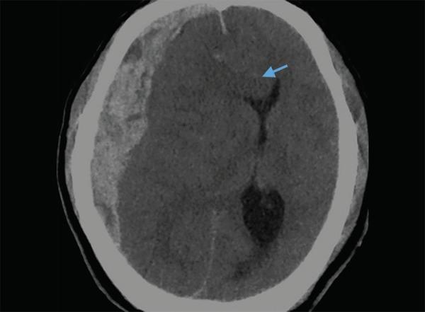
This diagnostic axial non-contrast CT scan of the brain demonstrates an acute subdural hematoma (SDH) in the right frontoparietal region. The visual features include a hyperdense, crescentic (concavo-convex) extra-axial collection extending along the inner table of the calvarium. The hematoma exerts significant mass effect on the underlying brain parenchyma, resulting in a marked leftward midline shift. A blue arrow identifies subfalcine herniation, where the cingulate gyrus is displaced across the midline beneath the falx cerebri. There is also evident effacement of the ipsilateral (right) lateral ventricle and cortical sulci, accompanied by surrounding hypodense areas consistent with cerebral edema. This image serves as an educational example of acute traumatic intracranial hemorrhage and secondary brain herniation syndromes, highlighting the critical radiological signs used in neurosurgical triage and emergency medicine.
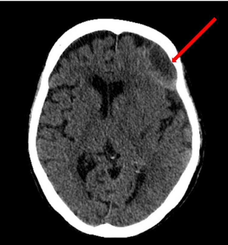
This diagnostic image is an axial non-contrast Computed Tomography (CT) scan of the brain demonstrating a large, multiloculated chronic subdural hematoma in the left frontal region. The hematoma is characterized by a crescent-shaped, predominantly hypodense collection relative to the brain parenchyma, indicated by a red arrow. The collection is situated between the inner table of the skull and the brain surface, exerting a significant mass effect. Visual evidence of this mass effect includes compression and distortion of the adjacent left frontal lobe and effacement of the nearby sulci. Furthermore, there is a distinct midline shift of approximately 7 mm toward the right side, as evidenced by the displacement of the falx cerebri and the lateral ventricles from the median plane. This image illustrates key radiological findings associated with significant intracranial volume expansion and secondary structural distortion. It is used in medical education to teach the diagnostic identification of subdural collections and the assessment of mass effect on intracranial anatomy.
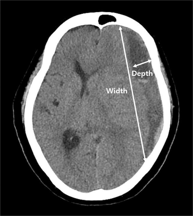
An axial non-contrast computed tomography (CT) scan of the head demonstrating a chronic subdural hematoma (SDH) located along the right convexity. The hematoma presents as a crescent-shaped, hypodense collection relative to the brain parenchyma, situated between the inner table of the skull and the brain surface. The image highlights the methodology for calculating depressed brain volume through two annotated measurements: 'Depth' and 'Width'. Depth is indicated by a short double-headed arrow representing the maximal transverse diameter between the inner skull table and the displaced brain surface. Width is indicated by a long double-headed arrow representing the linear distance between the superior and inferior margins where the hematoma meets the skull. The SDH causes visible mass effect, resulting in the effacement of cortical sulci and inward displacement of the underlying brain tissue. This visual serves as an educational reference for neuroradiological assessment of intracranial fluid collections and the quantitative evaluation of brain compression.
PMID: 40493076
Subdural Hematoma (SDH) - Management
Classification (by time to presentation)
| Type | Timeline | CT Appearance |
|---|---|---|
| Acute | <24 h (some use <2 wks) | Hyperdense (bright white), crescent-shaped |
| Subacute | 2-21 days | Isodense (can be missed on plain CT; use contrast or MRI) |
| Chronic | >2-3 weeks | Hypodense (dark), crescentic over convexity |
Imaging
General Principles
- SDH results from tearing of bridging veins traversing the subdural space (between inner dura leaf and arachnoid)
- Key rule: "It could always be a subdural" - image any comatose or confused patient even without focal signs
- Reverse anticoagulation promptly in patients on warfarin/NOACs - continued venous leakage is particularly dangerous
- Prognostic factors for acute SDH: age, time from injury to treatment, pupillary abnormalities, coma (immediate vs. lucid interval), hematoma volume
Acute SDH - Treatment
Conservative (observation)
- Thin crescentic clots without symptoms or signs of raised ICP
- Small SDH causing no symptoms - follow serial CT; most self-absorb
- Asymptomatic patient with small volume, no midline shift
Surgical - Indications
- SDH ≥10 mm thickness OR any midline shift with neurological deterioration
- Coma, hemiparesis, unilateral pupillary dilation (signs of herniation)
- Rapidly deteriorating/comatose patient = emergency craniotomy
- GCS <8 with acute SDH - surgical mortality is 40-60%
Surgical Technique
- Burr holes - sufficient for early clots before full organization; blood/clot extruded, gentle suction
- Craniotomy - required for larger hematomas, especially if several hours old with clot formation; also allows bleeding point control
- Craniectomy (decompressive) probably adds little benefit to craniotomy alone
Key: The interval between loss of consciousness and surgical drainage is the most important determinant of outcome. - Adams & Victor's Principles of Neurology, 12th Ed.
Chronic SDH - Treatment
Conservative
- Small collections causing no mass effect on imaging + minimal symptoms
- Serial imaging with clinical observation is a reasonable approach
- Oral corticosteroids (e.g., prednisone) - reduce vasogenic edema from the leaky hematoma membrane; can cause significant reduction and even resorption (as illustrated in Plum & Posner's series showing near-complete resorption at 2 months)
- Mild hydration + flat head of bed - encourages brain re-expansion
Surgical - Indications
- Chronic SDH >1 cm or any symptomatic SDH
- Neurological deterioration, increasing mass effect
Surgical Technique - Burr Hole Drainage (first-line)
- Single burr hole over the dependent edge of collection
- Copious irrigation until fluid runs clear
- Second anterior burr hole if collection not draining adequately (membrane compartmentalization)
- Convert to craniotomy if:
- SDH too congealed for irrigation drainage
- Complex membranes prevent effective drainage
- Persistent hemorrhage beyond reach of bipolar cautery
Post-operative measures
- Subdural drain left in place for 1-2 days - shown to reduce recurrence risk
- Flat head of bed + mild hydration to encourage brain expansion
- High inspired oxygen - helps draw nitrogen out of residual cavity
- Follow-up CT at 1 month to document resolution
- Note: corticosteroid use post-operatively has been associated with higher morbidity without benefit per Schwartz's Surgery
Anticoagulation Reversal (Critical Step)
| Agent | Reversal |
|---|---|
| Warfarin | Vitamin K + FFP or 4-factor PCC (prothrombin complex concentrate) |
| Heparin | Protamine sulfate |
| Dabigatran | Idarucizumab |
| Xa inhibitors | Andexanet alfa or 4-factor PCC |
Middle Meningeal Artery Embolization (MMAE) - Emerging Therapy
- In nonsurgical chronic SDH patients: MMAE significantly reduced progression (RR 0.36, 95% CI 0.22-0.60, p<0.001)
- In surgical patients: MMAE did not significantly reduce recurrence
- No effect on functional outcome
Tranexamic Acid (TXA) in Chronic SDH
Special Populations
- Elderly/atrophied brain: High risk; even minor or forgotten trauma causes SDH; bilateral in ~20%
- Anticoagulated patients: Highest priority reversal; acute SDH especially dangerous
- Alcoholics / hemodialysis patients: Chronic SDH risk factors; coagulopathy must be corrected
- Infants <2 years: High risk; bifrontal chronic SDH possible without significant trauma history (non-accidental injury must be considered)
- Posterior fossa SDH: Headache, vomiting, CN palsies, ataxia - often missed on CT; low threshold for MRI
non surgical rx
"subdural hematoma" AND "conservative" AND "non-surgical"
"chronic subdural hematoma" AND (dexamethasone OR tranexamic OR "middle meningeal")
PMID: 40652463
PMID: 39565989
Non-Surgical (Conservative) Management of Subdural Hematoma
When to Manage Conservatively
| Situation | Approach |
|---|---|
| Small SDH, no mass effect, minimal/no symptoms | Observation + serial imaging |
| Chronic SDH with headache only (no neurological deficit) | Corticosteroids ± observation |
| Patient unfit for surgery (high anaesthetic risk) | Medical optimisation + close monitoring |
| Post-anticoagulation reversal with stable neurology | Watch and wait |
1. Observation + Serial Imaging
- Clinical observation combined with serial CT is a reasonable approach for patients with few symptoms and small chronic subdural collections that do not cause mass effect
- Many small SDHs self-absorb; follow-up CT at 4-6 weeks documents resolution
- Any neurological deterioration on observation = triggers surgical re-evaluation
- Harrison's Internal Medicine 22e, Goldman-Cecil Medicine
2. Corticosteroids
- Oral prednisone rapidly and effectively reverses symptoms in chronic SDH
- Plum & Posner document a case series where bilateral SDH (11.5 mm + 8 mm) treated with oral prednisone showed near-complete resorption by 2 months on serial imaging
- The DUBS trial (Hutchinson et al., NEJM 2020) found dexamethasone reduced the need for surgery in chronic SDH but was associated with more adverse events (diabetes, infection)
- Current consensus: corticosteroids are not routinely recommended post-operatively (higher morbidity without benefit), but may be used for conservative management of small symptomatic chronic SDH
Note: Goldman-Cecil states corticosteroids are "not beneficial" as routine treatment, while Plum & Posner support their use for the edema component - the evidence is mixed, and patient selection matters.
3. Reversal of Anticoagulation / Antiplatelet Agents
| Drug | Reversal |
|---|---|
| Warfarin | Vitamin K (slow) + 4-factor PCC / FFP (urgent) |
| Heparin | Protamine sulfate |
| Dabigatran | Idarucizumab |
| Rivaroxaban / Apixaban | Andexanet alfa or 4-factor PCC |
| Aspirin / Clopidogrel | Delay surgery 7-10 days if clinically stable to allow platelet normalization |
- Coexisting electrolyte disturbance and infection must also be excluded and corrected
- Bailey & Love's Surgery 28e
4. ICP Management (Raised Intracranial Pressure)
- Head of bed elevation 30°
- Avoid hypotonic fluids - use isotonic/slightly hypertonic solutions
- Osmotic agents (mannitol, hypertonic saline) - used cautiously; note that rapid osmotic diuresis in elderly patients can paradoxically worsen SDH by causing brain shrinkage and further tearing of bridging veins
- Avoid hypercarbia - maintain normal PaCO₂; short-term hyperventilation as bridge therapy only
- Seizure prophylaxis - prophylactic AEDs are not routinely recommended in chronic SDH
5. Positioning
- Flat head of bed (after drainage, or in non-operative management) encourages brain re-expansion into the subdural space
- Mild hydration helps expand the brain
- High inspired oxygen may facilitate nitrogen washout from the residual cavity, promoting collapse and resorption
6. Tranexamic Acid (TXA) - Antifibrinolytic
- 4 RCTs, 148 patients
- Trend toward earlier and more complete resolution after burr hole drainage
- No statistically significant reduction in recurrence or post-operative volume
- Safe profile - no significant adverse events
7. Middle Meningeal Artery Embolization (MMAE) - Emerging
- MAGIC-MT trial (Liu et al., NEJM 2024, PMID 39565989): 722 patients - MMAE did not significantly reduce 90-day symptomatic recurrence/progression (6.7% vs 9.9%, p=0.10) but reduced serious adverse events (6.7% vs 11.6%, p=0.02)
- EMBOLISE trial (Fiorella et al., NEJM 2025, PMID 39565980): also assessed MMAE
- Meta-analysis (Gillespie et al., Acta Neurochir 2025, PMID 40493076): In non-surgical patients, MMAE significantly reduced progression (RR 0.36, 95% CI 0.22-0.60, p<0.001); in surgical patients - not significant
8. Statins
- Proposed to reduce neuroinflammation and membrane neovascularization
- Currently under investigation; not recommended for routine use
- Goldman-Cecil Medicine
Summary Table
| Modality | Evidence Level | Current Recommendation |
|---|---|---|
| Serial observation | Expert consensus | First-line for small, asymptomatic cSDH |
| Anticoagulation reversal | Standard of care | Mandatory |
| Corticosteroids (oral) | Moderate (mixed RCT data) | Consider for symptomatic cSDH not requiring surgery |
| Osmotic agents (mannitol) | Supportive | Use cautiously; risk of paradoxical worsening |
| Tranexamic acid | Low-moderate (meta-analysis 2025) | Safe but not yet routine |
| MMAE | High (3 RCTs, meta-analysis 2025) | Non-surgical cSDH; reduces progression |
| Statins | Investigational | Not recommended yet |
| Prophylactic AEDs | Negative evidence | Not recommended |