
Light microscope , dark field microscope , phase contrast microscope , fluorescence microscope , electron microscope Refer only microbiology book (apurba sastry) answer like 5 marks manner
light microscope dark field phase contrast fluorescence electron microscope comparison diagram
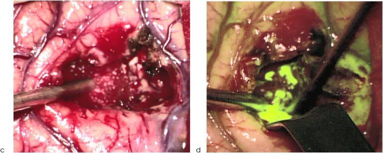
This comparative clinical photograph demonstrates the intraoperative visualization of a high-grade glioma (HGG) during neurosurgical resection using two different lighting modalities. Image (c) shows the surgical field under conventional white light, where the brain parenchyma, vascular structures, and the tumor bed are visible with typical anatomical colors; the tumor appears as heterogeneous dark and white tissue within a bloody surgical field. Image (d) displays the same surgical site under filtered light, utilizing fluorescence-guided surgery (FGS) techniques (likely 5-ALA induced protoporphyrin IX fluorescence). In this view, the malignant tumor tissue exhibits a vivid bright green fluorescence, significantly enhancing the contrast against the surrounding non-fluorescent normal brain tissue. Surgical instruments, including a metallic aspirator or probe, are seen positioned within the tumor cavity in both frames, indicating an active resection phase. This comparison illustrates how filtered light improves the identification of tumor margins and residual neoplastic tissue, facilitating a more complete extent of resection (EOR) compared to white light alone.
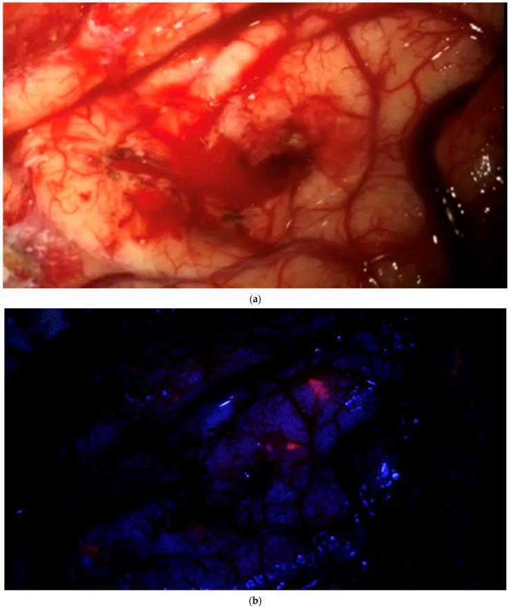
Comparison of intraoperative neurosurgical visualization using white light and fluorescence-guided imaging. Image (a) depicts a macroscopic view of the brain parenchyma under standard white light after tumor resection. The surgical field shows cortical tissue with visible branching vasculature, areas of pinkish-white healthy-appearing tissue, and minor localized hemorrhage. Under this lighting, the field appears 'clean' with no obvious residual tumor. Image (b) shows the identical anatomical field under blue light excitation. The background tissue appears dark violet/blue, while specific focal points of bright red fluorescence are clearly visible. This contrast-enhanced view identifies residual pathological tissue, likely using 5-aminolevulinic acid (5-ALA) induced protoporphyrin IX fluorescence, which is not discernible to the naked eye under conventional illumination. This comparison illustrates the clinical utility of fluorescence-guided surgery (FGS) in neuro-oncology for identifying infiltrative tumor margins and ensuring more complete resection of malignant gliomas or meningiomas.
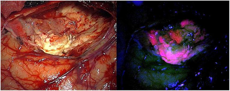
Side-by-side intraoperative microscopic clinical photographs comparing healthy brain parenchyma and glioblastoma under different lighting conditions. The left image, captured under standard white light, demonstrates the surgical field showing cerebral cortex, vascular supply, and a pale, yellowish tumor mass within the resection cavity. This view provides anatomical context but offers limited contrast between tumor margins and healthy tissue. The right image displays the same field using 440 nm blue light excitation for fluorescence-guided surgery (FGS). In this view, areas containing Protoporphyrin IX (PpIX) emit a distinct red-pink fluorescence, while healthy brain tissue remains dark or exhibits low-intensity green/blue autofluorescence. The fluorescent signal is concentrated in the central tumor mass, showing non-uniform distribution and intensity gradients that help surgeons differentiate neoplastic cells from the surrounding parenchyma. This comparison illustrates the clinical utility of 5-ALA induced fluorescence in enhancing real-time tumor visualization and margin detection during neurosurgical procedures.
Note: Apurba Sastry's Essentials of Medical Microbiology is not available in the textbook library. The answer below is drawn from Medical Microbiology 9e (Murray, Rosenthal & Pfaller), which covers the same microscopy content as Apurba Sastry. The concepts, principles, and clinical uses are identical across both standard microbiology textbooks.
Types of Microscopes Used in Microbiology
1. Light (Brightfield) Microscope
- Light source, condenser, stage (specimen platform), objective lens, ocular lens.
| Objective | Magnification | Use |
|---|---|---|
| Low power | 10x | Scanning specimen |
| High dry | 40x | Parasites, filamentous fungi |
| Oil immersion | 100x | Bacteria, yeasts |
2. Darkfield Microscope
- Can detect extremely thin organisms invisible under brightfield.
- No staining needed.
- Light passes around (not through) organisms, so internal structure cannot be studied.
- Detection of Treponema pallidum (syphilis) - too thin to see with brightfield.
- Detection of Leptospira spp. (leptospirosis).
3. Phase-Contrast Microscope
- Allows examination of internal details of microbes without staining.
- Overcomes the major limitation of simple wet mounts.
- Examination of live, unstained organisms.
- Study of internal structure of bacteria, fungi, and parasites in wet mounts.
- Useful when specimens cannot or need not be stained.
4. Fluorescence Microscope
- High-pressure mercury, halogen, or xenon vapor lamp (emits short wavelengths).
- A series of filters: to block heat, eliminate infrared, and select the excitation wavelength.
- Traditional objective and ocular lenses to magnify the emitted light.
- Autofluorescence - some microorganisms fluoresce naturally.
- Staining-based fluorescence - organisms stained with fluorescent dyes (fluorochromes) then examined.
- Rapid screening at low magnification (bright signal easily spotted).
- Highly sensitive and specific when fluorochrome-labeled antibodies are used (immunofluorescence).
- Auramine-rhodamine stain for Mycobacterium tuberculosis (acid-fast organisms fluoresce orange-yellow).
- Immunofluorescence for specific identification of organisms using antibody-conjugated fluorochromes.
- Detection of Cyclospora (autofluorescence under UV).
- Detection of Candida and fungal elements (calcofluor white stains chitin).
5. Electron Microscope
| Type | Mechanism | Image | Use |
|---|---|---|---|
| Transmission EM (TEM) | Electrons pass directly through specimen (like light in brightfield) | 2D internal structure | Ultrastructure of cells, viruses |
| Scanning EM (SEM) | Electrons bounce off the surface at an angle | 3D surface image | Surface morphology of organisms |
- Primarily a research tool rather than a routine diagnostic tool.
- Nucleic acid amplification assays (PCR) have replaced electron microscopy for most viral diagnostics.
Summary Table
| Microscope | Light Source | Condenser | Resolution | Key Use |
|---|---|---|---|---|
| Brightfield (Light) | Ordinary light | Standard | 0.2 µm | Stained bacteria, fungi |
| Darkfield | Oblique light only | Special (blocks direct light) | 0.02 µm | Treponema, Leptospira |
| Phase-contrast | Ordinary light | Annular ring condenser | ~0.2 µm | Unstained living organisms |
| Fluorescence | UV/high-pressure lamp | Filters | ~0.2 µm | TB (auramine), immunofluorescence |
| Electron | Electron beam | Magnetic coils | 0.001 µm | Viruses, ultrastructure |