
/generatehandwrittenimages A ckd stage 4 patient presents with severe hyperkalemia (6.8 mEq/ L) and metabolic acidosis. Gfr is 20 ml/min. Explain A) why renal potassium handling is impaired in ckd? (5 marks) B)describe the compensatory mechanisms that help mitigate hyperkalemia (5 marks) .- for md physiology 10 marks theory question. You can make many images and not just one for completing the answer
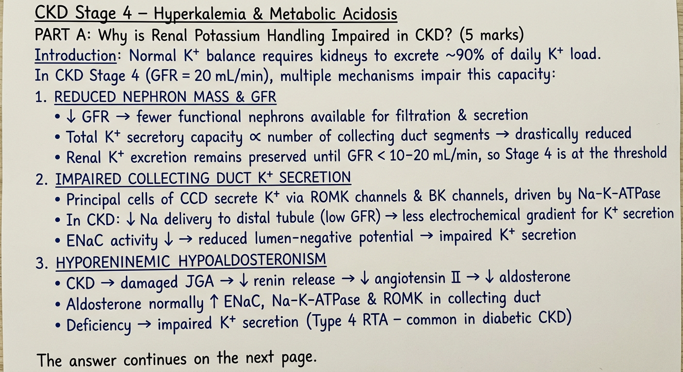
Handwritten medical exam answer on lined paper, blue ink, neat student handwriting, MD physiology exam style. The page shows: Title (underlined): CKD Stage 4 – Hyperkalemia & Metabolic Acidosis PART A: Why is Renal Potassium Handling Impaired in CKD? (5 marks) Introduction: Normal K+ balance requires kidneys to excrete ~90% of daily K+ load. In CKD Stage 4 (GFR = 20 mL/min), multiple mechanisms impair this capacity: 1. REDUCED NEPHRON MASS & GFR (underlined) • ↓ GFR → fewer functional nephrons available for filtration & secretion • Total K+ secretory capacity ∝ number of collecting duct segments → drastically reduced • Renal K+ excretion remains preserved until GFR < 10–20 mL/min, so Stage 4 is at the threshold 2. IMPAIRED COLLECTING DUCT K+ SECRETION (underlined) • Principal cells of CCD secrete K+ via ROMK channels & BK channels, driven by Na-K-ATPase • In CKD: ↓ Na delivery to distal tubule (low GFR) → less electrochemical gradient for K+ secretion • ENaC activity ↓ → reduced lumen-negative potential → impaired K+ secretion 3. HYPORENINEMIC HYPOALDOSTERONISM (underlined) • CKD → damaged JGA → ↓ renin release → ↓ angiotensin II → ↓ aldosterone • Aldosterone normally ↑ ENaC, Na-K-ATPase & ROMK in collecting duct • Deficiency → impaired K+ secretion (Type 4 RTA – common in diabetic CKD) The answer continues on the next page. Paper has faint blue horizontal lines, slightly aged look.
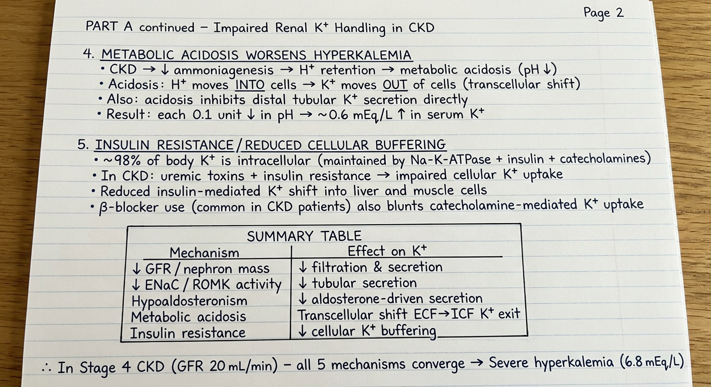
Handwritten medical exam answer on lined paper, blue ink, neat student handwriting, MD physiology exam style. This is page 2 of the answer. Content: PART A continued – Impaired Renal K+ Handling in CKD 4. METABOLIC ACIDOSIS WORSENS HYPERKALEMIA (underlined) • CKD → ↓ ammoniagenesis → H+ retention → metabolic acidosis (pH ↓) • Acidosis: H+ moves INTO cells → K+ moves OUT of cells (transcellular shift) • Also: acidosis inhibits distal tubular K+ secretion directly • Result: each 0.1 unit ↓ in pH → ~0.6 mEq/L ↑ in serum K+ 5. INSULIN RESISTANCE / REDUCED CELLULAR BUFFERING (underlined) • ~98% of body K+ is intracellular (maintained by Na-K-ATPase + insulin + catecholamines) • In CKD: uremic toxins + insulin resistance → impaired cellular K+ uptake • Reduced insulin-mediated K+ shift into liver and muscle cells • β-blocker use (common in CKD patients) also blunts catecholamine-mediated K+ uptake SUMMARY TABLE (drawn as a simple hand-drawn box): | Mechanism | Effect on K+ | | ↓ GFR / nephron mass | ↓ filtration & secretion | | ↓ ENaC / ROMK activity | ↓ tubular secretion | | Hypoaldosteronism | ↓ aldosterone-driven secretion | | Metabolic acidosis | Transcellular shift ECF→ICF K+ exit | | Insulin resistance | ↓ cellular K+ buffering | Bottom note: "∴ In Stage 4 CKD (GFR 20 mL/min) – all 5 mechanisms converge → Severe hyperkalemia (6.8 mEq/L)" Paper has faint blue horizontal lines.
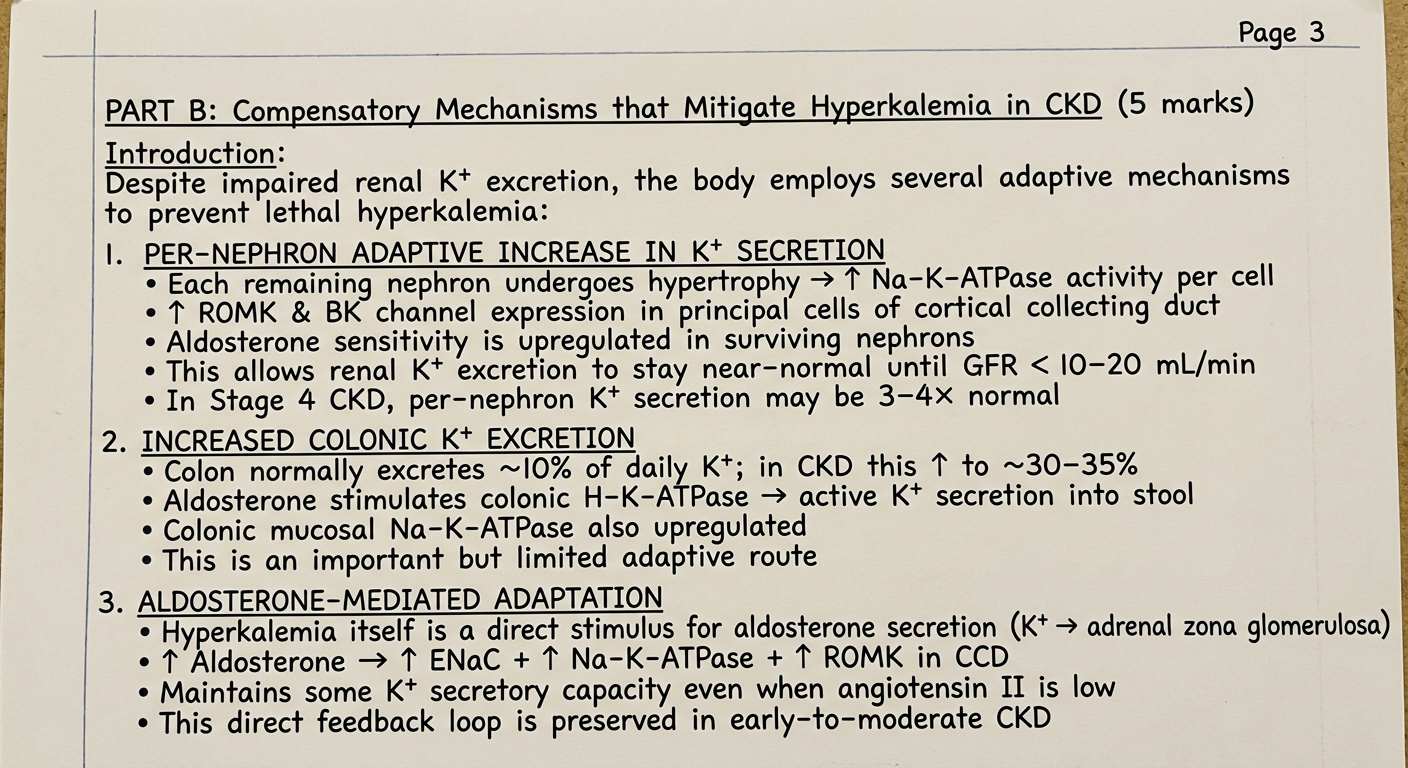
Handwritten medical exam answer on lined paper, blue ink, neat student handwriting, MD physiology exam style. This is page 3 of the answer. PART B: Compensatory Mechanisms that Mitigate Hyperkalemia in CKD (5 marks) Introduction: Despite impaired renal K+ excretion, the body employs several adaptive mechanisms to prevent lethal hyperkalemia: 1. PER-NEPHRON ADAPTIVE INCREASE IN K+ SECRETION (underlined) • Each remaining nephron undergoes hypertrophy → ↑ Na-K-ATPase activity per cell • ↑ ROMK & BK channel expression in principal cells of cortical collecting duct • Aldosterone sensitivity is upregulated in surviving nephrons • This allows renal K+ excretion to stay near-normal until GFR < 10–20 mL/min • In Stage 4 CKD, per-nephron K+ secretion may be 3–4× normal 2. INCREASED COLONIC K+ EXCRETION (underlined) • Colon normally excretes ~10% of daily K+; in CKD this ↑ to ~30–35% • Aldosterone stimulates colonic H-K-ATPase → active K+ secretion into stool • Colonic mucosal Na-K-ATPase also upregulated • This is an important but limited adaptive route 3. ALDOSTERONE-MEDIATED ADAPTATION (underlined) • Hyperkalemia itself is a direct stimulus for aldosterone secretion (K+ → adrenal zona glomerulosa) • ↑ Aldosterone → ↑ ENaC + ↑ Na-K-ATPase + ↑ ROMK in CCD • Maintains some K+ secretory capacity even when angiotensin II is low • This direct feedback loop is preserved in early-to-moderate CKD Paper has faint blue horizontal lines, slightly aged look.
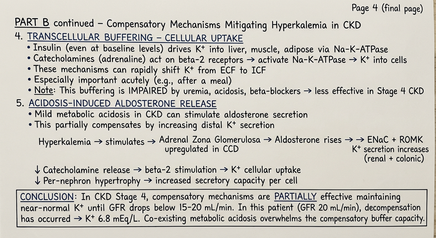
Handwritten medical exam answer on lined paper, blue ink, neat student handwriting, MD physiology exam style. This is page 4 (final page) of the answer. PART B continued – Compensatory Mechanisms Mitigating Hyperkalemia 4. TRANSCELLULAR BUFFERING – CELLULAR UPTAKE (underlined) • Insulin (even at baseline levels) drives K+ into liver, muscle, adipose via Na-K-ATPase • Catecholamines (adrenaline) → β2-receptor → activate Na-K-ATPase → K+ into cells • These mechanisms can rapidly shift K+ from ECF → ICF • Especially important acutely (e.g., after a meal) • Note: This buffering is IMPAIRED by uremia, acidosis, β-blockers → less effective in Stage 4 CKD 5. ACIDOSIS-INDUCED ALDOSTERONE RELEASE (underlined) • Mild metabolic acidosis in CKD can stimulate aldosterone secretion • This partially compensates by increasing distal K+ secretion FLOWCHART (hand-drawn with arrows): Hyperkalemia → direct stimulus → Adrenal Zona Glomerulosa → ↑ Aldosterone → CCD ENaC + ROMK ↑ → ↑ K+ secretion (renal + colonic) ↓ β2-adrenergic ↑ → K+ cellular uptake ↓ Per-nephron hypertrophy → ↑ secretory capacity CONCLUSION (underlined): "In CKD Stage 4, these compensatory mechanisms are PARTIALLY effective – maintaining near-normal K+ until GFR drops below ~15–20 mL/min. In our patient (GFR 20 mL/min), decompensation has occurred → K+ 6.8 mEq/L. Metabolic acidosis further overwhelms the compensatory buffer capacity." [End of answer] Paper has faint blue horizontal lines.
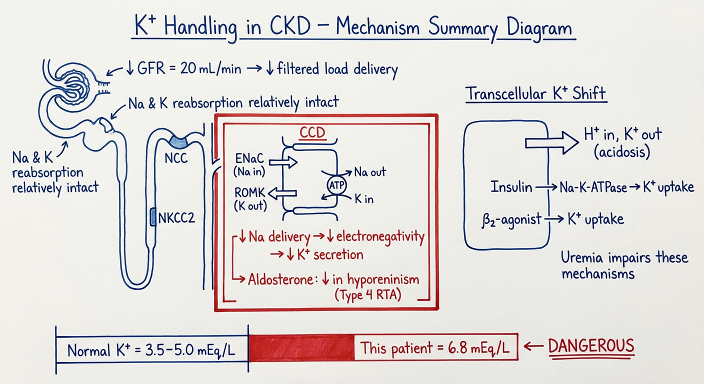
Medical physiology diagram on white paper, hand-drawn style with labels, blue and red ink. Title at top: "K+ Handling in CKD – Mechanism Summary Diagram" Show a simplified nephron diagram on the left side with: - Glomerulus at top labeled "↓ GFR = 20 mL/min → ↓ filtered load delivery" - Proximal tubule: "Na & K reabsorption relatively intact" - Thick ascending limb: labeled "NKCC2" - Distal convoluted tubule: "NCC" - Cortical Collecting Duct (CCD) highlighted in red box with arrows showing: → Principal cell: ENaC (Na in), ROMK (K out), Na-K-ATPase → "↓ Na delivery → ↓ electronegativity → ↓ K+ secretion" → "Aldosterone: ↓ in hyporeninism (Type 4 RTA)" On the right side, show a cell diagram labeled "Transcellular K+ Shift": - Arrow pointing OUT of cell: "H+ in, K+ out (acidosis)" - Arrow pointing INTO cell: "Insulin → Na-K-ATPase → K+ uptake" - Arrow pointing INTO cell: "β2-agonist → K+ uptake" - Text: "Uremia impairs these mechanisms" At the bottom: simple bar showing "Normal K+ = 3.5–5.0 mEq/L" vs "This patient = 6.8 mEq/L ← DANGEROUS" Clean, educational diagram with labels, arrows, boxes. Hand-drawn medical student style.
Page 1 - Part A: Introduction + Points 1-3
Page 2 - Part A: Points 4-5 + Summary Table
Page 3 - Part B: Compensatory Mechanisms Points 1-3
Page 4 - Part B: Points 4-5 + Flowchart + Conclusion

Page 5 - Mechanism Diagram (Bonus Visual)
Quick Content Reference (for revision):
- Reduced nephron mass/GFR - fewer collecting duct segments available for K+ secretion; threshold is GFR <10-20 mL/min (Stage 4 is right at this)
- Impaired collecting duct K+ secretion - low distal Na delivery → reduced lumen-negative potential → ↓ ROMK/BK channel-mediated K+ secretion
- Hyporeninemic hypoaldosteronism - damaged JGA → ↓ renin → ↓ aldosterone → ↓ ENaC + Na-K-ATPase activity (classic Type 4 RTA of diabetic CKD)
- Metabolic acidosis - H+ shifts into cells, K+ shifts out; acidosis also directly inhibits distal K+ secretion; each 0.1 pH unit fall raises K+ by ~0.6 mEq/L
- Insulin resistance + uremic toxins - impaired cellular K+ buffering by Na-K-ATPase; ↓ insulin-mediated K+ uptake into liver/muscle
- Per-nephron adaptive hypersecretion - surviving nephrons upregulate Na-K-ATPase, ROMK, BK channels; per-nephron K+ secretion 3-4x normal
- Increased colonic K+ excretion - rises from 10% to 30-35% of daily intake; aldosterone stimulates colonic H-K-ATPase; limited but real
- Direct aldosterone release by hyperkalemia - K+ directly stimulates adrenal zona glomerulosa even without renin/angiotensin II; preserved feedback
- Transcellular buffering - insulin + catecholamines shift K+ into cells acutely; blunted in CKD by uremia and acidosis
- Adaptations in distal tubular sensitivity - upregulation of mineralocorticoid receptors in surviving nephrons compensates for lower aldosterone levels
Part 4 image cannot be seen
Handwritten medical exam answer on lined paper, blue ink, neat student handwriting, MD physiology exam style. This is page 4 (final page) of the answer. PART B continued – Compensatory Mechanisms Mitigating Hyperkalemia in CKD 4. TRANSCELLULAR BUFFERING – CELLULAR UPTAKE (underlined) • Insulin (even at baseline levels) drives K+ into liver, muscle, adipose via Na-K-ATPase • Catecholamines (adrenaline) act on beta-2 receptors → activate Na-K-ATPase → K+ into cells • These mechanisms can rapidly shift K+ from ECF to ICF • Especially important acutely (e.g., after a meal) • Note: This buffering is IMPAIRED by uremia, acidosis, beta-blockers → less effective in Stage 4 CKD 5. ACIDOSIS-INDUCED ALDOSTERONE RELEASE (underlined) • Mild metabolic acidosis in CKD can stimulate aldosterone secretion • This partially compensates by increasing distal K+ secretion FLOWCHART (hand-drawn with arrows): Hyperkalemia → stimulates → Adrenal Zona Glomerulosa → Aldosterone rises → ENaC + ROMK upregulated in CCD → K+ secretion increases (renal + colonic) ↓ Catecholamine release → beta-2 stimulation → K+ cellular uptake ↓ Per-nephron hypertrophy → increased secretory capacity per cell CONCLUSION (box drawn around it): In CKD Stage 4, compensatory mechanisms are PARTIALLY effective maintaining near-normal K+ until GFR drops below 15-20 mL/min. In this patient (GFR 20 mL/min), decompensation has occurred → K+ 6.8 mEq/L. Co-existing metabolic acidosis overwhelms the compensatory buffer capacity. Paper has faint blue horizontal lines, clean neat handwriting, slightly aged paper look.
Page 4 - Part B: Points 4-5 + Flowchart + Conclusion
CCD full form in page 1. ?
/generatehandwrittenimages What are the various types of cell membrane proteins? (2 marks) Describe the role of these proteins in maintaining cellular function and homeostasis . (8 marks) - for md physiology 10 marks theory question. You can make many images and not just one for completing the answer
Handwritten medical exam answer on lined paper, blue ink, neat student handwriting, MD physiology 10-mark theory question style. Slightly aged paper with faint blue horizontal lines. Title (underlined, centered): Cell Membrane Proteins – Types and Functions PART A: Types of Cell Membrane Proteins (2 marks) (underlined) The plasma membrane is a phospholipid bilayer embedded with various proteins, mainly glycoproteins. There are TWO major types: 1. INTEGRAL (INTRINSIC) PROTEINS (underlined) • Penetrate all the way through the lipid bilayer (transmembrane proteins) • Amphipathic: hydrophobic core region + hydrophilic ends • Subtypes: a) Channel proteins – form water-filled pores (ion channels: Na+, K+, Ca2+, Cl-) b) Carrier/Transporter proteins – bind and transport specific molecules c) Pump proteins – use ATP for active transport (e.g., Na+-K+-ATPase) d) Receptor proteins – bind hormones/ligands on extracellular surface e) Enzyme proteins – catalyze membrane-bound reactions f) Structural proteins – maintain cell shape (e.g., spectrin linkage) 2. PERIPHERAL (EXTRINSIC) PROTEINS (underlined) • NOT embedded in lipid bilayer • Loosely attached to surface of integral proteins by ionic/electrostatic bonds • Found on cytoplasmic OR extracellular surface • Functions: act as enzymes, signal transducers, cytoskeletal anchors • Easily removed by mild conditions (salt solutions) Ref: Guyton & Hall Medical Physiology – Fig. 2.3
Hand-drawn medical diagram on white paper, blue and red ink, student style. Title at top underlined: "Types of Cell Membrane Proteins – Diagram" Draw a phospholipid bilayer (two rows of circles with tails pointing inward) running horizontally across the middle of the page. Label the following components with arrows: - A large protein spanning the entire bilayer: "Integral Protein (Transmembrane)" with arrow pointing to it - A protein only on the outer surface: "Peripheral Protein (extracellular side)" - A protein only on the inner cytoplasmic side: "Peripheral Protein (cytoplasmic side)" - An ion channel (pore visible through protein): "Channel Protein → ion diffusion (Na+, K+)" - A pump protein with ATP label: "Na+-K+-ATPase (Pump) → Active Transport" - A receptor with ligand binding: "Receptor Protein → hormone binding → signal transduction" - Carbohydrate chains on outer surface: "Glycocalyx (carbohydrate moieties)" At the bottom, a small legend box: Integral proteins = embedded through bilayer Peripheral proteins = surface-attached only Channel / Carrier / Pump / Receptor / Enzyme / Structural Clean educational diagram, labels with neat arrows, blue ink on white background.
Handwritten medical exam answer on lined paper, blue ink, neat student handwriting, MD physiology exam style. Faint blue horizontal lines, slightly aged paper. PART B: Role of Membrane Proteins in Cellular Function and Homeostasis (8 marks) (underlined, centered) Introduction: Cell membrane proteins perform diverse and indispensable functions in maintaining cellular homeostasis. These are described systematically below: 1. TRANSPORT FUNCTION – Channel Proteins (underlined) • Ion channels form water-filled pores selective for specific ions (Na+, K+, Ca2+, Cl-) • Gated channels: voltage-gated (nerve action potential), ligand-gated (e.g., ACh at NMJ), mechanically gated • Maintain resting membrane potential (RMP ≈ -70 mV in neurons) • Rapid ion flux underlies nerve conduction, muscle contraction, cardiac rhythm • Example: voltage-gated Na+ channels → depolarization phase of action potential • Aquaporins (AQP) – water channels → regulate water reabsorption in renal tubules 2. ACTIVE TRANSPORT – Pump Proteins (underlined) • Na+-K+-ATPase: pumps 3 Na+ out and 2 K+ in per ATP cycle • Maintains: high intracellular K+, low intracellular Na+, cell volume, RMP • Creates electrochemical gradient used for secondary active transport • Ca2+-ATPase: pumps Ca2+ out of cytoplasm → maintains Ca2+ homeostasis • H+-K+-ATPase: in gastric parietal cells → HCl secretion • Essential for cell volume regulation and preventing osmotic lysis Continued on next page...
Handwritten medical exam answer on lined paper, blue ink, neat student handwriting, MD physiology exam style. Faint blue horizontal lines, slightly aged paper. This is page 3 of the answer. PART B continued – Role of Membrane Proteins (8 marks) 3. CARRIER / TRANSPORTER PROTEINS (underlined) • Undergo conformational change to carry specific molecules across membrane • Facilitated diffusion: GLUT transporters (glucose uptake into cells, no ATP) - GLUT1: RBCs, brain; GLUT2: liver, pancreatic beta cells; GLUT4: muscle (insulin-stimulated) • Secondary active transport: - Na+-glucose co-transporter (SGLT) in renal tubule and gut → couples Na+ gradient to glucose uptake - Na+-H+ exchanger (NHE) → acid-base regulation • Amino acid transporters, neurotransmitter reuptake transporters (e.g., serotonin, dopamine) 4. RECEPTOR PROTEINS – Signal Transduction (underlined) • Allow cells to respond to extracellular signals (hormones, neurotransmitters, growth factors) • Types: a) G-protein coupled receptors (GPCRs): β-adrenergic, muscarinic – most abundant receptor type → ligand binding → G protein activation → adenylyl cyclase → cAMP → PKA b) Receptor Tyrosine Kinases (RTKs): insulin receptor, growth factor receptors → autophosphorylation → intracellular signaling cascades (MAPK, PI3K/Akt) c) Ligand-gated ion channels: nicotinic ACh receptor at NMJ → Na+ influx → muscle contraction d) Cytokine receptors: JAK-STAT pathway → immune regulation • Maintain hormonal homeostasis and coordinate organ function Continued on next page...
Error calling model 'gemini-3-pro-image' (RESOURCE_EXHAUSTED): 429 RESOURCE_EXHAUSTED. {'error': {'code': 429, 'message': 'Resource has been exhausted (e.g. check quota).', 'status': 'RESOURCE_EXHAUSTED'}}
We encountered an error while processing your request. Please try again or reach out to us for help.
Retry
Handwritten medical exam answer on lined paper, blue ink, neat student handwriting, MD physiology 10-mark theory question style. Slightly aged paper with faint blue horizontal lines. Title (underlined, centered): Cell Membrane Proteins – Types and Functions PART A: Types of Cell Membrane Proteins (2 marks) (underlined) The plasma membrane is a phospholipid bilayer embedded with various proteins, mainly glycoproteins. There are TWO major types: 1. INTEGRAL (INTRINSIC) PROTEINS (underlined) • Penetrate all the way through the lipid bilayer (transmembrane proteins) • Amphipathic: hydrophobic core region + hydrophilic ends • Subtypes: a) Channel proteins – form water-filled pores (ion channels: Na+, K+, Ca2+, Cl-) b) Carrier/Transporter proteins – bind and transport specific molecules c) Pump proteins – use ATP for active transport (e.g., Na+-K+-ATPase) d) Receptor proteins – bind hormones/ligands on extracellular surface e) Enzyme proteins – catalyze membrane-bound reactions f) Structural proteins – maintain cell shape 2. PERIPHERAL (EXTRINSIC) PROTEINS (underlined) • NOT embedded in lipid bilayer • Loosely attached to surface of integral proteins by ionic bonds • Found on cytoplasmic OR extracellular surface • Functions: act as enzymes, signal transducers, cytoskeletal anchors • Easily removed by mild salt solutions Ref: Guyton and Hall Medical Physiology, Chapter 2
Hand-drawn medical diagram on white paper, blue and red ink, neat student style. Title at top underlined: "Types of Cell Membrane Proteins – Diagram" Draw a phospholipid bilayer (two rows of circles with zigzag tails pointing inward) running horizontally across the middle of the page. Label these components with neat arrows: - Large protein spanning entire bilayer: arrow → "Integral Protein (Transmembrane)" - Protein only on outer surface: arrow → "Peripheral Protein (extracellular)" - Protein only on inner cytoplasmic side: arrow → "Peripheral Protein (cytoplasmic)" - Ion channel (open pore through protein): arrow → "Channel Protein: Na+, K+ diffusion" - Pump protein with small ATP label nearby: arrow → "Na+-K+-ATPase (Pump)" - Receptor protein with Y-shaped ligand attached: arrow → "Receptor Protein: hormone binding" - Carbohydrate chains sticking out on outer surface: arrow → "Glycocalyx" Bottom of page: small neat legend box: Integral = embedded through bilayer | Peripheral = surface-attached Subtypes: Channel / Carrier / Pump / Receptor / Enzyme / Structural
Handwritten medical exam answer on lined paper, blue ink, neat student handwriting, MD physiology exam style. Faint blue horizontal lines, slightly aged paper. PART B: Role of Membrane Proteins in Cellular Function and Homeostasis (8 marks) (underlined, centered) 1. TRANSPORT – Channel Proteins (underlined) • Ion channels form selective water-filled pores for Na+, K+, Ca2+, Cl- • Types: voltage-gated, ligand-gated, mechanically-gated • Maintain resting membrane potential (RMP = -70 mV in neurons) • Rapid ion flux underlies nerve conduction, muscle contraction, cardiac rhythm • Example: voltage-gated Na+ channels → depolarization of action potential • Aquaporins (AQP2) → water reabsorption in collecting duct (ADH-regulated) 2. ACTIVE TRANSPORT – Pump Proteins (underlined) • Na+-K+-ATPase: pumps 3 Na+ OUT and 2 K+ IN per ATP hydrolysis • Maintains: high ICF K+, low ICF Na+, RMP, cell volume • Creates electrochemical gradient for secondary active transport • Ca2+-ATPase (SERCA): pumps Ca2+ back into SR after muscle contraction → relaxation • H+-K+-ATPase: gastric parietal cells → HCl secretion (gastric acid) • H+-ATPase: renal alpha-intercalated cells → urinary acidification • Prevents osmotic swelling/lysis of cells 3. FACILITATED AND SECONDARY ACTIVE TRANSPORT – Carrier Proteins (underlined) • GLUT transporters (facilitated diffusion, no ATP): - GLUT1: brain, RBCs; GLUT2: liver, pancreatic B-cells; GLUT4: muscle/fat (insulin-dependent) • SGLT1/2 (Na+-glucose co-transporter): couples Na+ gradient to glucose reabsorption in PCT • Na+-H+ exchanger (NHE3): PCT acid-base regulation, bicarbonate reclamation • Neurotransmitter reuptake transporters: SERT (serotonin), DAT (dopamine) – terminate synaptic signals Continued next page...
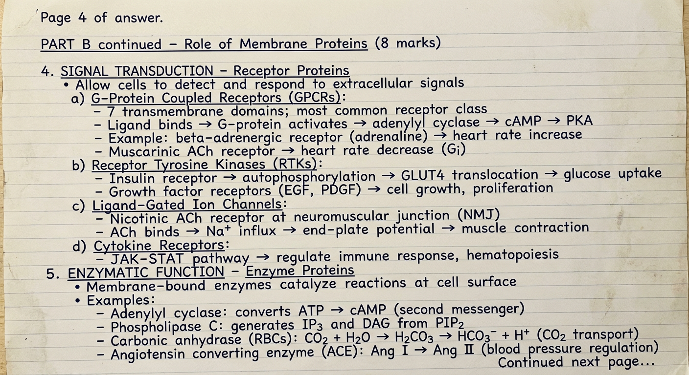
Handwritten medical exam answer on lined paper, blue ink, neat student handwriting, MD physiology exam style. Faint blue horizontal lines, slightly aged paper. Page 4 of answer. PART B continued – Role of Membrane Proteins (8 marks) 4. SIGNAL TRANSDUCTION – Receptor Proteins (underlined) • Allow cells to detect and respond to extracellular signals a) G-Protein Coupled Receptors (GPCRs): (underlined) - 7 transmembrane domains; most common receptor class - Ligand binds → G-protein activates → adenylyl cyclase → cAMP → PKA - Example: beta-adrenergic receptor (adrenaline) → heart rate increase - Muscarinic ACh receptor → heart rate decrease (Gi) b) Receptor Tyrosine Kinases (RTKs): (underlined) - Insulin receptor → autophosphorylation → GLUT4 translocation → glucose uptake - Growth factor receptors (EGF, PDGF) → cell growth, proliferation c) Ligand-Gated Ion Channels: (underlined) - Nicotinic ACh receptor at neuromuscular junction (NMJ) - ACh binds → Na+ influx → end-plate potential → muscle contraction d) Cytokine Receptors: (underlined) - JAK-STAT pathway → regulate immune response, hematopoiesis 5. ENZYMATIC FUNCTION – Enzyme Proteins (underlined) • Membrane-bound enzymes catalyze reactions at cell surface • Examples: - Adenylyl cyclase: converts ATP → cAMP (second messenger) - Phospholipase C: generates IP3 and DAG from PIP2 - Carbonic anhydrase (RBCs): CO2 + H2O → H2CO3 → HCO3- + H+ (CO2 transport) - Angiotensin converting enzyme (ACE): Ang I → Ang II (blood pressure regulation) Continued next page...
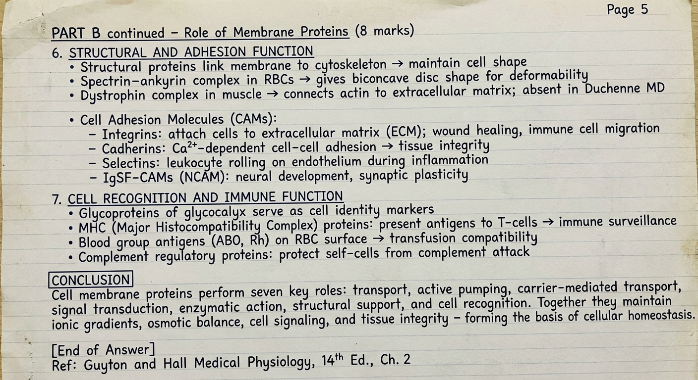
Handwritten medical exam answer on lined paper, blue ink, neat student handwriting, MD physiology exam style. Faint blue horizontal lines, slightly aged paper. Page 5 - final page of answer. PART B continued – Role of Membrane Proteins (8 marks) 6. STRUCTURAL AND ADHESION FUNCTION (underlined) • Structural proteins link membrane to cytoskeleton → maintain cell shape • Spectrin-ankyrin complex in RBCs → gives biconcave disc shape for deformability • Dystrophin complex in muscle → connects actin to extracellular matrix; absent in Duchenne MD • Cell Adhesion Molecules (CAMs): - Integrins: attach cells to extracellular matrix (ECM); wound healing, immune cell migration - Cadherins: Ca2+-dependent cell-cell adhesion → tissue integrity - Selectins: leukocyte rolling on endothelium during inflammation - IgSF-CAMs (NCAM): neural development, synaptic plasticity 7. CELL RECOGNITION AND IMMUNE FUNCTION (underlined) • Glycoproteins of glycocalyx serve as cell identity markers • MHC (Major Histocompatibility Complex) proteins: present antigens to T-cells → immune surveillance • Blood group antigens (ABO, Rh) on RBC surface → transfusion compatibility • Complement regulatory proteins: protect self-cells from complement attack CONCLUSION (box around it): Cell membrane proteins perform seven key roles: transport, active pumping, carrier-mediated transport, signal transduction, enzymatic action, structural support, and cell recognition. Together they maintain ionic gradients, osmotic balance, cell signaling, and tissue integrity – forming the basis of cellular homeostasis. [End of Answer] Ref: Guyton and Hall Medical Physiology, 14th Ed., Ch. 2
Page 1 - Part A: Types of Cell Membrane Proteins (2 marks)
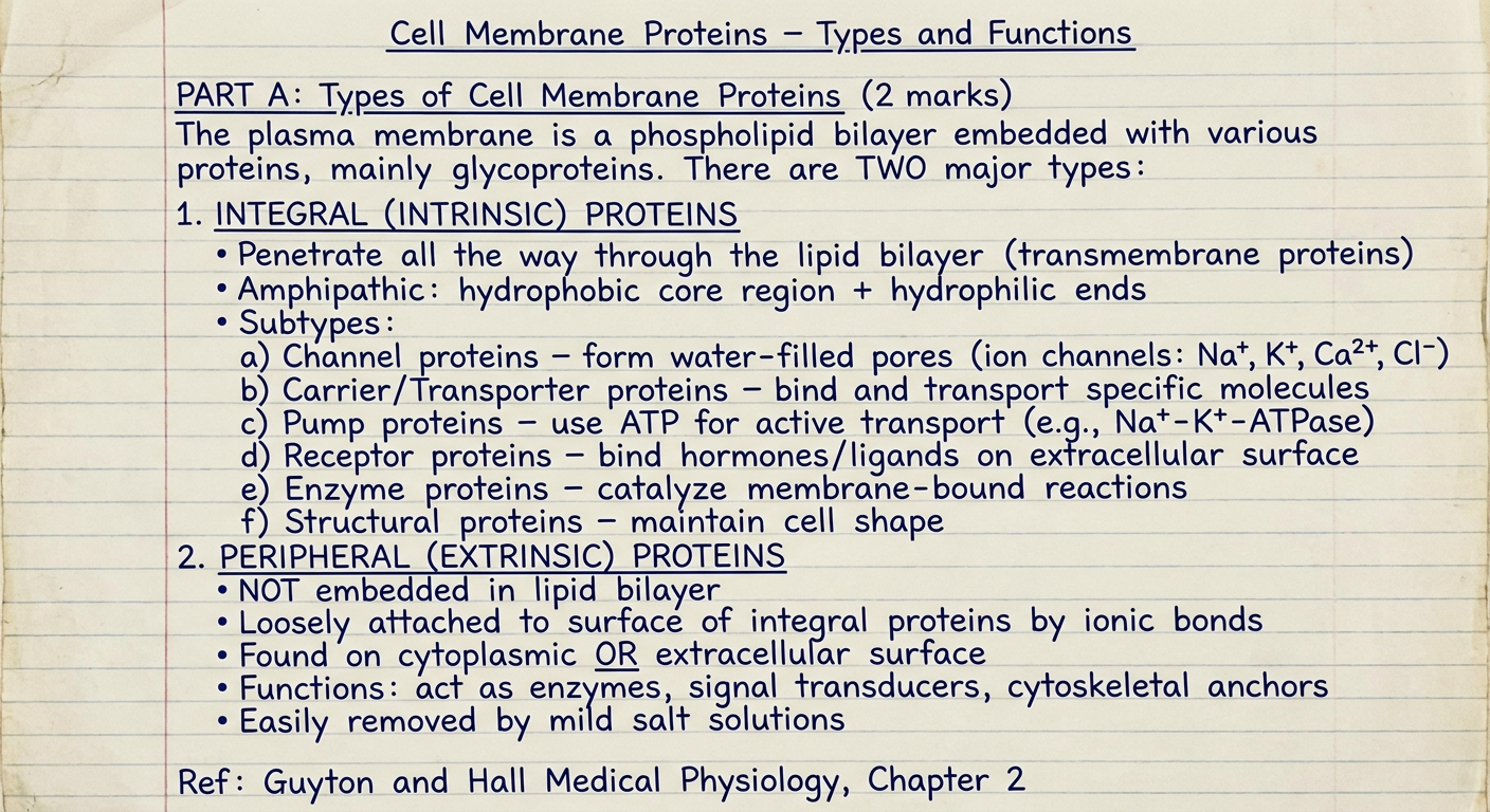
Page 2 - Diagram: Membrane Protein Structure
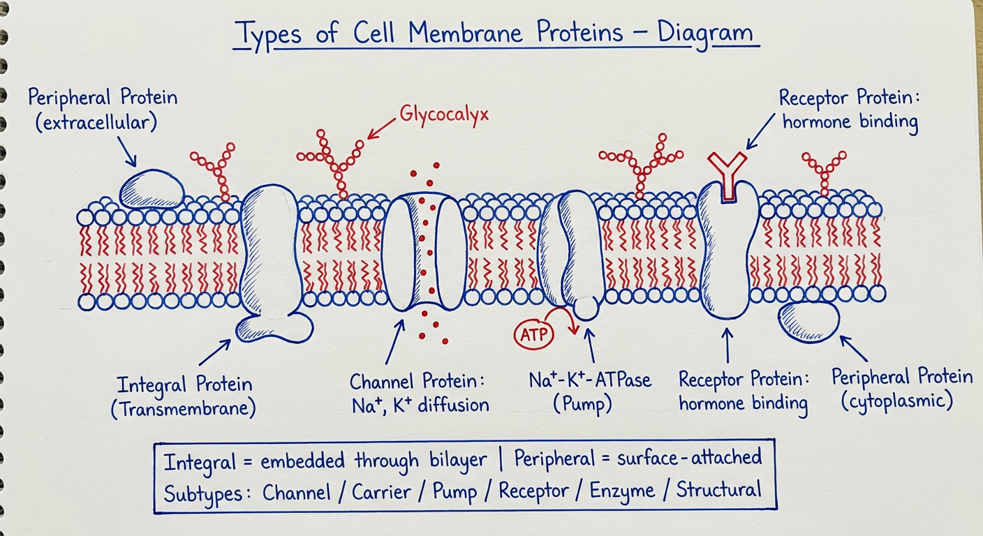
Page 3 - Part B: Points 1-3 (Channel, Pump, Carrier proteins)
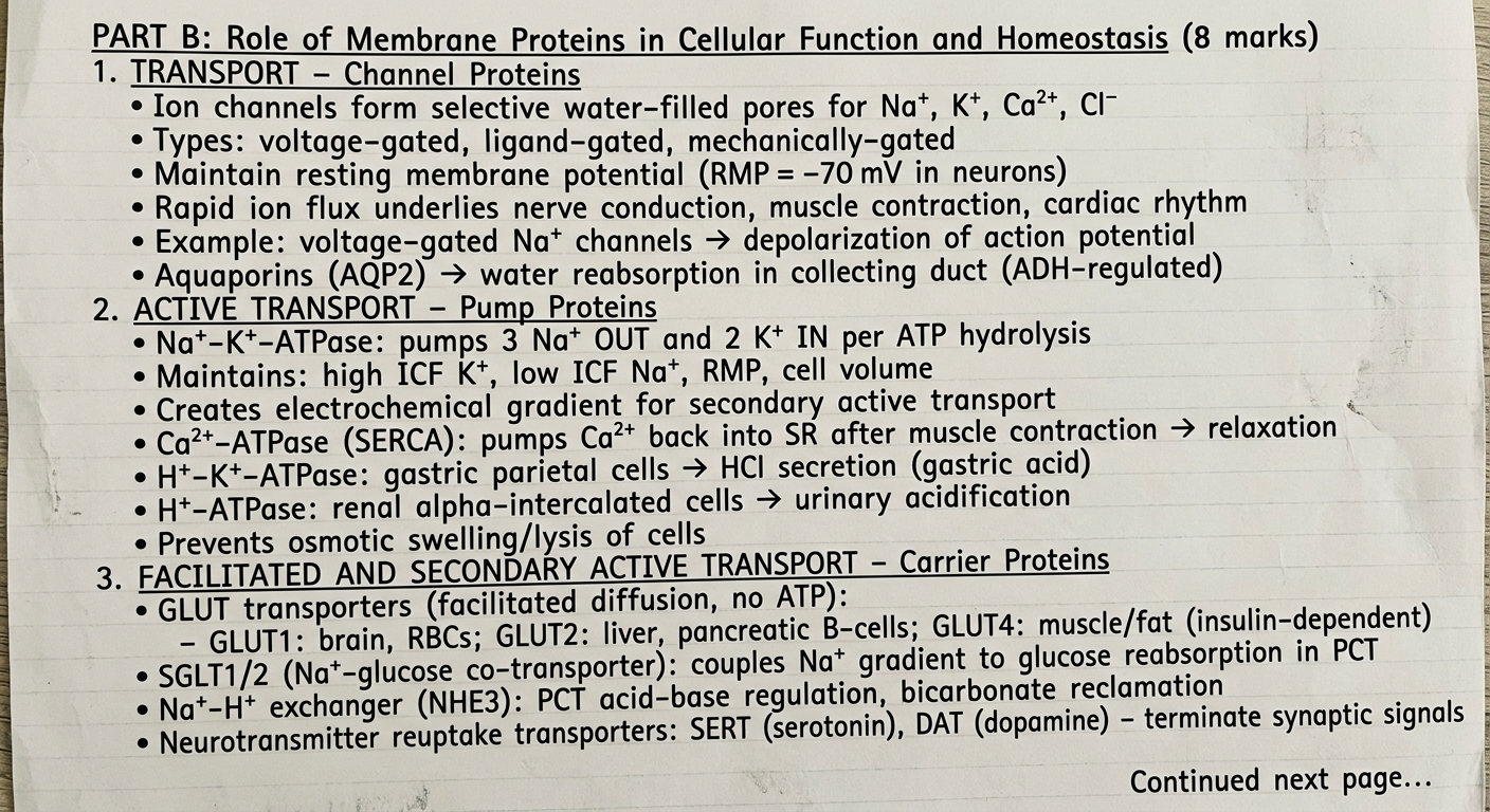
Page 4 - Part B: Points 4-5 (Receptor & Enzyme proteins)
Page 5 - Part B: Points 6-7 + Conclusion (Structural, Immune, Summary)
Answer Structure at a Glance:
| Part | Marks | Coverage |
|---|---|---|
| A | 2 | Integral (6 subtypes) + Peripheral proteins |
| B – Point 1 | ~1.5 | Channel proteins: ion channels, aquaporins |
| B – Point 2 | ~1.5 | Pump proteins: Na-K-ATPase, Ca-ATPase, H-K-ATPase |
| B – Point 3 | ~1 | Carrier proteins: GLUT, SGLT, NHE, neurotransmitter transporters |
| B – Point 4 | ~2 | Receptor proteins: GPCRs, RTKs, ligand-gated channels, cytokine receptors |
| B – Point 5 | ~1 | Enzyme proteins: adenylyl cyclase, carbonic anhydrase, ACE |
| B – Points 6-7 | ~1 | Structural (spectrin, dystrophin, integrins) + immune/cell recognition |