
How to differentiate between Aki and ckd and id cmd difference in ckd or aki in detail diagnosis
Reading File
Now let me search the medical library for comprehensive content on AKI vs CKD differentiation.
Finding Sources
Finding Sources
Searching PubMed
"acute kidney injury" AND "chronic kidney disease" AND differentiation diagnosis
Excellent - found a dedicated section. Let me now read the most relevant textbook content in parallel.
Reading File
Finding Sources
Finding Sources
Let me read more content from these key sections in parallel.
Reading File
Reading File
Finding Sources
Reading File
Finding Sources
Searching Images
acute kidney injury vs chronic kidney disease differentiation diagnosis
Reading File
Finding Sources
Finding Sources
Let me fetch the AKI investigation flowchart image and get the key table on urinary indices for complete diagnostic workup.
Loading Image
Reading File
I now have comprehensive data from authoritative textbooks. Let me compile the full detailed answer.
AKI vs CKD: Detailed Differentiation + AKI-on-CKD
Definitions First
| Feature | AKI | CKD |
|---|---|---|
| Definition | Abrupt rise in serum creatinine ≥0.3 mg/dL within 48 h, OR ≥1.5× baseline within 7 days, OR urine output <0.5 mL/kg/h for ≥6 h (KDIGO criteria) | eGFR <60 mL/min/1.73 m² persisting for >3 months, OR kidney damage markers persisting >3 months (proteinuria, hematuria, structural abnormality) |
| Time course | Hours to days | Months to years (by definition >3 months) |
| Reversibility | Potentially reversible with treatment | Usually irreversible; progressive |
1. History and Clinical Clues
Pointing to AKI
- Acute precipitating event: recent surgery, sepsis, hypotension, trauma, rhabdomyolysis, nephrotoxin exposure (NSAIDs, aminoglycosides, IV contrast, ACE inhibitors/ARBs, chemotherapy, herbal medicines)
- Sudden change in urine output (oliguria <500 mL/day or anuria)
- Recent hospitalization, burns, diarrhea, vomiting, bleeding
- Previously documented normal creatinine
Pointing to CKD
- Long-standing hypertension, diabetes mellitus, recurrent urinary tract infections, obstructive uropathy
- Family history of kidney disease (polycystic kidney disease)
- Prior abnormal creatinine or proteinuria on old records
- Symptoms of uraemia that have been building for months: fatigue, anorexia, nausea, pruritus, restless leg syndrome, peripheral neuropathy
"Knowledge of prior serum creatinine concentrations is required to determine the degree of potentially reversible AKI." - Comprehensive Clinical Nephrology, 7th Edition, p. 985
2. Urinary Output Pattern
| Pattern | Significance |
|---|---|
| Oliguria <500 mL/day | Common in AKI (especially ATN); also possible in severe CKD |
| Sudden anuria | Bilateral obstruction, bilateral renal artery occlusion - strongly AKI |
| Polyuria (large urine volumes despite rising creatinine) | Suggests CKD with tubular dysfunction; or recovering AKI |
| Nonoliguric AKI | AIN, contrast nephropathy, aminoglycosides |
3. Physical Examination
Favoring AKI
- Signs of acute volume depletion: low BP, tachycardia, dry mucous membranes, reduced JVP, weight loss
- Signs of acute cause: sepsis, bleeding, rhabdomyolysis bruising, toxin exposure
Favoring CKD
- Pallor (anaemia of chronic disease)
- Sallow/yellowish skin (uraemic pigmentation)
- Peripheral neuropathy (stocking-glove distribution)
- Uraemic frost (very late, rare)
- Hypertensive retinopathy, arteriovenous nicking on fundoscopy
- Cardiomegaly from long-standing hypertension
4. Laboratory Investigations - The Key Differentiators
A. Serum Creatinine & BUN Trajectory
| AKI | CKD | |
|---|---|---|
| Rise in creatinine | Abrupt (hours-days) | Gradual (months-years) |
| Previous creatinine records | Normal | Chronically elevated |
| BUN/Creatinine ratio | >20:1 in prerenal AKI | 10:1 to 15:1 in CKD (unless uremia is superimposed) |
B. Urinary Indices (distinguish prerenal AKI from intrinsic AKI)
From Comprehensive Clinical Nephrology, 7th Edition (Table 72.3):
| Parameter | Prerenal AKI | Intrinsic AKI (e.g., ATN) |
|---|---|---|
| FENa | <1% | >1% |
| FEUrea | <35% | >35% |
| Urine Na (UNa) | <20 mmol/L | >40 mmol/L |
| Urine osmolality | >500 mOsm/kg | <350 mOsm/kg |
| Urine sediment | Normal/few hyaline casts | Muddy brown granular casts |
| BUN/SCr | >20 | <20 |
Important caveat: FENa has limited utility when AKI is superimposed on CKD - interpret cautiously. Contrast nephropathy and rhabdomyolysis can also give a falsely low FENa (<1%) due to early vasoconstriction. - Comprehensive Clinical Nephrology, 7th Edition
C. Haematology
| Parameter | AKI | CKD |
|---|---|---|
| Anaemia | Usually absent (or acute from bleeding) | Normocytic normochromic anaemia (EPO deficiency) - hallmark of CKD |
| Platelet count | May be low in TMA-associated AKI | Normal to mildly reduced |
| Peripheral smear | Schistocytes if TMA | Usually normal |
"Normocytic anemia, hyperparathyroidism, peripheral neuropathy, and broad waxy casts in the urinary sediment suggest CKD." - Comprehensive Clinical Nephrology, 7th Edition, p. 985
D. Calcium-Phosphate-PTH Axis
| AKI | CKD | |
|---|---|---|
| Serum calcium | Low (in severe AKI) | Low |
| Serum phosphate | Elevated in severe AKI | Elevated (often >5 mg/dL) |
| PTH | Not elevated (or mildly elevated) | Elevated - secondary hyperparathyroidism (hallmark) |
| ALP/bone disease | Absent | Present (renal osteodystrophy) |
E. Urinalysis and Urine Sediment
| Finding | Suggests |
|---|---|
| Muddy brown granular casts | ATN (AKI) |
| Red cell casts | Glomerulonephritis (AKI cause) |
| White cell casts | Acute interstitial nephritis (AKI) |
| Broad waxy casts | CKD (form in dilated, widened tubules) |
| Hyaline casts | Prerenal AKI (benign, non-specific) |
| Proteinuria >1 g/day | Glomerular disease (can be AKI or CKD cause) |
| Eosinophiluria | AIN (AKI) |
5. Imaging - Ultrasound (Most Important Single Test)
Ultrasound is the first-line imaging for both AKI and CKD evaluation.
| Finding | Interpretation |
|---|---|
| Small kidneys (<9 cm), cortical thinning, increased echogenicity | CKD (irreversible scarring) |
| Normal or enlarged kidneys, normal echogenicity, hypoechoic medulla | AKI (interstitial edema, e.g., ATN) |
| Dilated pelvicalyceal system | Postrenal AKI (obstruction) |
| Large kidneys despite elevated creatinine | Can be CKD from diabetic nephropathy, amyloidosis, HIV nephropathy, ADPKD |
From National Kidney Foundation Primer on Kidney Diseases, 8th Edition:
"Small kidney size strongly supports a diagnosis of CKD, helping to differentiate acute from chronic kidney injury. Cortical echogenicity can be assessed, with a hyperechoic cortex present in most causes of CKD. In ATN, ultrasound usually shows enlarged kidneys with normal cortical echogenicity and a normal or hypoechoic medulla, primarily due to interstitial edema."
Renal resistive index (RI) on Doppler ultrasound: Normal ≤0.70. Elevated RI correlates with severity of AKI and predicts non-recovery. Emerging tool, operator-dependent.
6. Investigation and Management Flowchart
This is the standard diagnostic approach when elevated creatinine is found:
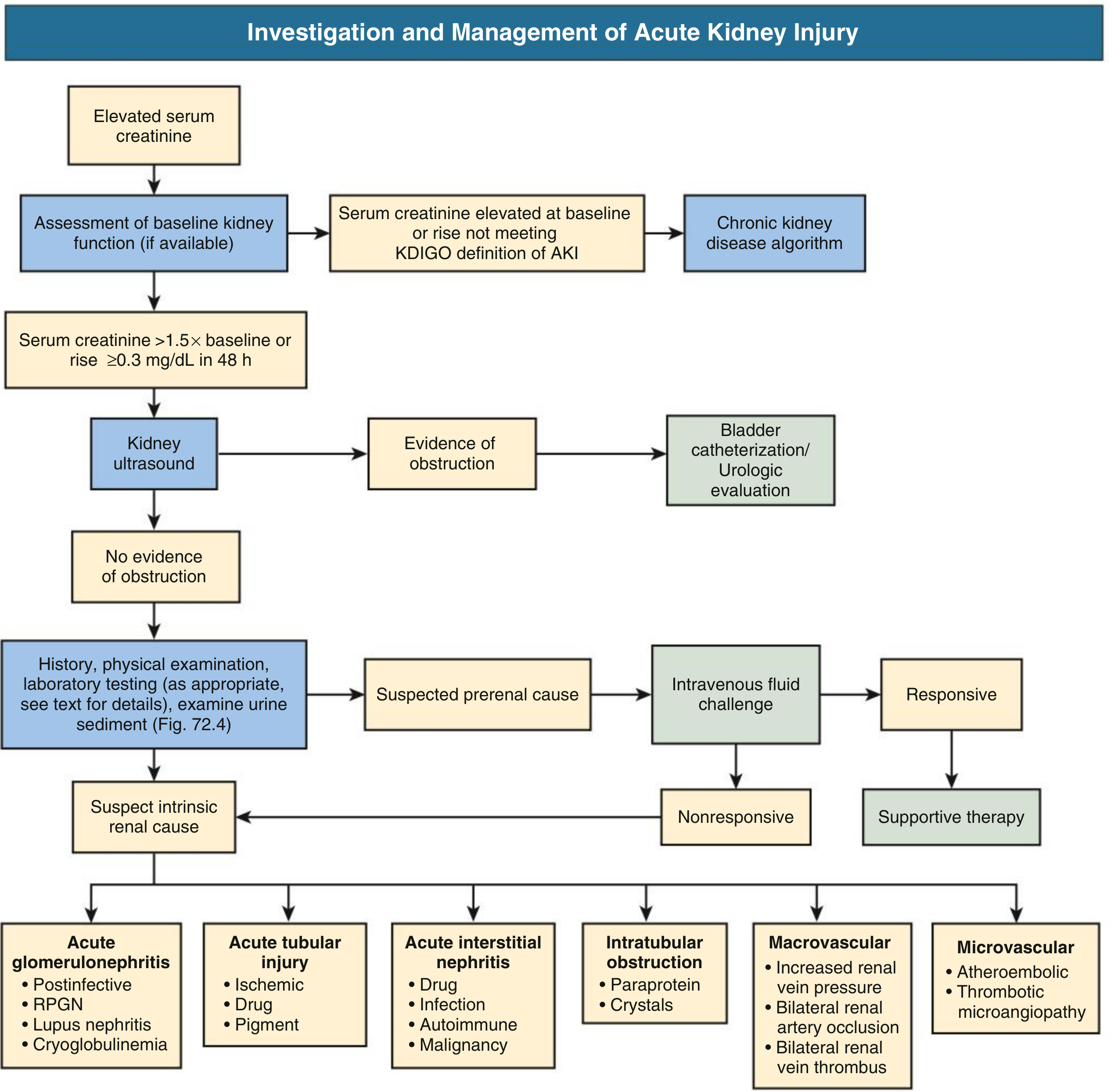
Fig. 72.2 - Comprehensive Clinical Nephrology, 7th Edition, p. 985
Key steps:
- Check baseline creatinine - if chronically elevated → CKD algorithm
- If creatinine >1.5× baseline or rises ≥0.3 mg/dL in 48 h → AKI confirmed
- Renal ultrasound to exclude obstruction
- History + physical + urine sediment to identify AKI cause
- If prerenal suspected → IV fluid challenge
- If intrinsic cause suspected → classify (ATN, AIN, GN, vascular)
- Biopsy if cause remains unclear after 2-3 weeks
7. Kidney Biopsy
Indicated in AKI when:
- Cause is not obvious
- Prolonged course >2-3 weeks without recovery
- AKI accompanied by heavy proteinuria and/or persistent hematuria
- Suspected GN, vasculitis, or systemic disease
- Biopsy needed to guide specific therapy (immunosuppression)
Biopsy in CKD: More dangerous when kidneys are small; findings often show non-specific end-stage changes (interstitial fibrosis, tubular atrophy).
8. Novel Biomarkers
These help identify AKI earlier than creatinine (which rises late):
| Biomarker | Role |
|---|---|
| NGAL (neutrophil gelatinase-associated lipocalin) | Rises within 2 hours of kidney injury; serum or urine |
| KIM-1 (kidney injury molecule-1) | Urine tubular injury marker |
| Cystatin C | More sensitive than creatinine for GFR estimation |
| IGFBP7 × TIMP-2 | Cell cycle arrest markers; predict AKI 12 hours before creatinine rise |
9. AKI on CKD (the third entity)
This is arguably the most clinically challenging scenario.
Definition
AKI superimposed on pre-existing CKD. The patient has a chronically impaired kidney that sustains an acute insult.
Why it matters
- CKD is one of the strongest risk factors for developing AKI
- Recovery is worse - the "creatinine return to baseline" standard breaks down when baseline itself is already reduced
- Risk of non-recovery and progression to end-stage kidney disease (ESKD) is substantially higher
Clues to AKI on CKD
- Known prior CKD (eGFR <60 for >3 months) with acute additional rise in creatinine
- Small kidneys on ultrasound (confirming CKD) + acute precipitant (confirming AKI component)
- Both anaemia of CKD (normocytic) AND muddy brown casts (ATN)
- Both elevated PTH AND evidence of an acute nephrotoxin/haemodynamic insult
- FENa has reduced utility in this setting - interpret with caution
- Background proteinuria from CKD + new oliguria from AKI
Risk Factors for CKD After AKI (Box 73.2, Comprehensive Clinical Nephrology 7th Ed.)
- Age
- Low baseline eGFR
- Congestive heart failure
- Hypertension
- Recurrent AKI episodes
- Low serum albumin during hospitalisation
- Severity of AKI (stage 3, need for dialysis)
- Low eGFR at hospital discharge
10. Summary Comparison Table
| Feature | AKI | CKD | AKI on CKD |
|---|---|---|---|
| Onset | Hours to days | Months to years | Acute rise on chronic baseline |
| Prior creatinine | Normal | Chronically elevated | Chronically elevated with new jump |
| Kidney size | Normal/enlarged | Small, echogenic | Small (CKD) + edematous |
| Anaemia | Absent or acute | Normocytic (EPO deficiency) | Normocytic + may worsen acutely |
| PTH | Normal | Elevated | Elevated |
| Urine casts | Muddy brown, RBC, WBC casts | Broad waxy casts | Mixed |
| FENa | <1% (prerenal), >1% (ATN) | Not typically used | Limited utility |
| Reversibility | Potentially fully reversible | Not reversible | Partial recovery; high ESKD risk |
| Ultrasound | Normal/enlarged kidneys | Small, scarred kidneys | Small kidneys + may show obstruction |
| PTH/bone disease | Absent | Present | Present |
| Biopsy | If cause unclear | If diagnosis unclear | Higher risk; may show combined picture |
Sources:
- Comprehensive Clinical Nephrology, 7th Edition, Chapter 72 - AKI Diagnosis and Chapter 73 - AKI Outcomes, pp. 985-999
- National Kidney Foundation Primer on Kidney Diseases, 8th Edition, Chapter on Imaging in AKI
- Brenner and Rector's The Kidney, 2-Volume Set - AKI Markers and AKI-CKD Transition
- Recent reviews: AKI-to-CKD transition mechanisms (PMID: 39298548, Am J Physiol Renal Physiol 2024) and AKI-CKD transition update (PMID: 38653563, Yonsei Med J 2024)
This is a shared conversation. Sign in to Orris to start your own chat.