
Heart
Reading File
The query "Heart" is broad. Let me clarify what you're looking for before diving in.
Asking for Preferences
What aspect of the heart are you interested in?
The heart is a four-chambered muscular pump in the mediastinum, divided into right and left sides separated by the septum. It has specialized chambers (atria and ventricles), valves (tricuspid, pulmonary, mitral, aortic), a conduction system, and its own blood supply via the coronary arteries.
Searching sources...
Finding Sources
Finding Sources
Finding Sources
Finding Sources
Good - there are anatomy and cardiology books. Let me search those directly.
Finding Sources
Finding Sources
Reading File
Reading File
Reading File
Reading File
Reading File
Loading Image
Loading Image
Reading File
I now have rich textbook content from two authoritative anatomy sources. Here is a comprehensive overview of heart anatomy and structure.
Heart Anatomy & Structure
Location and Orientation
The heart is a fibromuscular, hollow organ with a rounded, conical (pyramidal) shape. It sits in the mediastinum of the thorax, positioned obliquely so that:
- The apex points forward, downward, and to the left - lying deep to the left 5th intercostal space, 8-9 cm from the midsternal line
- The base (posterior surface) faces right, upward, and posteriorly
- One-third of the heart is to the right of the midline; two-thirds are to the left
- Gray's Anatomy for Students, p. 226; Color Atlas of Human Anatomy Vol 2, p. 37
Surfaces
| Surface | Formed by |
|---|---|
| Anterior (sternocostal) | Mostly right ventricle; some right atrium (right), some left ventricle (left) |
| Diaphragmatic (inferior) | Left ventricle + small portion of right ventricle |
| Left pulmonary | Left ventricle + portion of left atrium |
| Right pulmonary | Right atrium |
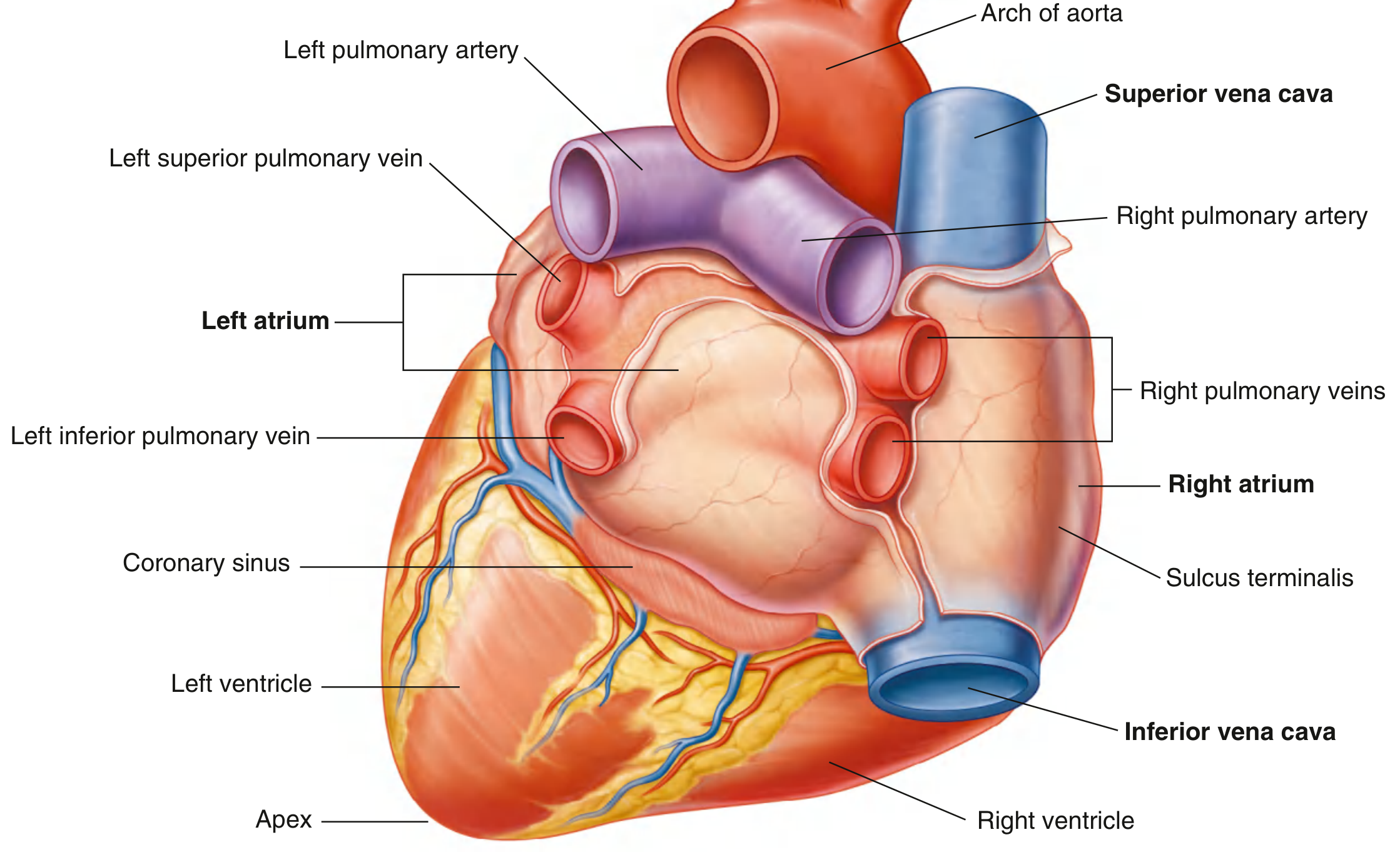
| Base (posterior) | Left atrium + small portion of right atrium + proximal great veins |
Radiological borders (PA chest X-ray):
- Right border: Superior vena cava, right atrium, inferior vena cava
- Left border: Aortic arch, pulmonary trunk, left auricle, left ventricle
- Inferior border: Right ventricle and left ventricle at apex
- Gray's Anatomy for Students, p. 228
External Grooves (Sulci)
External grooves on the heart surface correspond to internal partitions:
- Coronary sulcus - circles the heart, separating atria from ventricles; contains the coronary vessels and adipose tissue
- Anterior interventricular sulcus - on the sternocostal surface between the two ventricles; contains the anterior interventricular artery (LAD) and vein
- Posterior interventricular sulcus - on the diaphragmatic surface between ventricles; contains the posterior interventricular artery and middle cardiac vein
The Four Chambers
Right Atrium
- Consists of two parts: a smooth-walled posterior sinus (receives the superior and inferior venae cavae) and the true atrium (with pectinate muscles projecting into the cavity)
- The crista terminalis is a muscular ridge separating the smooth and trabeculated portions
- The fossa ovalis (remnant of the fetal foramen ovale) is present on the interatrial septum
- The coronary sinus opens into the right atrium, draining most venous blood returning from the heart wall itself
Right Ventricle
- Has thin walls relative to the left ventricle
- Divided into an inflow tract (posterior) and outflow tract (conus arteriosus/infundibulum, anterior) by two muscular ridges: the supraventricular crest and the septomarginal trabecula
- Contains trabeculae carneae (muscular ridges) in the inflow tract
- Blood exits through the pulmonary valve into the pulmonary trunk
Left Atrium
- Receives four pulmonary veins (two on each side) carrying oxygenated blood from the lungs
- Smooth-walled posteriorly; has a small left auricle anteriorly
Left Ventricle
- Has thick walls (approximately 3x thicker than the right ventricle) to generate the higher pressures needed for systemic circulation
- Has an inflow portion and an outflow portion (aortic vestibule)
- The apex of the heart is formed by the inferolateral part of the left ventricle
- Contains two large papillary muscles (anterior and posterior) supporting the mitral valve
- Color Atlas of Human Anatomy Vol 2, pp. 43-50
Valves
Atrioventricular Valves (close during systole)
- Tricuspid valve (right side): 3 leaflets - anterior (largest), posterior, and septal cusps. Chordae tendineae anchor leaflets to 3 papillary muscles, preventing prolapse.
- Mitral (bicuspid) valve (left side): 2 leaflets - anterior and posterior cusps. Supported by anterior and posterior papillary muscles.
Semilunar Valves (open during systole)
- Pulmonary valve: 3 semilunar cusps (anterior, right, left). Located between the right ventricular infundibulum and pulmonary trunk.
- Aortic valve: 3 semilunar cusps (posterior, right, left). Located between the left ventricular outflow tract and aorta. The coronary arteries arise from the aortic sinuses (sinuses of Valsalva) adjacent to the right and left cusps.
- Color Atlas of Human Anatomy Vol 2, pp. 55-56
Coronary Circulation
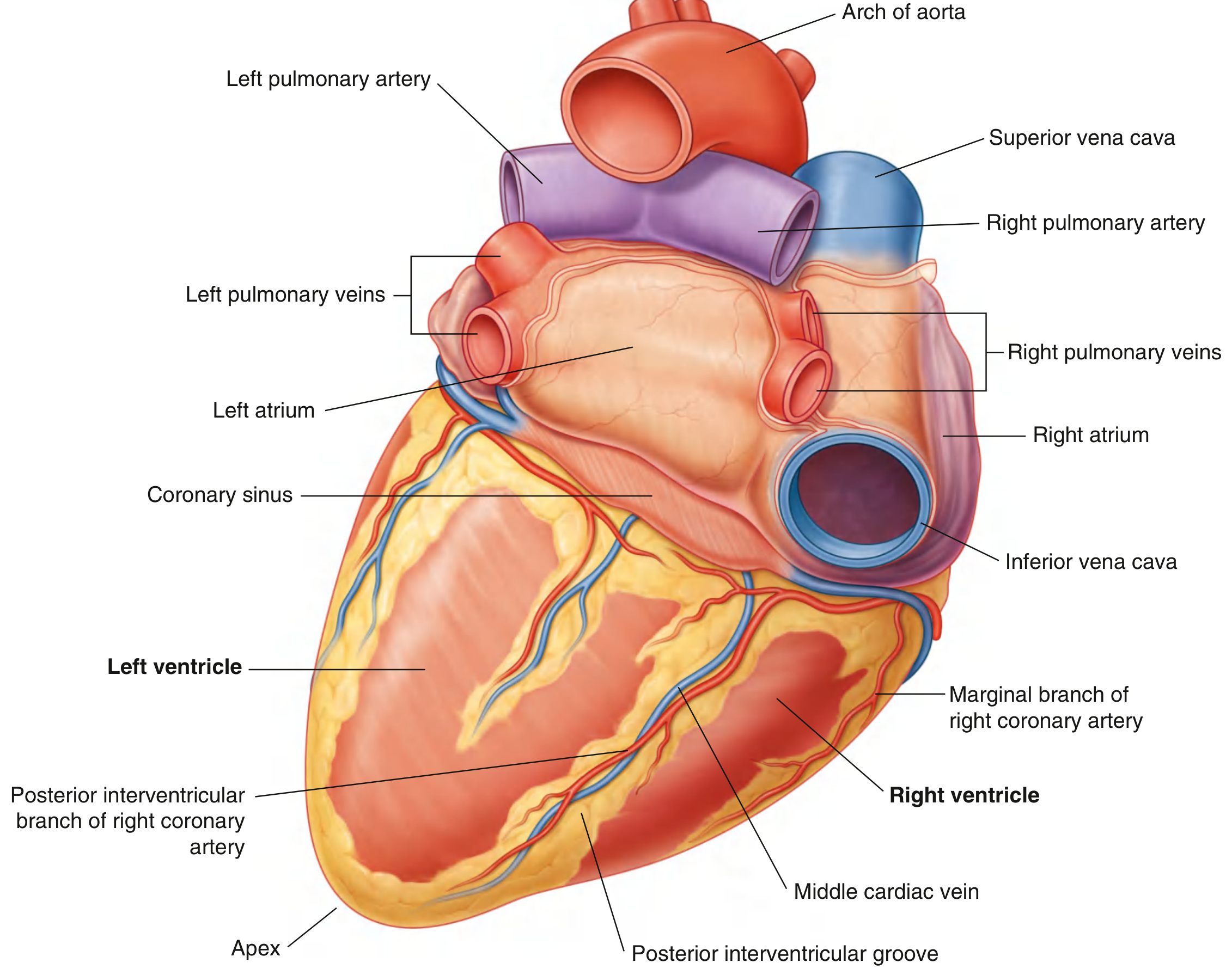
The coronary arteries are the first branches of the aorta, arising from the aortic sinuses:
Right Coronary Artery (RCA):
- Travels in the right coronary sulcus
- Gives off the right marginal artery, then continues posteriorly to become the posterior interventricular artery (PDA)
- Typically supplies: right atrium, most of the right ventricle, conducting system, posterior interventricular septum, diaphragmatic surface
Left Coronary Artery (LCA):
- Short main stem, then divides into:
- Anterior Interventricular Artery (LAD) - travels in the anterior interventricular sulcus; supplies the anterior left ventricle and anterior interventricular septum
- Circumflex artery - runs posteriorly in the left coronary sulcus; supplies the left atrium and posterior left ventricle
Coronary Veins: The great cardiac vein, middle cardiac vein, and small cardiac vein drain into the coronary sinus, which empties into the right atrium. The coronary sinus lies in the posterior part of the coronary sulcus.
Clinical note: Coronary arteries function as end arteries - their anastomoses are insufficient to develop adequate collateral circulation if one becomes occluded. Over 90% of acute myocardial infarctions result from coronary thrombosis on a ruptured atherosclerotic plaque. - Color Atlas of Human Anatomy Vol 2, p. 58
Layers of the Heart Wall
| Layer | Description |
|---|---|
| Epicardium | Visceral layer of the pericardium; outermost layer, contains coronary vessels in subepicardial fat |
| Myocardium | Middle muscular layer; cardiac muscle; thickest in left ventricle |
| Endocardium | Inner lining of the heart chambers; continuous with the intima of blood vessels |
The Pericardium
The heart is enclosed in the pericardium, a fibroserous sac consisting of:
- Fibrous pericardium (outer) - tough connective tissue anchoring the heart to the diaphragm and great vessels
- Serous pericardium - has a parietal layer (lines the fibrous pericardium) and a visceral layer (= epicardium). Between them is the pericardial cavity containing a thin film of serous fluid reducing friction during cardiac movement.
Sources: Color Atlas of Human Anatomy Vol 2 (Internal Organs); Gray's Anatomy for Students
This is a shared conversation. Sign in to Orris to start your own chat.