

Diagram of above mentioned name
"Meckel Diverticulum"[MeSH Terms]
Meckel diverticulum vitelline duct embryology anatomy authoritative
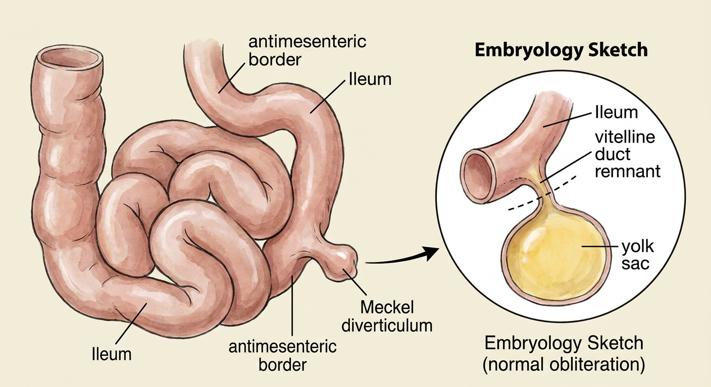
A clean, accurate labeled medical anatomy diagram of Meckel diverticulum in an infant. Show a simple loop of distal ileum, with a blind-ending true diverticulum arising from its antimesenteric border. Include an inset embryology sketch showing the vitelline (omphalomesenteric) duct from ileum to yolk sac normally obliterating, with persistence at the ileal end forming Meckel diverticulum. Use clear labels: Ileum, Meckel diverticulum, antimesenteric border, vitelline duct remnant, yolk sac. Educational textbook diagram.
Meckel diverticulum
Umbilical sinus, polyp, fistula diagram to draw
diagram persistent omphalomesenteric duct umbilical fistula umbilical sinus umbilical polyp
"Omphalomesenteric Duct" AND umbilical
omphalomesenteric duct remnant umbilical sinus polyp fistula diagram
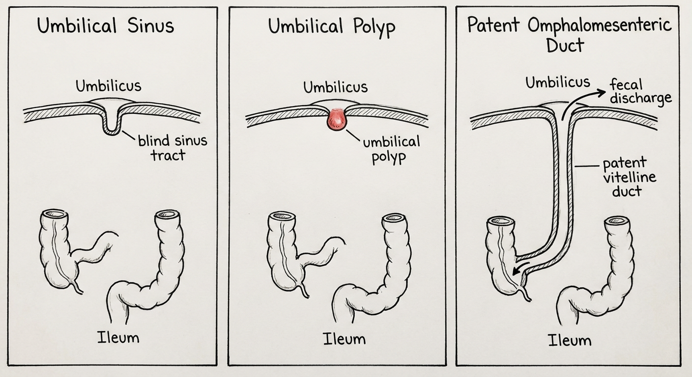
A clean, hand-drawable medical embryology exam diagram containing three separate labeled panels of omphalomesenteric (vitelline) duct remnants. Panel 1: Umbilical sinus, showing an ileal loop at bottom, a short blind-ending tract extending inward from the umbilicus but not reaching ileum. Panel 2: Umbilical polyp, showing a small red mucosal nodule at the umbilicus with no tract to ileum. Panel 3: Patent omphalomesenteric duct or umbilical-enteric fistula, showing a continuous open tract from ileum to umbilicus with arrow showing fecal discharge. Include one consistent orientation in every panel: umbilicus at top, ileum at bottom. Simple black line educational textbook illustration, labels only: Umbilicus, Ileum, blind sinus tract, umbilical polyp, patent vitelline duct, fecal discharge.
Vitelline duct remnants: diagrams to draw
-
Umbilical sinus
- Draw umbilicus at the top and ileum at the bottom.
- Draw a short blind tract going inward from the umbilicus.
- It does not reach the ileum.
- Label: Umbilical sinus, blind sinus tract, ileum.
-
Umbilical polyp
- Draw only a small rounded red/pink nodule at the umbilicus.
- No connection to ileum.
- Label: Umbilical polyp.
-
Umbilical fistula / patent vitelline duct
- Draw a continuous tube from umbilicus to ileum.
- Add an outward arrow at the umbilicus: fecal discharge.
- Label: Patent vitellointestinal (omphalomesenteric) duct / umbilical fistula.