
Dislocation of tmj
Reading File
Now let me search the medical library for TMJ dislocation content in parallel.
Finding Sources
Finding Sources
Searching PubMed
temporomandibular joint dislocation management
Good results. Now let me read the key sections from Rosen's Emergency Medicine and Tintinalli's (both excellent EM references) and S Das (surgery) in parallel.
Reading File
Reading File
Reading File
Excellent content. Let me now read the reduction technique section from Rosen's and the rest of Tintinalli's mandible dislocation content, plus fetch the anatomy image.
Reading File
Reading File
Loading Image
Let me also read more of the management and get the reduction diagram.
Reading File
Reading File
Loading Image
I now have comprehensive information from multiple authoritative sources. Let me compile the full answer.
Dislocation of the Temporomandibular Joint (TMJ)
Anatomy & Mechanism
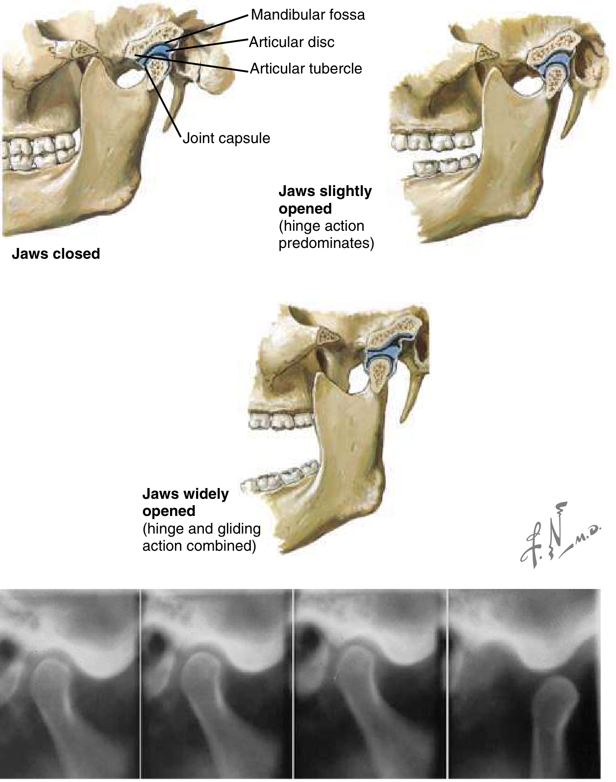
The TMJ is the articulation between the squamous portion of the temporal bone and the condyle of the mandible. It functions as both a hinge and a sliding (gliding) joint. With slight mouth opening, hinge action predominates; with wide opening, translational movement occurs as the articular disc and condyle complex slide anteriorly and inferiorly along the articular eminence.
Dislocation occurs when the condyle travels anteriorly past the articular eminence and becomes locked in the anterosuperior position. The masseter, internal pterygoid, and temporalis muscles then go into spasm attempting to close the mouth - this trismus prevents spontaneous reduction.
Types of Dislocation
| Type | Mechanism | Notes |
|---|---|---|
| Anterior | Condyle moves in front of articular eminence | Most common |
| Posterior | Blow forcing condyle backward toward mastoid | Rare; condylar head may prolapse into external auditory canal |
| Lateral | Often associated with mandibular fracture | Condyle forced laterally then superiorly |
| Superior | Blow to partially open mouth forcing condyle upward | May cause cerebral contusion, facial nerve palsy, deafness |
Dislocations are usually bilateral but can be unilateral.
Predisposing Factors
- Anatomic disharmonies between the fossa and articular eminence
- Weakness or laxity of the joint capsule and temporomandibular ligaments
- Torn ligaments
- Dystonic drug reactions (e.g., antipsychotics)
- Prolonged wide mouth opening (dental procedures, endotracheal intubation, yawning, seizures)
- Prior dislocation (one episode strongly predisposes to recurrence)
Clinical Features
Bilateral anterior dislocation:
- Mouth is open and fixed (cannot close)
- Prognathous deformity (jaw thrust forward)
- Difficulty speaking and swallowing
- Preauricular depression visible and palpable
- Pain anterior to the tragus
Unilateral anterior dislocation:
- Jaw deviates to the opposite side (key sign)
- Partially open mouth
- A small hollow can be felt just behind the dislocated condyle
S. Das clinical test: Insert the little finger into the external ear with the pulp directed forward - on the dislocated side, condylar movement will not be felt when the patient attempts to open and close the mouth.
Differential Diagnosis
- Mandibular fracture
- Traumatic hemarthrosis
- Acute closed locking of the TMJ meniscus (disc displacement without reduction)
- TMJ dysfunction
- Trismus from odontogenic causes (wisdom tooth, dental abscess, parotitis)
- Tetanus (risus sardonicus)
Imaging
- Straightforward atraumatic dislocation: No imaging required - diagnosis is clinical
- Traumatic dislocation: Panoramic view (Panorex) or CT of facial bones to exclude fracture before manipulation
- CT is the preferred ED modality as Panorex is usually unavailable
Reduction Techniques
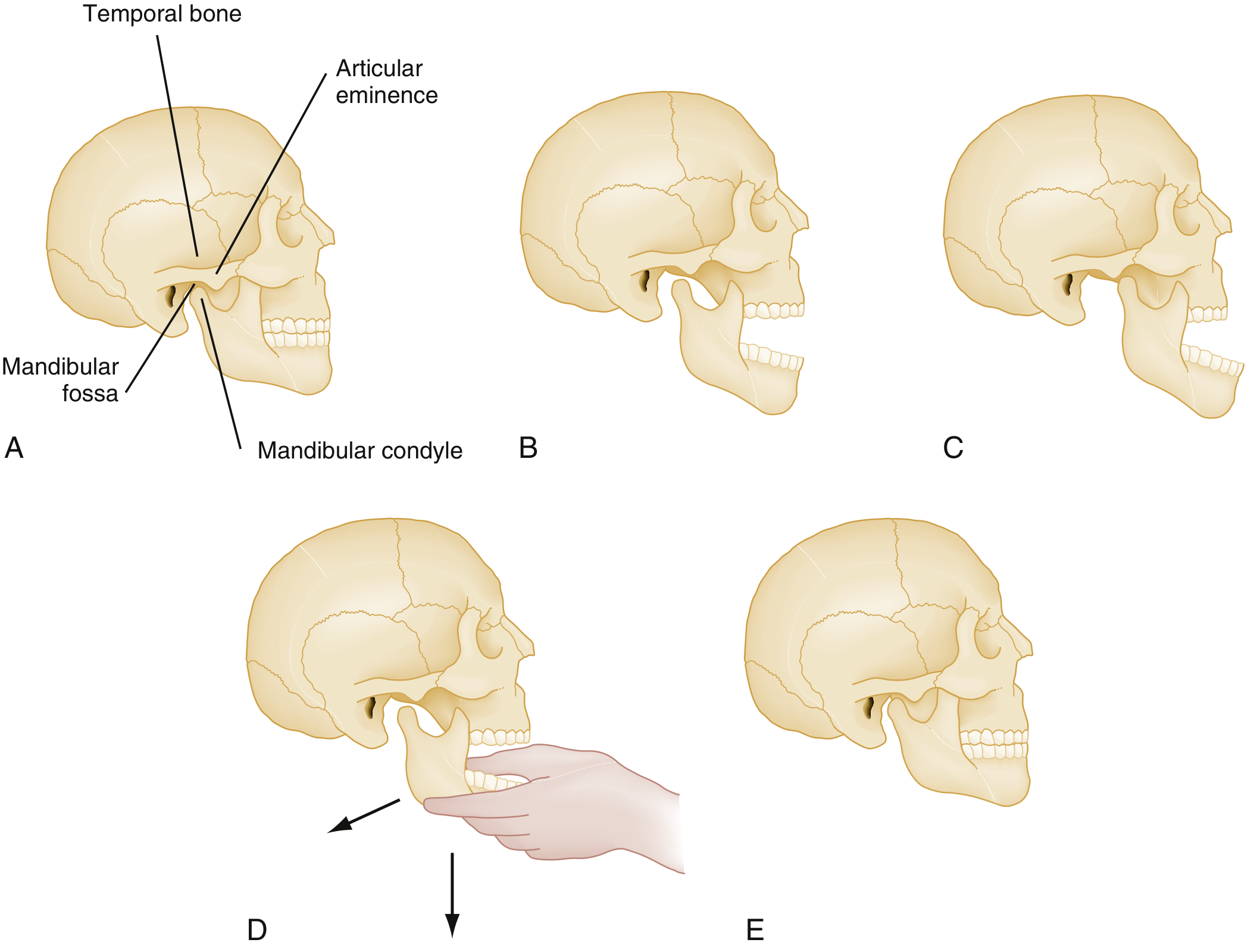
Pre-procedure: Procedural sedation/analgesia is often necessary to overcome masseter spasm. Options:
- IV short-acting muscle relaxant (e.g., midazolam)
- Local anesthetic: 2 mL of 2% lidocaine injected into the preauricular depression just anterior to the tragus using a 21-gauge needle
1. Conventional Method (Intraoral)
- Patient seated firmly with head against a wall
- Wrap thumbs in gauze (protection from snapping jaw on reduction)
- Place thumbs over the occlusal surfaces of the lower molars, as far back as possible
- Fingers curl beneath the angle and body of the mandible
- Apply downward then backward pressure with the thumbs
- When the condyle clears the eminence, masseter contraction returns it to the fossa
2. Wrist Pivot Method
- Patient and operator both seated
- Thumbs placed on the mentum (chin) applying upward force
- Fingers apply downward force on lower molars, forcing the mandibular body inferiorly
- Flexing the wrist rotates the mandible, the condyle moves inferiorly and slips back into the fossa
- A single RCT of 90 patients found the wrist pivot method had the highest success rate (96.7%) vs. conventional intraoral (86.7%) vs. extraoral (66.7%) - (Tintinalli's Emergency Medicine)
3. Extraoral Method
- Operator stands in front of seated patient
- Thumb placed on the cheek over the mandibular ramus/coronoid process
- Apply persistent posterior pressure; fingers behind the angle of the mandible stabilize the grip
- Contralateral hand pulls the angle anteriorly - this further dislocates the ipsilateral side and facilitates contralateral reduction
- Once one side reduces, the other usually follows spontaneously
4. Syringe Technique (Hands-Free)
- A 5-10 mL syringe placed between the posterior upper and lower molars
- Patient gently bites down and rolls the syringe back and forth
- The syringe acts as a rolling fulcrum to help the condyle slip back into position
- Useful for nontraumatic acute dislocations
Post-Reduction Care
- Patient should be able to close the mouth immediately after successful reduction
- Post-reduction radiographs only needed if difficult procedure, traumatic, or significant residual pain
- Soft diet for 1 week
- Avoid extreme mouth opening (yawning, wide laughing) for 2 weeks - patient should support the mandible with a hand when yawning
- Limit mouth opening to <2 cm for 2 weeks
- Warm compresses to the TMJ area
- NSAIDs and muscle relaxants
- Barton bandage (elastic bandage wrapping over head and mandible) for chronic/recurrent cases
- Elective referral to oral maxillofacial surgeon
Disposition & Referrals
Discharge home: Successfully reduced uncomplicated dislocations
Urgent specialty consultation (oral maxillofacial surgeon/ENT):
- Irreducible dislocation
- Open dislocation
- Superior dislocation
- Dislocation with associated fracture
- Dislocation with nerve injury
- Chronic recurrent dislocation (may require surgical intervention - e.g., eminectomy, capsulorrhaphy, or sclerosing injections)
Complications of Reduction
- Iatrogenic mandibular fracture
- Avulsion of articular cartilage
- Operator thumb injury from snapping jaw (protect with gauze)
Sources: ROSEN's Emergency Medicine, 9th ed. - Tintinalli's Emergency Medicine - S Das: A Manual on Clinical Surgery, 13th ed.
This is a shared conversation. Sign in to Orris to start your own chat.