

1000071044.jpg
Micro
Reading File
The image shows a Microbiology notes page titled HERPES VIRUSES with 11 topics. Let me search the textbook library for all these topics simultaneously.
Finding Sources
Finding Sources
Finding Sources
Finding Sources
Finding Sources
Finding Sources
I have good sources. Let me now read the most relevant microbiology-focused sections covering herpes virus classification, HSV infections, VZV, CMV, EBV, and Roseola.
Reading File
Reading File
Reading File
Reading File
Finding Sources
Finding Sources
Excellent - Jawetz Melnick is the core microbiology textbook. Let me read the VZV pathogenesis section, Ramsay Hunt section, and search for CMV/EBV/Roseola from Jawetz.
Reading File
Reading File
Reading File
Reading File
Reading File
Reading File
Reading File
Reading File
Reading File
Reading File
Finding Sources
Reading File
Here is a comprehensive answer to all 11 topics on your Herpes Viruses notes page, drawn from Jawetz Melnick & Adelberg's Medical Microbiology, Tintinalli's Emergency Medicine, Cummings Otolaryngology, and Sherris & Ryan's Medical Microbiology.
HERPES VIRUSES - Complete Notes
1. Classification of Herpes Viruses
Herpesviruses are large, enveloped, double-stranded DNA viruses. They are classified into 3 subfamilies based on cell tropism, site of latency, and replication characteristics:
| Subfamily | Virus | Common Name |
|---|---|---|
| Alphaherpesvirinae | HSV-1, HSV-2 | Herpes simplex 1 & 2 |
| VZV (HHV-3) | Varicella-zoster virus | |
| Betaherpesvirinae | CMV (HHV-5) | Cytomegalovirus |
| HHV-6A, HHV-6B | Roseola (6B) | |
| HHV-7 | Roseola (minor) | |
| Gammaherpesvirinae | EBV (HHV-4) | Epstein-Barr virus |
| KSHV (HHV-8) | Kaposi sarcoma-associated herpesvirus |
Key shared properties of all herpesviruses:
- dsDNA, icosahedral capsid, enveloped
- Establish lifelong latency after primary infection
- Can undergo reactivation (symptomatic or asymptomatic)
- All produce intranuclear inclusion bodies (Cowdry type A)
2. Infections Caused by HSV-1 and HSV-2
Both serotypes are morphologically and clinically indistinguishable. Incubation period: 2-14 days.
HSV-1 infections (typically orofacial):
- Herpes labialis ("cold sores") - vesicles at vermillion border
- Primary herpetic gingivostomatitis - erythematous, bleeding gums; painful ulcers throughout oral cavity
- Herpetic keratoconjunctivitis - dendritic corneal ulcers (ophthalmic emergency)
- Herpes encephalitis - most common cause of sporadic fatal encephalitis; involves temporal lobes
- Eczema herpeticum - extensive vesicular eruption in areas of pre-existing eczema; can be life-threatening; treatment = IV acyclovir 80 mg/kg/day q6h for 10 days
- Herpetic whitlow - infection of distal finger (autoinoculation from oral lesion)
HSV-2 infections (typically genital):
- Genital herpes - painful vesicles/ulcers on genitalia; recurrences common
- Neonatal herpes - contracted during delivery through infected birth canal (HSV-2 > HSV-1)
- Herpes meningitis (Mollaret's meningitis - recurrent benign lymphocytic meningitis)
- Sacral radiculopathy (urinary retention, constipation)
Both types cause:
- Asymptomatic viral shedding (important for transmission)
- CNS disease (encephalitis, meningitis)
- Disseminated disease in immunocompromised/neonates
3. Lab Diagnosis of HSV Infections
| Method | Details |
|---|---|
| Tzanck smear | Scrape base of vesicle; Giemsa/Wright stain; shows multinucleated giant cells with intranuclear inclusions (positive in both HSV and VZV - not specific) |
| Viral culture | Gold standard for active infection; cytopathic effect visible in 24-48 hours; HSV spreads rapidly in culture |
| PCR | Most sensitive; method of choice for CSF (herpes encephalitis), keratitis, and atypical cases |
| Direct fluorescent antibody (DFA) | Rapid; uses fluorescent-labeled anti-HSV antibodies on vesicle scrapings |
| Serology (ELISA) | Type-specific IgG antibodies (gG1 for HSV-1, gG2 for HSV-2) distinguish serotypes; useful for epidemiology, not acute diagnosis |
| Histopathology | Cowdry type A intranuclear inclusions; multinucleated giant cells |
For herpes encephalitis: CSF PCR for HSV DNA is the method of choice - highly sensitive and specific.
4. Pathogenesis of Chickenpox (Varicella)
VZV is inhaled and the following sequence occurs:
- Entry - VZV enters via mucosa of upper respiratory tract or conjunctiva
- Primary viremia - Virus replicates in regional lymph nodes → enters bloodstream
- Visceral replication - Replicates in liver and spleen → secondary viremia
- Secondary viremia - Infected mononuclear cells transport virus to skin
- Skin manifestations - Swelling of epithelial cells, ballooning degeneration, accumulation of tissue fluids → vesicle formation
- Latency - Virus gains access to trigeminal and dorsal root ganglia → lifelong latency
- Immune clearance - Humoral + cellular immunity + interferon terminate primary infection
Incubation period: 10-21 days
Rash characteristics: Successive crops - macules → papules → vesicles → crusts ("dewdrop on rose petal" vesicle on erythematous base); all stages present simultaneously; starts on trunk, spreads centrifugally; palms/soles spared.
Complications: Pneumonia (most common in adults), encephalitis, hepatitis, Reye syndrome (with aspirin use), bacterial superinfection (Staph/Strep).
5. Neonatal Varicella / Congenital Varicella
Neonatal varicella:
- Occurs when mother develops chickenpox in the perinatal period (5 days before to 2 days after delivery)
- Infant has no time to receive maternal IgG antibodies via placenta
- No passive immunity → severe, disseminated disease
- Can be fatal (pneumonia, hepatitis, DIC)
- Management: VZIG (Varicella-Zoster Immune Globulin) to the neonate immediately; IV acyclovir if disease develops
Congenital varicella syndrome (CVS):
- Occurs when mother has chickenpox during 1st-2nd trimester (especially weeks 8-20)
- Features (LIMB mnemonic):
- Limb hypoplasia (scarring, atrophy of limbs)
- Intrauterine growth retardation
- Microcephaly, cortical atrophy, mental retardation
- Body - skin scarring (cicatrix in dermatomal distribution), eye anomalies (chorioretinitis, cataracts, microphthalmia)
- Risk of CVS is ~2% if primary infection occurs in first 20 weeks
6. Ramsay Hunt Syndrome
Definition: Reactivation of VZV in the geniculate ganglion of the facial nerve (CN VII), producing a triad of:
- Otalgia (severe pain in and around the ear)
- Herpetic vesicles on the pinna and external auditory canal (herpes zoster oticus)
- Peripheral facial palsy (resembling Bell's palsy)
Additional features:
- SNHL (sensorineural hearing loss), tinnitus, and vestibular symptoms in up to 50% of patients
- Cranial nerves V, IX-XII may also be involved
- Incidence: ~30 cases per 100,000 persons
Pathophysiology: VZV latent in geniculate ganglion reactivates (usually with declining immunity) → inflammation of sensory AND motor cranial nerves
Diagnosis: Usually clinical; confirmed by VZV PCR from vesicular fluid or MRI (enhancement along intratemporal facial nerve)
Treatment:
- Oral corticosteroids (proven benefit by Cochrane review)
- Oral antiviral (acyclovir/valacyclovir) - data less certain but routinely given
- Combination therapy recommended in most guidelines
7. Lab Diagnosis of Chickenpox (VZV)
| Method | Details |
|---|---|
| Clinical diagnosis | Usually sufficient based on characteristic rash (dewdrop vesicles, multiple stages simultaneously, centrifugal spread) |
| Tzanck smear | Multinucleated giant cells with intranuclear inclusions; does NOT distinguish VZV from HSV |
| Direct fluorescent antibody (DFA) | More specific than Tzanck; detects VZV antigen in vesicle scrapings |
| PCR | Most sensitive and specific; method of choice for atypical/immunocompromised cases; can be done on vesicular fluid, CSF, blood |
| Viral culture | Technically difficult - VZV remains cell-associated, spreads slowly; rarely done |
| Serology - FAMA test | Fluorescent Antibody to Membrane Antigen - gold standard for detecting VZV immunity; checks for past infection/vaccination; not readily available |
| ELISA / complement fixation | Can detect rising antibody titers; useful for confirming immunity |
8. Immunoprophylaxis of Chickenpox
Active immunization:
- Live attenuated varicella vaccine (Oka strain): Approved in USA since 1995; 2 doses recommended (12-15 months and 4-6 years); produces immunity lasting at least 20 years; also available as MMRV (measles-mumps-rubella-varicella)
- Zoster vaccine (Shingrix - recombinant): For adults >50 years; highly effective in preventing shingles and postherpetic neuralgia; 2 doses given 2-6 months apart
- Old live zoster vaccine (Zostavax): For adults >60 years; being phased out
Passive immunization:
- Varicella-Zoster Immune Globulin (VariZIG): For post-exposure prophylaxis in high-risk seronegative individuals (immunocompromised, neonates, pregnant women); administered within 10 days of exposure; does not prevent infection but modifies severity
- Standard immune globulin (IVIG): Low titer of VZV antibodies; generally not effective as VZV prophylaxis
High-risk groups requiring prophylaxis: Immunocompromised patients, seronegative pregnant women, neonates of mothers with perinatal varicella, premature infants.
9. Infections Caused by Cytomegalovirus (CMV)
CMV (HHV-5) is the largest human herpesvirus (240 kbp genome). Infections depend heavily on host immune status:
In normal (immunocompetent) hosts:
- Usually asymptomatic (most common)
- CMV mononucleosis - heterophil-negative mononucleosis; fever, malaise, fatigue, mild hepatitis; self-limited; accounts for 20-50% of heterophil-negative mono cases; distinguish from EBV by absence of heterophil antibodies, exudative pharyngitis, lymphadenopathy less prominent
In immunocompromised hosts:
- CMV pneumonia (interstitial pneumonitis) - most common in bone marrow transplant recipients
- CMV colitis - bloody diarrhea, abdominal pain
- CMV retinitis - "pizza pie" appearance on fundoscopy; progressive blindness if untreated; common in AIDS (CD4 <50)
- CMV hepatitis
- CMV encephalitis
- Post-transplant: leukopenia, obliterative bronchiolitis (lung Tx), graft atherosclerosis (heart Tx), renal allograft rejection
Congenital CMV (most common congenital viral infection - 1% of live births in USA):
- Most from primary maternal infection (1/3 transmit to fetus)
- Features of cytomegalic inclusion disease: intrauterine growth retardation, microcephaly, periventricular calcifications, jaundice, hepatosplenomegaly, thrombocytopenic purpura ("blueberry muffin baby"), chorioretinitis, sensorineural hearing loss
- Mortality ~20%; survivors often develop mental retardation, deafness, ocular defects
- Key pathology: "owl eye" intranuclear inclusions in massively enlarged cells
Treatment: Ganciclovir, valganciclovir (first-line); foscarnet, cidofovir (alternatives for resistance/toxicity)
10. Infections Caused by Epstein-Barr Virus (EBV)
EBV (HHV-4) infects B lymphocytes via CD21 (CR2 receptor - the C3d receptor). It is the most oncogenic human herpesvirus.
Infectious Mononucleosis ("kissing disease"):
- Incubation period: 30-50 days
- Transmission: oropharyngeal secretions (saliva)
- Classic triad: fever, sore throat (exudative pharyngitis), lymphadenopathy (especially posterior cervical)
- Splenomegaly (50-60%); hepatitis; maculopapular rash (especially with ampicillin/amoxicillin use)
- Atypical lymphocytes (large T cells reacting against EBV-infected B cells) = Downey cells
- Heterophil antibodies (agglutinate sheep/horse RBCs) - detected by monospot test
- Duration: 2-4 weeks (self-limited in normal hosts)
- Complications: splenic rupture, airway obstruction, hemolytic anemia, thrombocytopenia
EBV-associated malignancies:
- Burkitt lymphoma (B cell; jaw tumor in African children; >90% contain EBV DNA; associated with t(8;14) c-myc translocation + malaria as cofactor)
- Nasopharyngeal carcinoma (common in Chinese/Southeast Asian males; EBNA1 + LMP1 expressed)
- Hodgkin and non-Hodgkin lymphoma
- Post-transplant lymphoproliferative disorder (PTLD) in immunosuppressed
Lab diagnosis:
- Monospot test (heterophil agglutination)
- EBV-specific serology: VCA-IgM (current infection), VCA-IgG (past infection/immunity), EA antibodies (active replication), EBNA antibodies (appear weeks after acute infection; persist for life)
- Atypical lymphocytosis on CBC
11. Roseola Infantum (Exanthem Subitum)
Causative agent: Primarily HHV-6B (Human Herpesvirus 6B); less commonly HHV-7
Age group: Infants and children 6 months to 4 years
Alternative name: Exanthem subitum = "sudden rash"
Clinical course (classic):
- Phase 1 (3-5 days): Abrupt onset of high fever (39-40°C), irritability; child looks relatively well despite high fever; may cause febrile convulsions (seizures)
- Defervescence: Fever drops suddenly
- Phase 2 (24-48 hours): Macular/maculopapular rash appears on trunk and spreads centrifugally; faint and transient; disappears within 1-3 days
Key features:
- Leukopenia during febrile phase
- Rash appears as fever resolves (pathognomonic pattern)
- Usually mild and self-limited
- No specific treatment; symptomatic (antipyretics, hydration)
- Diagnosis is clinical
Mechanism: HHV-6B primary infection in early childhood; virus establishes latency in monocytes/macrophages and salivary glands.
Sources: Jawetz Melnick & Adelberg's Medical Microbiology 28th Ed, Tintinalli's Emergency Medicine, Cummings Otolaryngology Head & Neck Surgery, Sherris & Ryan's Medical Microbiology 8th EdAll 11 topics from your Herpes Viruses notes are covered above. Here is a quick orientation to the notes:
Quick Summary of Key Exam Points:
- Classification - Alpha (HSV-1/2, VZV), Beta (CMV, HHV-6/7), Gamma (EBV, KSHV)
- HSV-1 vs HSV-2 - HSV-1 = orofacial (cold sores, gingivostomatitis, encephalitis); HSV-2 = genital herpes + neonatal herpes
- Lab Dx of HSV - PCR (best), culture, Tzanck smear (multinucleated giant cells), DFA, type-specific serology
- VZV pathogenesis - Respiratory entry → primary viremia (lymph nodes) → secondary viremia (liver/spleen) → skin (vesicles); latency in dorsal root ganglia
- Neonatal vs Congenital varicella - Neonatal = perinatal exposure (severe, fatal); Congenital = 1st/2nd trimester (limb hypoplasia, microcephaly, cicatrix)
- Ramsay Hunt - VZV reactivation in geniculate ganglion; triad: otalgia + ear vesicles + facial palsy; treat with steroids + antivirals
- Lab Dx of VZV - Clinical diagnosis usually sufficient; FAMA test is gold standard for immunity; PCR for atypical cases; Tzanck smear
- Immunoprophylaxis VZV - Live attenuated vaccine (Oka strain, 2 doses); VariZIG for high-risk post-exposure
- CMV infections - Congenital (owl-eye inclusions, microcephaly, IUGR); CMV mono (heterophil-negative); immunocompromised: retinitis, pneumonia, colitis; treat with ganciclovir
- EBV infections - Infectious mono (fever + pharyngitis + lymphadenopathy + atypical lymphocytes + heterophil antibodies); associated with Burkitt lymphoma, NPC, Hodgkin's
- Roseola infantum - HHV-6B; ages 6mo-4yr; high fever 3-5 days → sudden defervescence → macular rash; febrile seizures possible; self-limited
Don't need the summery full explanation
Understood! All the detailed notes for all 11 topics are already written above — just scroll up and use them directly. No summary at the end next time. Let me know which topic you want to go deeper on or if you have more questions!
Give again I cant see above
HERPES VIRUSES - Complete Notes
1. Classification of Herpes Viruses
Herpesviruses are large, enveloped, double-stranded DNA viruses. Classified into 3 subfamilies:
| Subfamily | Virus | Common Name |
|---|---|---|
| Alphaherpesvirinae | HSV-1 (HHV-1) | Herpes simplex 1 |
| HSV-2 (HHV-2) | Herpes simplex 2 | |
| VZV (HHV-3) | Varicella-zoster virus | |
| Betaherpesvirinae | CMV (HHV-5) | Cytomegalovirus |
| HHV-6A, HHV-6B | Roseola (6B causes disease) | |
| HHV-7 | Roseola (minor cause) | |
| Gammaherpesvirinae | EBV (HHV-4) | Epstein-Barr virus |
| KSHV (HHV-8) | Kaposi sarcoma herpesvirus |
Key shared properties:
- dsDNA, icosahedral capsid, enveloped
- Establish lifelong latency after primary infection
- Can reactivate (symptomatic or asymptomatic)
- All produce intranuclear inclusion bodies (Cowdry type A)
- All alphaherpesviruses are neurotropic (latency in sensory ganglia)
2. Infections Caused by HSV-1 and HSV-2
Incubation period: 2-14 days. Both serotypes are clinically indistinguishable.
HSV-1 (orofacial predominance):
- Herpes labialis - "cold sores"; vesicles at vermillion border; reactivation from trigeminal ganglion
- Primary herpetic gingivostomatitis - erythematous bleeding gums, painful vesicles throughout oral cavity
- Herpetic keratoconjunctivitis - dendritic corneal ulcers; ophthalmic emergency
- Herpes encephalitis - most common cause of sporadic fatal encephalitis; temporal lobe involvement
- Eczema herpeticum - life-threatening vesicular eruption on pre-existing eczema skin; treat with IV acyclovir
- Herpetic whitlow - infection of distal finger by autoinoculation
HSV-2 (genital predominance):
- Genital herpes - painful vesicles/ulcers on genitalia; frequent recurrences
- Neonatal herpes - acquired during vaginal delivery; can be disseminated, CNS, or skin/eye/mouth (SEM) type
- Herpes meningitis - recurrent benign lymphocytic meningitis (Mollaret's)
- Sacral radiculopathy - urinary retention, constipation
Both types cause:
- Asymptomatic viral shedding (major source of transmission)
- Disseminated disease in immunocompromised/neonates
- Latency in sensory ganglia (trigeminal for HSV-1, sacral for HSV-2)
3. Lab Diagnosis of HSV Infections
| Method | Details |
|---|---|
| Tzanck smear | Scrape base of vesicle; Giemsa stain; shows multinucleated giant cells with intranuclear inclusions; does NOT distinguish HSV from VZV |
| Viral culture | Gold standard for active lesions; CPE visible in 24-48 hours; HSV spreads rapidly |
| PCR | Most sensitive and specific; method of choice for CSF (encephalitis), atypical cases |
| DFA (Direct Fluorescent Antibody) | Rapid; fluorescent-labeled antibodies on vesicle scrapings; distinguishes HSV-1 from HSV-2 |
| Type-specific serology (ELISA) | gG-1 antibody = HSV-1; gG-2 antibody = HSV-2; useful for epidemiology, not acute diagnosis |
| Histopathology | Cowdry type A intranuclear inclusions; multinucleated giant cells |
- For herpes encephalitis: CSF PCR is the method of choice
- For ocular herpes: Slit lamp + fluorescein staining shows dendritic ulcers
4. Pathogenesis of Chickenpox (Varicella)
Step-by-step:
- Entry - VZV inhaled; infects mucosal cells of upper respiratory tract/conjunctiva
- Primary viremia - Replication in regional lymph nodes → enters bloodstream
- Visceral replication - Virus replicates in liver and spleen
- Secondary viremia - Infected mononuclear cells transport virus to skin and respiratory mucosa
- Rash formation - Swelling of epithelial cells, ballooning degeneration, tissue fluid accumulation → vesicle formation
- Immune response - Humoral + cellular immunity + interferon terminate infection
- Latency - Virus establishes latency in trigeminal and dorsal root ganglia
- Reactivation - If immunity wanes → herpes zoster (shingles)
Incubation period: 10-21 days
Rash: Successive crops - macules → papules → vesicles → crusts; ALL stages present simultaneously; starts on trunk, spreads centrifugally; palms/soles spared; "dewdrop on rose petal" appearance
Complications: Pneumonia (most common in adults/immunocompromised), encephalitis, hepatitis, Reye syndrome (with aspirin), bacterial superinfection (Staph/Strep)
5. Neonatal Varicella / Congenital Varicella
Neonatal Varicella:
- Mother develops chickenpox 5 days before to 2 days after delivery
- Infant receives no protective maternal IgG (insufficient time for transplacental transfer)
- Leads to severe, disseminated disease - pneumonia, hepatitis, DIC; can be fatal
- Management: VariZIG to neonate immediately; IV acyclovir if disease develops
Congenital Varicella Syndrome (CVS):
- Mother infected during 1st-2nd trimester (highest risk: weeks 8-20)
- Risk of CVS: approximately 2% of primary infections in first 20 weeks
Features of CVS:
- Skin: cicatricial (zigzag) scarring in dermatomal distribution
- Limb: hypoplasia, atrophy
- Eye: chorioretinitis, cataracts, microphthalmia
- CNS: microcephaly, cortical atrophy, mental retardation, seizures
- IUGR (intrauterine growth retardation)
6. Ramsay Hunt Syndrome
Definition: Reactivation of VZV in the geniculate ganglion of CN VII
Classic Triad:
- Otalgia - severe pain in and around the ear
- Herpetic vesicles - on the pinna, external auditory canal (herpes zoster oticus)
- Peripheral facial palsy - lower motor neuron facial nerve palsy (resembles Bell's palsy)
Additional features:
- SNHL, tinnitus, and vestibular symptoms in up to 50% of patients
- Cranial nerves V, IX, X, XI, XII may also be affected
- Incidence: ~30 per 100,000 persons
Pathophysiology: VZV reactivates in geniculate ganglion → inflammation of sensory + motor cranial nerves → facial palsy + ear vesicles + pain
Diagnosis: Clinical; confirm by VZV PCR from vesicular fluid or MRI (enhancement along intratemporal facial nerve)
Treatment: Oral corticosteroids + oral antivirals (acyclovir/valacyclovir); combination therapy recommended
7. Lab Diagnosis of Chickenpox (VZV)
| Method | Details |
|---|---|
| Clinical diagnosis | Usually sufficient; characteristic rash (multiple stages simultaneously, centrifugal spread) |
| Tzanck smear | Multinucleated giant cells; does NOT distinguish VZV from HSV |
| DFA | More specific; detects VZV antigen in vesicle scrapings; distinguishes from HSV |
| PCR | Most sensitive and specific; method of choice for immunocompromised, atypical cases, CSF |
| Viral culture | Technically difficult; VZV is cell-associated, spreads slowly; rarely performed |
| FAMA test | Fluorescent Antibody to Membrane Antigen; gold standard for detecting VZV immunity (past infection or vaccine response) |
| ELISA / complement fixation | Rising antibody titers confirm infection; useful for immunity screening |
8. Immunoprophylaxis of Chickenpox
Active immunization:
- Live attenuated varicella vaccine (Oka strain): Approved 1995; 2 doses (12-15 months + 4-6 years); also available as MMRV
- Zoster vaccine - Shingrix (recombinant subunit): Adults ≥50 years; 2 doses 2-6 months apart; highly effective against shingles and postherpetic neuralgia
- Zoster vaccine - Zostavax (live attenuated): Adults ≥60 years; being phased out in favour of Shingrix
Passive immunization:
- VariZIG (Varicella-Zoster Immune Globulin): Post-exposure prophylaxis for high-risk seronegative individuals; given within 10 days of exposure; modifies but may not prevent infection
- Standard IVIG: Low VZV antibody titer; generally not effective for VZV prophylaxis
High-risk groups requiring VariZIG:
- Immunocompromised seronegative patients
- Seronegative pregnant women
- Neonates of mothers with perinatal varicella
- Premature infants <28 weeks
9. Infections Caused by Cytomegalovirus (CMV)
CMV (HHV-5) has the largest genome of all human herpesviruses (240 kbp). Key pathology: "owl eye" intranuclear inclusions in massively enlarged (cytomegalic) cells.
In immunocompetent hosts:
- Usually asymptomatic
- CMV mononucleosis - heterophil-negative mono; fever, malaise, fatigue, mild hepatitis; self-limited; accounts for 20-50% of heterophil-negative mono cases
In immunocompromised hosts (transplant, AIDS):
- CMV retinitis - "pizza pie" fundus; leading cause of blindness in AIDS (CD4 <50)
- CMV pneumonitis - interstitial pneumonia; most common in bone marrow transplant recipients
- CMV colitis - bloody diarrhea, abdominal pain
- CMV hepatitis, encephalitis
- Post-transplant complications: leukopenia, obliterative bronchiolitis (lung Tx), graft atherosclerosis (heart Tx)
Congenital CMV (most common congenital viral infection):
- ~1% of live births in USA; ~5-10% of those develop severe disease
- Features: microcephaly, periventricular calcifications, IUGR, jaundice, hepatosplenomegaly, thrombocytopenic purpura ("blueberry muffin baby"), chorioretinitis, SNHL
- Mortality ~20%; survivors: mental retardation, deafness, ocular defects
- Even subclinical congenital CMV → deafness in ~10%
Treatment: Ganciclovir / valganciclovir (first-line); foscarnet, cidofovir (resistance/toxicity)
10. Infections Caused by Epstein-Barr Virus (EBV)
EBV (HHV-4) infects B lymphocytes via CD21 receptor (CR2, the C3d complement receptor). Establishes latency in B cells.
Infectious Mononucleosis ("kissing disease" / "mono"):
- Incubation: 30-50 days
- Transmission: oropharyngeal secretions (saliva)
- Classic triad: Fever + Exudative pharyngitis + Lymphadenopathy (especially posterior cervical)
- Splenomegaly (50-60%), hepatitis, maculopapular rash (especially with ampicillin/amoxicillin)
- Atypical lymphocytes (Downey cells) on blood smear - large reactive T cells
- Heterophil antibodies (agglutinate sheep/horse RBCs) - detected by Monospot test
- Self-limited: 2-4 weeks
- Complications: splenic rupture, airway obstruction, hemolytic anemia, thrombocytopenia
EBV-associated malignancies:
- Burkitt lymphoma - B cell lymphoma; jaw tumor in African children; >90% contain EBV DNA; t(8;14) c-myc translocation; malaria = cofactor
- Nasopharyngeal carcinoma - epithelial cancer; common in Chinese/Southeast Asian males; EBNA1 + LMP1 expressed
- Hodgkin's lymphoma (mixed cellularity subtype)
- Post-transplant lymphoproliferative disorder (PTLD)
- Non-Hodgkin lymphoma in AIDS
Lab diagnosis:
- Monospot test (heterophil agglutination)
- EBV serology:
- VCA-IgM = current/acute infection
- VCA-IgG = past infection (persists for life)
- EA (early antigen) antibodies = active viral replication
- EBNA antibodies = appear weeks after acute infection, persist for life (absence = recent primary infection)
11. Roseola Infantum (Exanthem Subitum)
Causative agent: Primarily HHV-6B; less commonly HHV-7
Alternative name: Exanthem subitum = "sudden rash"
Age group: Infants and children 6 months to 4 years
Clinical course:
| Phase | Duration | Features |
|---|---|---|
| Febrile phase | 3-5 days | Abrupt high fever (39-40°C); irritable; febrile convulsions possible; leukopenia |
| Defervescence | Sudden | Fever drops abruptly |
| Rash phase | 24-48 hours | Faint macular/maculopapular rash on trunk → spreads outward; disappears in 1-3 days |
Key point: Rash appears as fever resolves - this pattern is pathognomonic
Features:
- Child appears relatively well despite high fever
- Febrile seizures are a recognized complication
- Leukopenia during febrile phase
- Virus establishes latency in monocytes/macrophages and salivary glands
Treatment: None specific; symptomatic only (antipyretics, hydration); self-limited
Sources: Jawetz Melnick & Adelberg's Medical Microbiology 28th Ed | Tintinalli's Emergency Medicine | Cummings Otolaryngology | Sherris & Ryan's Medical Microbiology 8th Ed
Plz in easy language tell about latency and reactivation and a picture of rash
Loading Image
Loading Image
Latency and Reactivation - Simple Language
🦠 What is Latency?
Think of it like this:
The virus enters your body, causes disease, then HIDES inside your nerve cells and goes to SLEEP.
- After the first infection (primary infection), your immune system fights the virus
- But the virus is smart - it hides inside nerve cells (neurons) where the immune system cannot kill it
- Inside the nerve cell, the virus stops replicating - it just sits there quietly doing nothing
- This "sleeping" state is called LATENCY
- The virus stays there FOR LIFE - you can never fully get rid of it
Where does each virus hide (latency site)?
| Virus | Hiding Place |
|---|---|
| HSV-1 | Trigeminal ganglion (nerve near face) |
| HSV-2 | Sacral ganglion (nerve near genitals) |
| VZV | Dorsal root ganglia + trigeminal ganglia |
| EBV | B lymphocytes |
| CMV | Monocytes / macrophages |
⚡ What is Reactivation?
When the sleeping virus "wakes up" and causes disease again.
This happens when something weakens your immune system:
| Trigger | Example |
|---|---|
| Stress | Exam stress, emotional stress |
| Fever / illness | Cold, flu |
| Sunlight (UV) | For HSV-1 cold sores |
| Immunosuppression | HIV, steroids, chemotherapy |
| Old age | Immunity weakens naturally |
| Menstruation | Hormonal changes |
| Surgery / trauma | Physical stress |
What happens during reactivation?
- Virus wakes up inside the nerve cell
- Starts replicating again
- Travels down the nerve to the skin
- Causes vesicles (blisters) in that nerve's area of skin
- This is why shingles appears in a stripe/band - follows one nerve!
Simple Analogy 🏠
Think of the virus like a criminal hiding in your house basement.
- Police (immune system) fought the criminal during the first attack
- Criminal couldn't escape so hid in the basement (nerve cell)
- When police are strong - criminal stays hidden (latency)
- When police are weak (stress/illness/old age) - criminal comes out and attacks again (reactivation)
Latency vs Reactivation - Quick Table
| Latency | Reactivation | |
|---|---|---|
| Virus state | Sleeping, not replicating | Active, replicating |
| Symptoms | None | Disease returns |
| Immune system | Strong, keeping virus in check | Weakened |
| Location | Inside nerve cell | Travels to skin/mucosa |
| Example | Person had chickenpox as child, no symptoms for 40 years | Same person gets shingles at age 60 |
Chickenpox Rash - Real Photo
This is the classic varicella (chickenpox) rash - notice it starts on the trunk first:
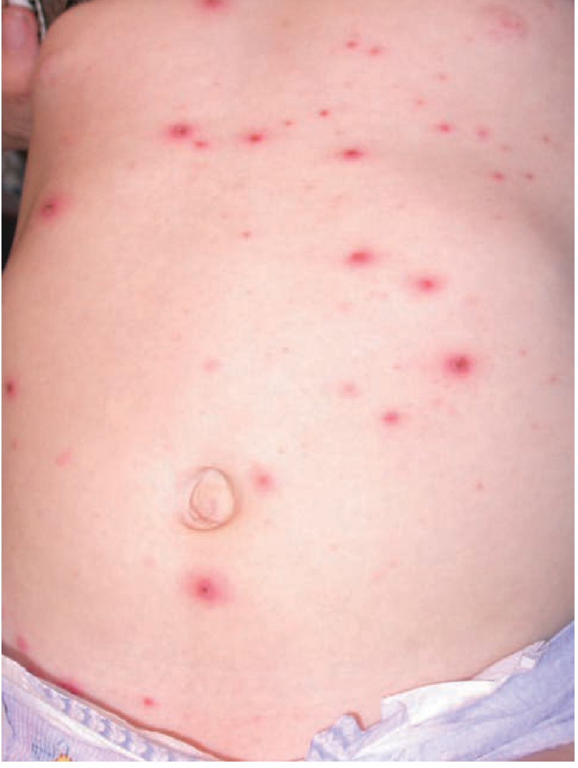
What you see in this photo:
- Multiple red spots scattered on the trunk (abdomen area)
- Different sizes - some small red dots, some larger with a central vesicle
- This is the early stage - spots start on trunk before spreading to face and limbs
- Later these become fluid-filled vesicles ("dewdrop on rose petal") then crust over
The Full Story of VZV - Primary → Latency → Reactivation
Child breathes in VZV
        ↓
Gets CHICKENPOX (primary infection)
        ↓
Immune system fights it - rash heals
        ↓
But virus HIDES in dorsal root ganglia (LATENCY)
        ↓
Person lives normally for decades
        ↓
At age 60-70, immunity weakens
        ↓
Virus WAKES UP (REACTIVATION)
        ↓
Travels down nerve to skin
        ↓
Causes SHINGLES (painful rash in a band/stripe)
Key exam point: The same VZV virus causes TWO different diseases:
- 1st time = Chickenpox (widespread rash, child)
- Reactivation = Shingles/Herpes Zoster (band-like painful rash, elderly/immunocompromised)
Bro give me lab diagnosis in step wise as in co bhaveha
Lab Diagnosis of HSV - Step by Step
🧪 SPECIMENS COLLECTED
First, decide what to collect based on the type of infection:
| Type of Infection | Specimen |
|---|---|
| Skin/genital lesion | Vesicle fluid, swab from base of ulcer |
| Encephalitis | CSF (cerebrospinal fluid) |
| Eye infection | Conjunctival swab |
| Neonatal herpes | Skin swab, blood, CSF |
| Oral lesion | Saliva / throat swab |
STEP 1 - DIRECT EXAMINATION (Fastest, done first)
🔬 Tzanck Smear
- Scrape the base of a fresh vesicle
- Smear on glass slide
- Stain with Giemsa or Wright stain
- See under microscope
What you see: Multinucleated giant cells with intranuclear inclusions
⚠️ This is POSITIVE in both HSV and VZV - cannot tell the difference between them
STEP 2 - VIRAL CULTURE (Gold Standard for active lesion)
- Take vesicle fluid or swab
- Inoculate into human diploid fibroblast cell line
- Incubate and observe for CPE (cytopathic effect)
What you see in culture:
- Rounding and clumping of cells
- CPE appears within 24-48 hours (HSV spreads fast in culture)
✅ Gold standard for active lesions ❌ Slow, not used for encephalitis
STEP 3 - PCR (Most Sensitive & Specific)
- Take specimen (CSF, vesicle fluid, swab)
- Amplify HSV DNA using PCR
Uses:
- Herpes encephalitis → CSF PCR = METHOD OF CHOICE
- Atypical cases, immunocompromised patients
- Can differentiate HSV-1 from HSV-2
✅ Most sensitive and specific ✅ Best for CNS infection ✅ Can be done even when culture is negative
STEP 4 - ANTIGEN DETECTION (Quick, Specific)
Direct Fluorescent Antibody (DFA)
- Smear vesicle scraping on slide
- Apply fluorescent-labeled anti-HSV antibodies
- View under fluorescent microscope
What you see: Bright apple-green fluorescence in infected cells
✅ Faster than culture ✅ Can distinguish HSV-1 from HSV-2 ✅ More specific than Tzanck
STEP 5 - SEROLOGY (For past infection / epidemiology)
- Take blood sample
- Detect HSV-specific antibodies by ELISA
| Antibody | Meaning |
|---|---|
| IgM | Current / recent infection |
| IgG (gG-1) | Past HSV-1 infection |
| IgG (gG-2) | Past HSV-2 infection |
⚠️ NOT useful for diagnosing acute active infection ✅ Useful for screening, epidemiology, partner counselling
STEP 6 - HISTOPATHOLOGY (Biopsy / Post mortem)
- Tissue biopsy stained with H&E
- See Cowdry type A intranuclear inclusions (eosinophilic inclusions surrounded by halo)
- Multinucleated giant cells
Used in encephalitis brain biopsy, post mortem cases
🗂️ SUMMARY TABLE
SPECIMEN COLLECTED
      ↓
┌─────────────────────────────────────────┐
│  STEP 1: Tzanck smear                   │
│  (Quick, cheap, not specific)           │
├─────────────────────────────────────────┤
│  STEP 2: Viral Culture                  │
│  (Gold standard - active skin lesion)   │
├─────────────────────────────────────────┤
│  STEP 3: PCR                            │
│  (Best for CSF/encephalitis)            │
├─────────────────────────────────────────┤
│  STEP 4: DFA                            │
│  (Rapid antigen detection)              │
├─────────────────────────────────────────┤
│  STEP 5: Serology                       │
│  (Past infection / epidemiology)        │
├─────────────────────────────────────────┤
│  STEP 6: Histopathology                 │
│  (Biopsy / post mortem)                 │
└─────────────────────────────────────────┘
🎯 WHICH TEST TO USE WHEN?
| Situation | Best Test |
|---|---|
| Fresh skin vesicle | Culture (gold standard) |
| Herpes encephalitis | CSF PCR |
| Quick bedside test | Tzanck smear |
| Distinguish HSV-1 vs HSV-2 | PCR or DFA |
| Check if person had past infection | Serology (IgG) |
| Neonatal herpes | PCR (blood + CSF + skin) |
| Eye herpes | Fluorescein staining (dendritic ulcer) + DFA |
🔑 EXAM POINTS
- Tzanck smear → multinucleated giant cells → positive in BOTH HSV and VZV
- Culture → gold standard for skin lesion → CPE in 24-48 hrs
- PCR → method of choice for encephalitis (CSF)
- DFA → rapid, distinguishes HSV-1 from HSV-2
- Cowdry type A inclusions → seen in histopathology
- Serology NOT useful for acute diagnosis
Lab diagnosis of chickenpox
Lab Diagnosis of Chickenpox (VZV) - Step by Step
🧪 SPECIMENS COLLECTED
| Type of Infection | Specimen |
|---|---|
| Skin lesion | Vesicle fluid, scraping from base of vesicle |
| Pneumonia | Bronchoalveolar lavage (BAL) |
| Encephalitis | CSF |
| Blood (viremia) | Blood / buffy coat |
| Congenital VZV | Amniotic fluid, neonatal blood |
⚠️ Important: Collect specimen from fresh vesicle (not crusted lesion - virus dies as vesicle crusts over)
STEP 1 - CLINICAL DIAGNOSIS (Most Common in Practice)
In a typical case, no lab test is needed - diagnosis is made clinically!
Clinical features that confirm diagnosis:
- Successive crops of rash (macule → papule → vesicle → crust)
- All stages present simultaneously on the body
- Starts on trunk → spreads to face and limbs
- Palms and soles spared
- "Dewdrop on rose petal" appearance of vesicle
- Mild fever, itching
✅ In a normal healthy child with classic rash → clinical diagnosis is enough ❌ Lab tests needed only in atypical cases, immunocompromised, or complications
STEP 2 - TZANCK SMEAR (Quick Bedside Test)
- Scrape the base of a fresh vesicle
- Smear on glass slide
- Stain with Giemsa stain
- View under microscope
What you see:
- Multinucleated giant cells
- Intranuclear inclusions (Cowdry type A)
⚠️ POSITIVE in both VZV and HSV - cannot distinguish between the two ✅ Quick, cheap, bedside test
STEP 3 - DIRECT FLUORESCENT ANTIBODY TEST (DFA)
- Scrape vesicle base
- Apply fluorescent-labeled anti-VZV antibodies on slide
- View under fluorescent microscope
What you see: Bright green fluorescence in VZV-infected cells
✅ More specific than Tzanck ✅ Distinguishes VZV from HSV (unlike Tzanck) ✅ Quick result (few hours)
STEP 4 - PCR (Most Sensitive & Specific)
- Take vesicle fluid / CSF / blood
- Amplify VZV DNA
Uses:
- Immunocompromised patients (atypical rash)
- VZV encephalitis / meningitis → CSF PCR
- Congenital varicella → amniotic fluid PCR
- Disseminated disease
✅ Most sensitive and specific ✅ Method of choice for CNS involvement ✅ Can confirm diagnosis when rash is atypical
STEP 5 - VIRAL CULTURE (Rarely Done)
- Inoculate vesicle fluid into human embryonic cell line
- Observe for CPE
What you see:
- Focal CPE (spreads slowly - unlike HSV)
- Intranuclear inclusion bodies
❌ Technically very difficult ❌ VZV is strongly cell-associated - very little free virus in fluid ❌ Spreads slowly (takes weeks) ❌ Rarely performed in clinical practice
STEP 6 - SEROLOGY (For Immunity Testing)
FAMA Test (Fluorescent Antibody to Membrane Antigen)
- Patient blood taken
- Detects VZV-specific antibodies against membrane antigens
✅ Gold standard for detecting VZV immunity ✅ Used to check if vaccine worked ✅ Used for post-exposure screening (is this person immune?) ❌ Not readily available everywhere
ELISA / Complement Fixation
- Detects rising antibody titers (compare acute + convalescent serum)
- 4-fold rise in titer = confirms recent infection
✅ Widely available ✅ Useful for confirming past infection or immunity
What Serology Tells You:
| Finding | Meaning |
|---|---|
| IgM positive | Active / recent infection |
| IgG positive | Past infection or vaccinated (immune) |
| IgG negative | Susceptible (not immune) |
| Rising IgG titer (4-fold) | Confirms recent primary infection |
STEP 7 - HISTOPATHOLOGY (Biopsy)
- Skin biopsy stained with H&E
- See Cowdry type A intranuclear inclusions
- Multinucleated giant cells
- Ballooning degeneration of epidermal cells
Used in atypical cases, immunocompromised, or post mortem
🗂️ SUMMARY TABLE
SPECIMEN COLLECTED (fresh vesicle fluid/scraping)
              ↓
┌──────────────────────────────────────────────┐
│ STEP 1: Clinical Diagnosis                   │
│ (Sufficient in typical cases)                │
├──────────────────────────────────────────────┤
│ STEP 2: Tzanck Smear                         │
│ (Quick - multinucleated giant cells)         │
│ Cannot distinguish VZV from HSV              │
├──────────────────────────────────────────────┤
│ STEP 3: DFA Test                             │
│ (Rapid, specific - distinguishes VZV/HSV)    │
├──────────────────────────────────────────────┤
│ STEP 4: PCR                                  │
│ (Best for CNS/immunocompromised/atypical)    │
├──────────────────────────────────────────────┤
│ STEP 5: Viral Culture                        │
│ (Rarely done - technically difficult)        │
├──────────────────────────────────────────────┤
│ STEP 6: Serology                             │
│ FAMA = Gold standard for immunity            │
│ ELISA = Widely available                     │
├──────────────────────────────────────────────┤
│ STEP 7: Histopathology                       │
│ (Cowdry type A inclusions - biopsy)          │
└──────────────────────────────────────────────┘
🎯 WHICH TEST TO USE WHEN?
| Situation | Best Test |
|---|---|
| Normal child, classic rash | Clinical diagnosis only |
| Quick bedside test | Tzanck smear |
| Distinguish VZV from HSV rapidly | DFA |
| Encephalitis / CNS involvement | CSF PCR |
| Immunocompromised / atypical rash | PCR |
| Check if person is immune (post vaccine) | FAMA test / ELISA IgG |
| Congenital varicella diagnosis | PCR (amniotic fluid / neonatal blood) |
🔑 EXAM POINTS
- Clinical diagnosis is sufficient in typical cases
- Tzanck smear → multinucleated giant cells → positive in BOTH VZV and HSV (not specific)
- DFA → distinguishes VZV from HSV
- PCR → method of choice for CNS/atypical/immunocompromised
- Viral culture → rarely done (cell-associated virus, spreads slowly)
- FAMA test → gold standard for detecting VZV immunity
- Fresh vesicle = best specimen (not crusted lesion)
This is a shared conversation. Sign in to Orris to start your own chat.