
Explain heart pathology with pictures
Reading File
Now let me search the library for heart pathology content with images, running multiple parallel searches:
Finding Sources
Finding Sources
Finding Sources
Robbins Pathology is the definitive source. Let me search for heart disease chapters in Robbins and the full Pathologic Basis of Disease:
Finding Sources
Finding Sources
Excellent. Now let me read the main heart pathology block from both Robbins books - the content-rich sections:
Reading File
Reading File
The content is very rich. Let me now search for image URLs in these blocks - particularly for ischemic heart disease, valvular disease, cardiomyopathy, and congenital heart disease sections:
Finding Sources
Finding Sources
I found an image URL. Let me now read the key sections and find more images:
Reading File
Reading File
I have rich content. Now let me fetch images for the key topics - I need to search for image URLs in the pathology blocks:
Reading File
Reading File
I found images. Let me now read the cardiomyopathy section and valvular disease section, and fetch the images I've found:
Reading File
Loading Image
Loading Image
Excellent images found. Let me now fetch more images from the cardiomyopathy section and valvular disease, and also read the MI morphology section:
Loading Image
Reading File
I now have excellent visual and textual content. Let me fetch the MI patterns image and the valvular disease section:
Loading Image
Reading File
I have a wealth of content and images. Let me now look for more images in the valvular disease and MI morphology sections:
Reading File
Reading File
Excellent - I have great images and detailed content. Let me fetch two more key images:
Loading Image
Loading Image
Excellent! I now have all the images and content I need to write a comprehensive, well-illustrated explanation of heart pathology. Let me compose the full response now.
Heart Pathology - A Comprehensive Overview with Pictures
Source: Robbins & Kumar Basic Pathology and Robbins, Cotran & Kumar Pathologic Basis of Disease
Overview
Heart disease is the leading cause of death globally, responsible for over 7.5 million deaths per year. The major categories of cardiac pathology are:
- Ischemic Heart Disease (IHD)
- Hypertensive Heart Disease
- Valvular Heart Disease
- Cardiomyopathies
- Congenital Heart Disease
- Pericardial Disease / Myocarditis
1. Ischemic Heart Disease (IHD)
IHD is caused by an imbalance between myocardial oxygen supply and demand. Over 90% of cases are caused by atherosclerotic coronary artery disease (CAD). The clinical spectrum includes:
| Syndrome | Mechanism |
|---|---|
| Stable angina | Fixed atherosclerotic narrowing; pain on exertion |
| Unstable angina | Plaque rupture + partial thrombosis; pain at rest |
| Myocardial Infarction (MI) | Complete occlusion → cardiomyocyte necrosis |
| Sudden cardiac death | Lethal ventricular fibrillation from ischemia |
| Chronic IHD / CHF | Progressive pump failure from accumulated ischemic damage |
Pathogenesis pathway:
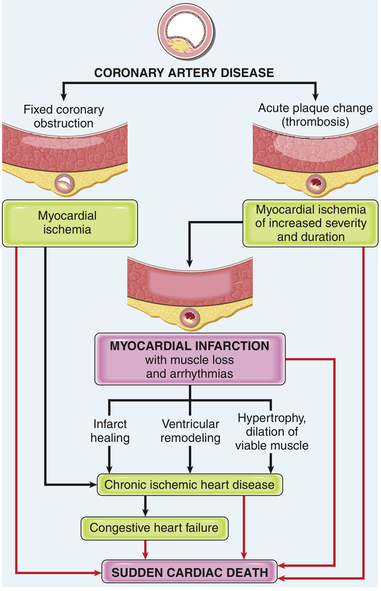
FIG. 9.15 - Robbins Basic Pathology: Pathways in the progression of ischemic heart disease.
Myocardial Infarction (MI)
MI = necrosis of heart muscle from ischemia. The sequence of events:
- Atheromatous plaque erodes or ruptures, exposing subendothelial collagen
- Platelet aggregation + thromboxane A2 release → vasospasm
- Coagulation cascade activation → growing thrombus
- Within minutes, the thrombus completely occludes the coronary lumen
Patterns of infarction depend on which vessel is occluded and whether flow is restored:
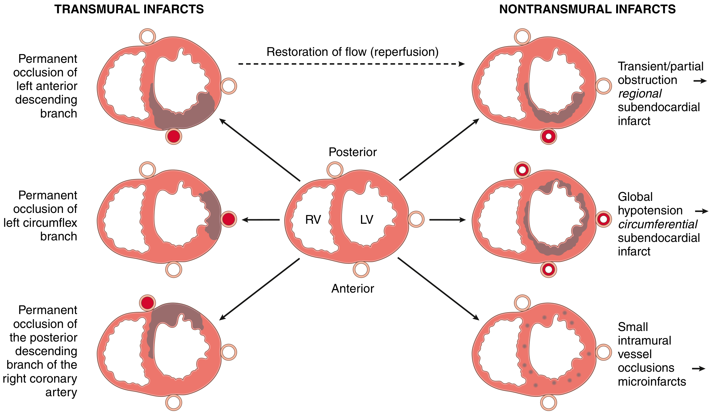
FIG. 9.9 - Robbins Basic Pathology: Dependence of MI on location and nature of diminished perfusion. Left = transmural infarcts from complete occlusion. Right = nontransmural patterns from partial or transient occlusion.
Morphologic evolution of MI (gross and microscopic):
| Time | Gross | Microscopy |
|---|---|---|
| 0-4 hours | None visible | Waviness of fibers at border |
| 4-12 hours | Occasional dark mottling | Coagulation necrosis begins; edema, hemorrhage |
| 12-24 hours | Dark mottling | Coagulation necrosis, pyknosis of nuclei, neutrophilic infiltrate |
| 1-3 days | Yellow-tan center | Ongoing necrosis, dense neutrophil infiltrate |
| 3-7 days | Hyperemic border; soft yellow-tan center | Macrophage phagocytosis at infarct border |
| 7-10 days | Maximally soft, yellow | Granulation tissue with neovascularization |
| 2-8 weeks | Gray-white fibrosis progressing | Scar collagen deposition |
| >2 months | Dense white scar | Dense fibrosis - complete scar |
Gross pathology specimen - acute MI stained with triphenyl tetrazolium chloride (TTC):
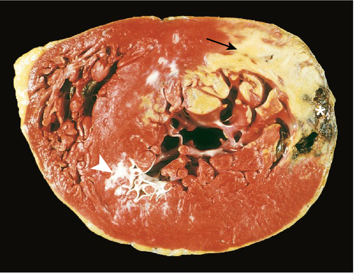
FIG. 9.10 - Robbins Basic Pathology: Acute MI of the posterolateral LV demonstrated by lack of TTC staining in areas of necrosis (arrow = acute infarct, pale/yellow). White arrowhead = old scar from remote infarction. Asterisk = myocardial hemorrhage from ventricular rupture (cause of death).
Complications of MI include:
- Contractile dysfunction - cardiogenic shock
- Papillary muscle rupture → acute mitral regurgitation
- Ventricular wall rupture (3-7 days, peak risk) → cardiac tamponade
- Ventricular septal defect (rupture of necrotic septum)
- Mural thrombus → systemic emboli
- Ventricular aneurysm → paradoxical bulging, arrhythmias
- Arrhythmias → VF is the most common cause of death in the first 24 hours
2. Hypertensive Heart Disease
Sustained hypertension imposes pressure overload on the left ventricle, driving concentric hypertrophy. Key features:
- LV wall thickness >2.0 cm (normal 1.2-1.4 cm)
- Heart weight >500 g (normal ~320-360 g in a 60-70 kg person)
- Microscopy: increased myocyte transverse diameter, "boxcar nuclei" (prominent nuclear enlargement), interstitial fibrosis
- Diastolic dysfunction (stiff LV) precedes systolic dysfunction
- Late-stage: LV dilation and congestive heart failure
Right-sided hypertensive heart disease = cor pulmonale, caused by pulmonary hypertension from diseases like COPD, interstitial fibrosis, or recurrent PE.
3. Valvular Heart Disease
Valves can fail in two ways:
- Stenosis - valve fails to open fully → obstructs forward flow
- Insufficiency / Regurgitation - valve fails to close fully → backflow
Most common causes of acquired valve disease:
| Valve | Common Causes |
|---|---|
| Mitral stenosis | Rheumatic heart disease |
| Mitral regurgitation | MVP, endocarditis, ischemic papillary muscle dysfunction |
| Aortic stenosis | Senile calcification, bicuspid AV, rheumatic disease |
| Aortic regurgitation | Infective endocarditis, Marfan syndrome, rheumatic disease |
Calcific Degenerative Valve Disease (gross pathology):
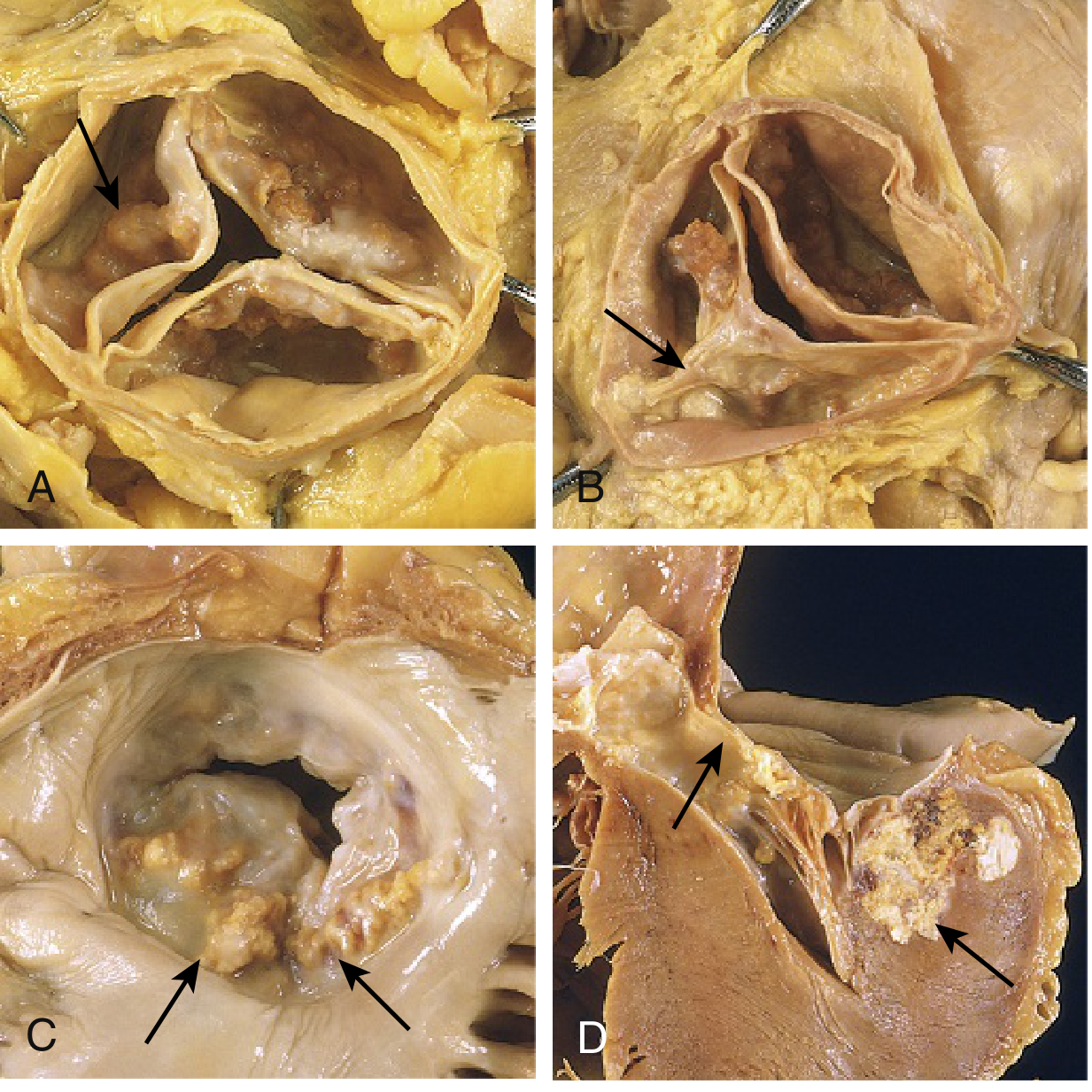
FIG. 9.17 - Robbins Basic Pathology: Calcific valvular degeneration. (A) Aortic stenosis - calcific nodules in sinuses of Valsalva, commissures NOT fused (unlike rheumatic). (B) Bicuspid aortic valve with raphe (arrow). (C-D) Mitral annular calcification, with extension into myocardium (can impinge on the conduction system).
Rheumatic heart disease is caused by cross-reactive antibodies after Group A Streptococcal pharyngitis (molecular mimicry). The mitral valve is most commonly affected. Acute features include:
- Aschoff bodies (foci of fibrinoid necrosis with multinucleated giant cells) - pathognomonic
- Small verrucous vegetations along valve closure line
Chronic changes: leaflet thickening, commissural fusion, chordal shortening → "fish-mouth" or "buttonhole" mitral stenosis orifice.
4. Cardiomyopathies
Cardiomyopathies are diseases of the myocardium itself. There are three major patterns:
Side-by-side comparison of the three types:
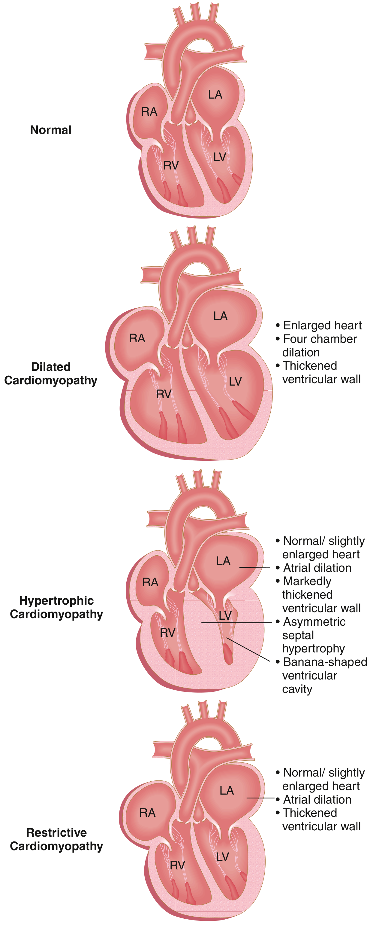
FIG. 9.23 - Robbins Basic Pathology: Three major forms of cardiomyopathy. DCM = systolic dysfunction. HCM and Restrictive = diastolic dysfunction.
Dilated vs Hypertrophic - Causes, Pathology, and Clinical Features:
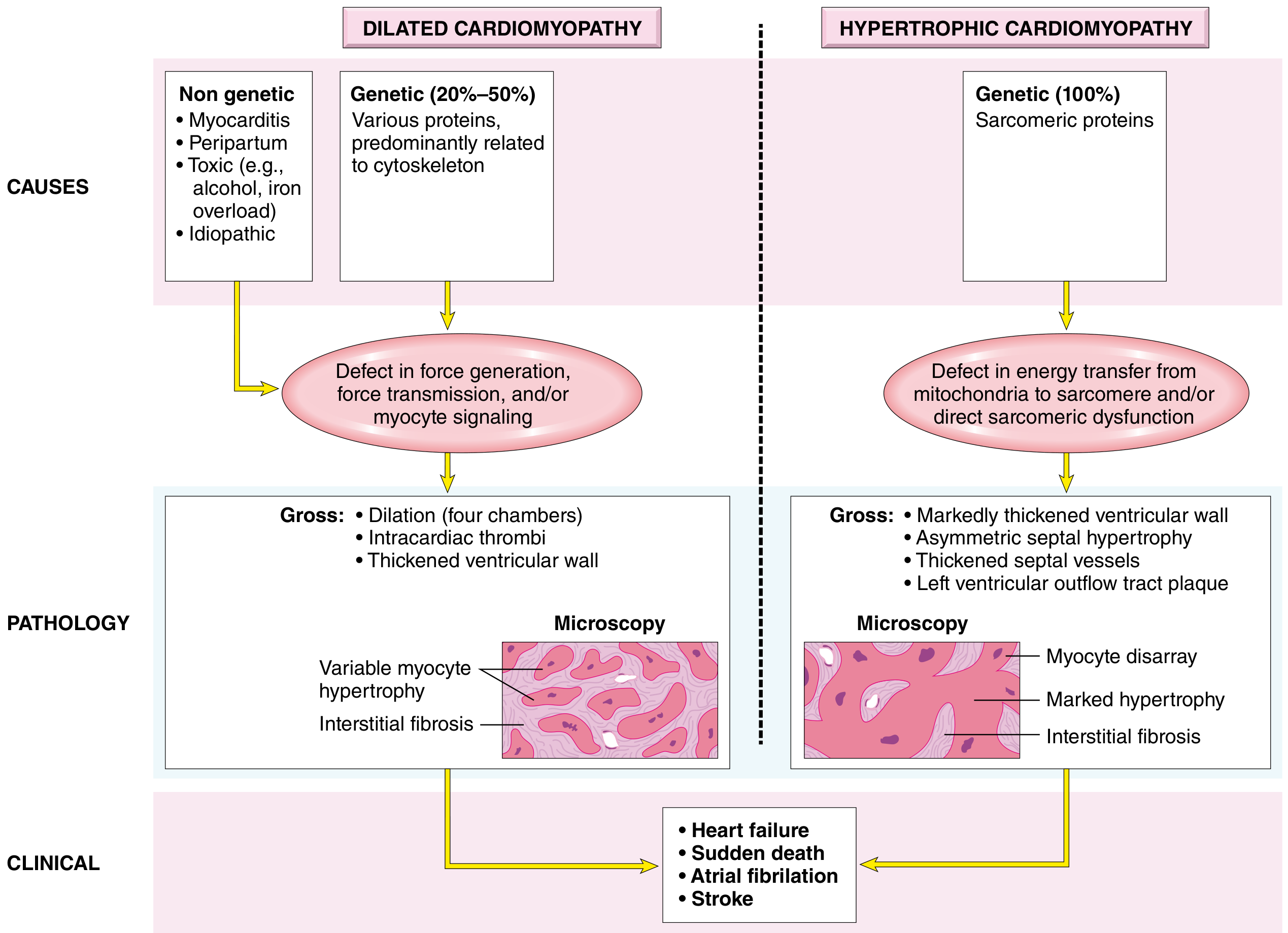
FIG. 12.33B - Robbins Pathologic Basis of Disease: Dilated vs Hypertrophic Cardiomyopathy.
Dilated Cardiomyopathy (DCM)
- Most common (90% of all cardiomyopathies)
- Progressive four-chamber dilation + systolic (contractile) dysfunction
- Causes: genetic (titin, dystrophin, β-myosin mutations), myocarditis (viral), alcohol, peripartum, doxorubicin toxicity
- Gross: flabby, enlarged heart; mural thrombi common → embolic stroke risk
- Micro: variable myocyte hypertrophy + interstitial fibrosis
- Treatment: heart failure therapy; eventual transplant
Hypertrophic Cardiomyopathy (HCM)
- 100% genetic - mutations in sarcomeric proteins (β-MHC, cardiac troponin T, myosin binding protein C)
- Massive asymmetric septal hypertrophy → banana-shaped LV cavity → dynamic outflow obstruction
- Micro: myocyte disarray (chaotically arranged myocytes) - pathognomonic
- Most common cause of sudden cardiac death in young athletes (<35 years)
- Diastolic dysfunction (stiff, non-compliant LV)
Restrictive Cardiomyopathy
- Least common; ventricular walls are stiff, impeding diastolic filling
- Causes: amyloidosis, sarcoidosis, post-radiation fibrosis, hemochromatosis
- Normal/near-normal systolic function; severely impaired diastolic filling
- Bilateral atrial dilation is characteristic
5. Congenital Heart Disease
Congenital heart disease (CHD) affects ~1% of live births. The top 12 lesions account for 85% of all CHD:
| Malformation | Incidence per million live births | % of CHD |
|---|---|---|
| Ventricular septal defect (VSD) | 4482 | 42% |
| Atrial septal defect (ASD) | 1043 | 10% |
| Pulmonary stenosis | 836 | 8% |
| Patent ductus arteriosus (PDA) | 781 | 7% |
| Tetralogy of Fallot | 577 | 5% |
| Coarctation of aorta | 492 | 5% |
| AV septal defect | 396 | 4% |
| Aortic stenosis | 388 | 4% |
| Transposition of great arteries | 388 | 4% |
Key Lesions Explained
Left-to-right shunts (acyanotic initially; may become cyanotic late via Eisenmenger syndrome):
- VSD - most common; harsh holosystolic murmur; spontaneous closure common in small VSDs
- ASD - fixed split S2; pulmonary overcirculation; thromboembolism risk
- PDA - continuous "machinery" murmur; closes with indomethacin (inhibits prostaglandin)
Right-to-left shunts (cyanotic from birth):
- Tetralogy of Fallot (TOF): 4 components - (1) VSD, (2) pulmonary stenosis, (3) aorta overriding the VSD, (4) right ventricular hypertrophy. Classic "boot-shaped heart" on CXR. Presents with cyanosis, "tet spells," squatting (increases SVR).
- Transposition of the great arteries (TGA): Aorta arises from RV, pulmonary artery from LV → two parallel circuits incompatible with life unless ASD/VSD/PDA exists. "Egg on a string" CXR.
Obstructive lesions:
- Coarctation of the aorta: Segmental narrowing, usually postductal (near ligamentum arteriosum). Presents with upper-body hypertension, weak femoral pulses, rib notching on CXR from dilated intercostal collaterals.
6. Myocarditis
Myocarditis = inflammatory disease of the myocardium.
- Most common cause: Viral (Coxsackievirus B, parvovirus B-19, HIV, COVID-19)
- Chagas disease (Trypanosoma cruzi) - most common cause worldwide in South America
- Autoimmune: giant cell myocarditis (rapidly fatal without transplant), sarcoidosis
- Presents with: sudden onset heart failure in a previously healthy young person, chest pain mimicking MI, arrhythmias
- Diagnosis: cardiac MRI (late gadolinium enhancement) + endomyocardial biopsy
- Dallas criteria (biopsy): myocyte necrosis + adjacent inflammatory infiltrate
Summary Table
| Category | Hallmark Pathology | Key Feature |
|---|---|---|
| Ischemic / MI | Coagulation necrosis → scar | Pale/yellow area on TTC stain |
| Hypertensive | LV concentric hypertrophy | Boxcar nuclei, wall >2 cm |
| Valvular (calcific) | Calcium in cusps/annulus | Aortic > mitral; commissures not fused |
| Valvular (rheumatic) | Aschoff bodies, commissural fusion | "Fish-mouth" mitral orifice |
| DCM | 4-chamber dilation, interstitial fibrosis | Systolic dysfunction |
| HCM | Asymmetric septal hypertrophy, myocyte disarray | #1 cause SCD in young athletes |
| Restrictive | Stiff ventricle, atrial dilation | Amyloidosis most common cause |
| TOF | VSD + PS + overriding aorta + RVH | Cyanosis, tet spells, boot-shaped heart |
| Myocarditis | Myocyte necrosis + inflammation | Coxsackie B virus most common |
Sources: Robbins & Kumar Basic Pathology (9780323790185) and Robbins, Cotran & Kumar Pathologic Basis of Disease (9780443264528)
This is a shared conversation. Sign in to Orris to start your own chat.